Polarization - linearly polarized light
Microscopeparts and functions
Eyepiece design and construction have evolved over time to improve the quality and comfort of the viewing experience. Modern eyepieces are typically designed with multiple lens elements to minimize aberrations and distortions, resulting in a clearer and more accurate image. Some eyepieces also incorporate advanced coatings to reduce glare and improve contrast.

Function ofstagein microscope
The eyepiece on a microscope, also known as an ocular lens, is the part of the microscope that is looked through to view the magnified specimen. It is located at the top of the microscope and is the lens closest to the eye of the observer. The eyepiece is designed to magnify the image produced by the objective lens, which is the lens closest to the specimen being observed.
Glasses as listed above except clear tint. Clear tint gives excellent visibility but provides less blue light hazard protection than amber glasses above.
2. Ramsden eyepiece: This design features two plano-convex lenses with the convex sides facing away from each other. It offers a wider field of view compared to the Huygenian eyepiece and is commonly used in modern microscopes.
Function ofbasein microscope
Function ofnosepiecein microscope
For patient use during UV treatment; provides good visibility. To prevent cross-contamination, many clinics purchase these goggles and require that each patient purchase a set. Amber colour allows viewing of red timer displays found on many devices and efficiently blocks ultraviolet (200-400 nm) and blue light (400-500 nm).
In addition to magnification, the eyepiece also helps to focus the light rays coming from the objective lens and to direct them into the viewer's eye. This helps to create a clear and sharp image of the specimen under observation. The eyepiece also often contains a reticle or a graticule, which is a grid or scale that can be used to measure the size or dimensions of the specimen.
1. Huygenian eyepiece: This is a simple eyepiece design that consists of two plano-convex lenses with the convex sides facing each other. It provides a relatively narrow field of view and is commonly used in older microscopes.
The latest point of view on eyepiece design emphasizes the importance of ergonomic design to reduce eye strain and improve user comfort during extended periods of use. This includes features such as adjustable eye relief and eyecups to accommodate different users and provide a more comfortable viewing experience. Additionally, advancements in materials and manufacturing techniques have allowed for the production of lightweight yet durable eyepieces that are well-suited for various applications.
Function ofbody tubein microscope
The eyepiece on a microscope, also known as the ocular lens, is the lens at the top of the microscope through which the viewer looks. It is the lens closest to the eye when using the microscope. The primary function of the eyepiece is to magnify the image produced by the objective lens, which is the lens closest to the object being observed. This magnification allows the viewer to see a larger and more detailed image of the specimen.
Structure andfunction of eyepiece in microscope
3. Wide-field eyepiece: This type of eyepiece is designed to provide a larger and more comfortable viewing area, allowing the viewer to see more of the specimen at once. It is particularly useful for applications that require prolonged observation.
From a modern perspective, the eyepiece on a microscope may also be designed to reduce eye strain and provide a comfortable viewing experience. Some eyepieces are equipped with adjustable diopter settings to accommodate individual differences in vision, and others may incorporate anti-glare or anti-reflection coatings to improve image clarity.
What iseyepiece in microscope
Function ofarmin microscope

An eyepiece on a microscope, also known as an ocular lens, is the lens at the top of the microscope that you look through to view the specimen. It is the part of the microscope that is closest to your eye and is responsible for magnifying the image of the specimen. The eyepiece typically contains a set of lenses that work together to magnify the image produced by the objective lens, which is the lens closest to the specimen.
The magnification power of the eyepiece is a measure of how much the image is enlarged when viewed through the microscope. This is usually expressed as a number followed by an "x" (e.g., 10x, 20x), which indicates the number of times the image is magnified. For example, if the eyepiece has a magnification power of 10x and the objective lens has a magnification power of 40x, the total magnification of the microscope would be 400x (10x multiplied by 40x).
In summary, the eyepiece on a microscope is a crucial component that contributes to the overall quality of the viewing experience. Its design and construction have evolved to prioritize optical performance, user comfort, and versatility, making it an essential part of modern microscopy.
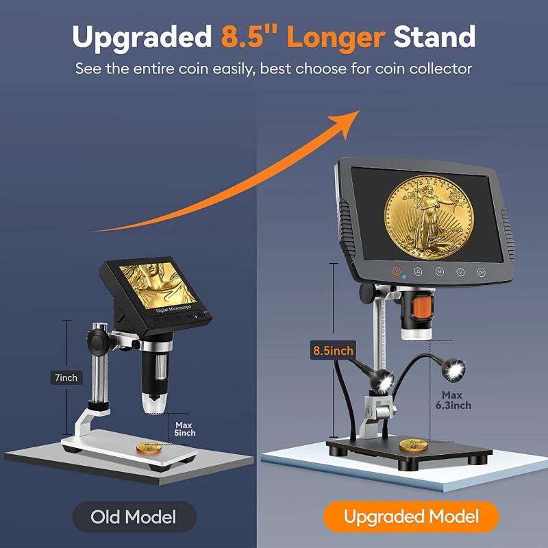
In recent years, there has been a growing interest in digital eyepieces, which incorporate digital imaging technology to capture and display the magnified image on a computer or other digital device. This allows for easier sharing of images and facilitates analysis and documentation of the specimens. Additionally, there has been a focus on ergonomic designs to improve user comfort and reduce eye strain during prolonged use. These advancements aim to enhance the overall microscopy experience and make it more accessible to a wider range of users.
An eyepiece on a microscope is a lens that is positioned at the top of the microscope and is used to view the magnified image of the specimen. It is also known as an ocular lens and is an essential component of the microscope's optical system. The eyepiece typically contains a set of lenses that further magnify the image produced by the objective lens, allowing the viewer to see a highly detailed and enlarged image of the specimen.
The headband and nosepiece are adjustable. The headband is made from white FDA rubber (non-latex). Includes clear storage tube (lower picture) so patient can easily carry in a jacket or purse. Good for an unlimited number of uses providing that they are not exposed to solvents or other damaging chemicals. These are the same goggles supplied with our SolRx devices. UL listed. Made in USA. These phototherapy goggles meet the following standards:
From the latest point of view, advancements in microscope technology have led to the development of eyepieces with variable magnification power, allowing users to adjust the level of magnification based on their specific needs. Additionally, some modern microscopes are equipped with digital eyepieces that can capture and display images on a computer screen, enabling users to easily share and analyze the microscopic images. These digital eyepieces often come with software that allows for further image enhancement and analysis, expanding the capabilities of traditional eyepieces.

Overall, the eyepiece on a microscope plays a crucial role in magnifying and enhancing the image of the specimen, as well as providing a comfortable and effective viewing experience for the user.
The eyepiece, also known as the ocular lens, is the lens at the top of the microscope that you look through to view the specimen. It typically contains a magnifying lens that further enlarges the image produced by the objective lens. The eyepiece is usually removable and interchangeable, allowing for different magnifications to be achieved depending on the specific needs of the user.
For use by phototherapy staff only. Not recommended for patient use during treatment because they do not fully seal to the face – use patient goggles instead. Amber tint provides improved blue light hazard protection and will fit over most prescription glasses. Meets ANSI Z136.1 Laser Protection Standard and ANSI Z87.1 Impact Standard. Supplied with black plastic storage sleeve shown. These glasses are also known as “UV Shields”. Made in USA.


 Ms.Cici
 8618319014500