
FC Connectors and Adapters | OEM Optical ... - fc fc connector
Not all products or services are approved or offered in every market, and approved labelling and instructions may vary between countries. Please contact your local representative for further information.
Leica microscopes offer phase contrast for the study of cells or tissues concerning various life-science and forensic applications. Phase contrast can also be useful for certain material and earth-science applications.
Typesof microscope objectives
Microscopes are usually complex assemblies that include an array of lenses, filters, polarizers, and beamsplitters. Illumination is arranged to provide enough light for a clear image, and sensors are used to ‘see’ the object.
Choosing the right microscope objective is pivotal for optimal imaging performance. Consider your specific application requirements, utilize the provided guide, and explore Avantier’s diverse objective offerings to ensure accurate and reliable results in your microscopy endeavors.
In the following content, we delve intensively into the various components and features of microscope objective lenses, exploring their construction, functionality, and specialized designs that enable researchers to gain deeper insights into the microscopic world.
A phase contrast microscope is similar to a conventional brightfield microscope, except it uses an annular aperture in front of the light source and a quarter-wave phase plate after the objective lens. For more information, refer to the article: Phase Contrast
Numerical aperture, magnification, optical tube length, degree of aberration correction, and other important characteristics are typically imprinted or engraved on the external portion of the barrel for easy reference. These specifications help researchers select the appropriate objective for their experiments, ensuring optimal performance and total magnification when combined with the ocular lens. Specifications like numerical aperture and magnification are typically labeled on the barrel for easy reference. These lenses are indispensable in scientific research providing high powered optics essential for research.
In modern microscopes, neither the eyepiece nor the microscope objective is a simple lens. Instead, a combination of carefully chosen optical components work together to create a high quality magnified image. A basic compound microscope can magnify up to about 1000x. If you need higher magnification, you may wish to use an electron microscope, which can magnify up to a million times.

For brightfield illumination to be effective, there needs to be a variation in opacity across the object. Without this variation, the illumination creates a dark blur around the object, resulting in an image with relative contrast between the object’s parts and the light source. Typically, brightfield illumination allows clear visualization of each part of the object unless it is extremely transparent. In cases where transparency hinders feature distinction, darkfield illumination becomes useful.
Both the objective lens and the eyepiece also contribute to the overall magnification of the system. If an objective lens magnifies the object by 10x and the eyepiece by 2x, the microscope will magnify the object by 20 times. If the microscope lens magnifies the object by 10x and the eyepiece by 10x, the microscope will magnify the object by 100x. This multiplicative relationship is the key to the power of microscopes, and the prime reason they perform so much better than simply magnifying glasses.
Function ofstagein microscope
Lasers find widespread applications, commonly employed to either (1) heat material onto a base or (2) ablate material off of a base. Laser ablation systems necessitate the integration of microscope components due to the precise manipulation of the laser beam, including focusing, bending, and reducing scattering. Typically, a laser ablation setup incorporates custom optics instead of off-the-shelf components, with the laser intricately designed into the system, as illustrated in Figure 14. The laser is strategically oriented in an epi-illumination design to leverage the microscope objective’s capacity to focus light at the object plane, generating exceptionally small spot sizes with minimal aberrations. Additionally, an eyepiece enables the user to visually locate the laser and ensure proper functionality. Filters are indispensable in shielding the user’s eyes from potential laser damage. Laser ablation setups, known for their superior precision compared to traditional surgical methods, find applications in medical and biological contexts.
The majority of microscope objective specifications are conveniently displayed on the objective’s body, including information such as the objective design/standard, magnification, numerical aperture, working distance, lens to image distance, and cover slip thickness correction. Refer to Figure 5 for guidance on interpreting microscope objective specifications. This direct placement of specifications on the objective facilitates a clear understanding of its characteristics, a crucial aspect when integrating multiple objectives into an application. Any additional specifications, like focal length, field of view (FOV), and design wavelength, can be readily calculated or obtained from the vendor or manufacturer’s provided specifications.
There are two major specifications for a microscope: the magnification power and the resolution. The magnification tells us how much larger the image is made to appear. The resolution tells us how far away two points must be to be distinguishable. The smaller the resolution, the larger the resolving power of the microscope. The highest resolution you can get with a light microscope is 0.2 um, but this depends on the quality of both the objective and eyepiece.
The phase contrast method for microscopy was developed in the 1930s by the Dutch physicist Frits Zernike. After 1942, it became a widely used microscopy technique. In 1953, Zernike was awarded the Nobel Prize for Physics. For more details, refer to the articles: A Brief History of Light Microscopy – From the Medieval Reading Stone to Super-Resolution & Phase Contrast
Leica microscopes providing phase contrast are commonly used in life science research for the visualization, analysis, and documentation of biological structures and cellular processes.
Infrared microscopy, alternatively referred to as infrared microspectroscopy, is a form of light microscopy that employs a light source transmitting infrared wavelengths to observe a sample’s image. In contrast to conventional optical microscopes utilizing absorbent glass optics, an infrared microscope incorporates reflective optics, enabling it to encompass the complete spectral range of infrared light.
What isobjective lensin microscope
Leica microscopes capable of phase contrast make a difference for the study of transparent and colorless minerals, crystals, and polymers.
Fluorescence microscopy is a powerful imaging technique used primarily in biomedical research to visualize and study samples labeled with fluorescent dyes or proteins at the microscopic level. The method relies on the phenomenon of fluorescence, where materials absorb light at a specific wavelength (excitation light) and then emit light at a longer wavelength (emission wavelength). A focused light source, such as a laser, is used to selectively excite fluorescent molecules within the sample. The emitted fluorescence is captured to form detailed images, providing valuable information about the sample’s internal structure and composition.
In many microscopes, backlight illumination is favored over traditional direct light illumination due to the latter’s tendency to over-saturate the object under inspection. One specific backlight illumination technique employed in microscopy is Koehler illumination. This method involves flooding the object with light from behind using incident light from a source like a light bulb (see Figure 2). Koehler illumination utilizes two convex lenses, the collector lens and the condenser lens(or called field lens) , to ensure even and bright illumination on both the object and image planes. This design prevents imaging the light bulb filament, a common issue with direct light illumination. Backlight illumination is also commonly referred to as brightfield illumination.
Darkfield illumination directs light rays obliquely onto the object, avoiding direct entry into the objective. Despite this oblique angle, the rays still illuminate the object plane. The resulting darkfield illumination image achieves high contrast between the transparent object and the light source. In a darkfield setup, a light source forms an inverted cone of light that blocks central rays but allows oblique rays to illuminate the object (see Figure 3). This design effectively forces light to illuminate the object without entering the optical system, making darkfield illumination particularly suitable for transparent objects. In contrast, no rays are blocked in a brightfield illumination setup.
This is the most common method, but if you focus on the center of the field of view, the periphery becomes blurred, so it is not suitable for inspection photography.
A microscope is an optical device designed to magnify the image of an object, enabling details indiscernible to the human eye to be differentiated. A microscope may project the image onto the human eye or onto a camera or video device.
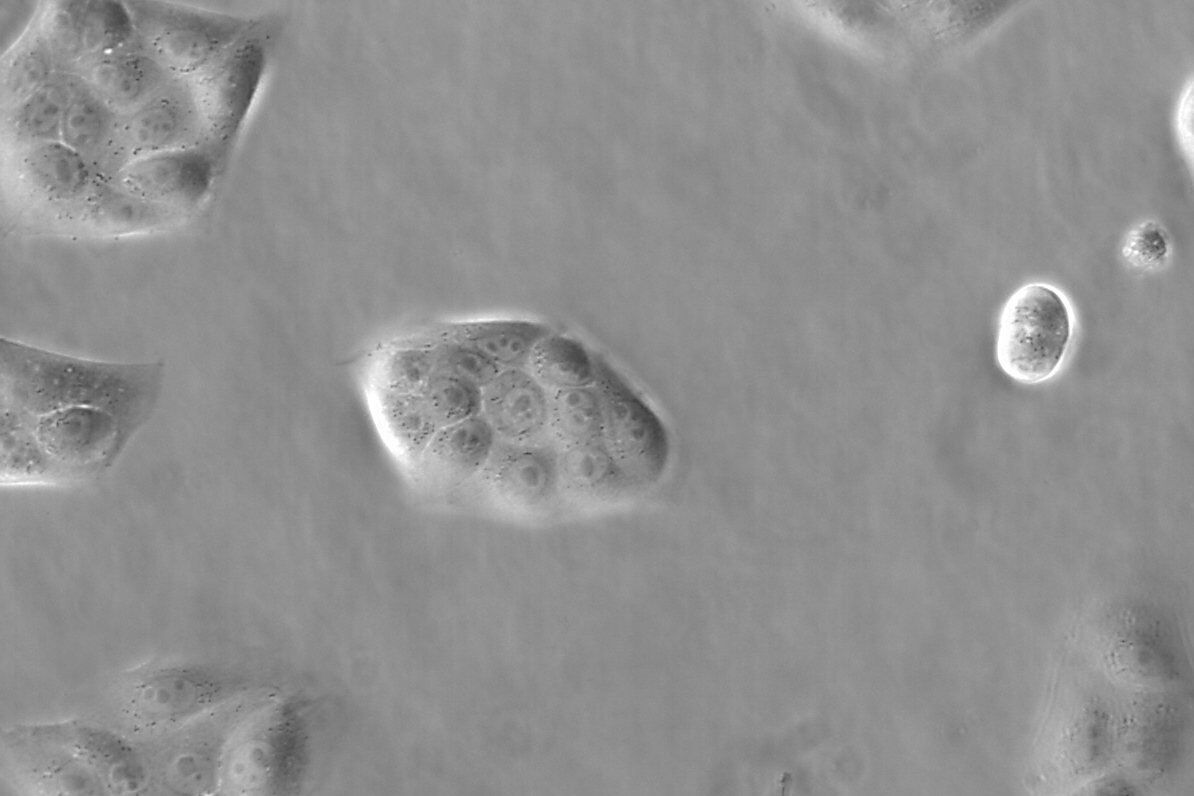
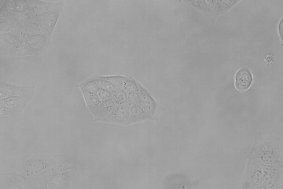
Brightfield microscopy normally only provides a low-contrast image of many transparent biological specimens where few details are distinguished. One way to enhance contrast with brightfield microscopy is to use selective stains, but such stains are often toxic to living cells. A phase contrast light microscope offers a way to view the structures of many types of biological specimens in greater contrast without the need of stains. The contrast method exploits differences in optical density between structures of a specimen that lead to a phase shift of the light that interacts with the specimen and its structures.
Microscope objectives are pivotal components in optical microscopy, especially in influencing image quality and resolution. Selecting the right objective is crucial for achieving optimal results in your microscopy applications. To guide you through the selection process, consider the following factors:
Confocal microscopy offers the capability to capture sharp images from a slender slice of a dense sample, minimizing background noise and reducing out-of-focus disturbances. Optical sectioning, widely employed in biomedical science and materials science, involves placing a sample on the microscope stage. An image is initially acquired at the primary focal plane, and subsequently, the stage or objective is adjusted vertically to capture images at successive focal planes.
Function ofcondenserin microscope
It is suitable for inspection photography because it focuses not only on the center of the field of view but also on the periphery, producing a flat image.
A phase contrast microscope is similar to a conventional widefield microscope, except it uses an aperture in the shape of an annulus and a quarter-wave (λ/4) phase plate. The annular aperture is placed between the light source and condenser lens and the phase plate after the objective inside the microscope optics. Ring-shaped light that passes through the aperture is focused by the condenser onto the biological specimen to be observed.
Objective lensfunction
Adding to these features, long working distance objectives allow ample space between the lens and the specimen, facilitating the manipulation of samples without compromising image quality. Infinity correction objectives utilize infinity-corrected optical systems, providing flexibility and compatibility with various microscopy accessories.
Stage clipsmicroscope function
If you’re interested in acquiring in-stock microscope objective lenses, please visit our ‘Stock – Microscope Objective‘ page.
The knowledge portal of Leica Microsystems offers scientific research and teaching material on the subjects of microscopy. The content is designed to support beginners, experienced practitioners and scientists alike in their everyday work and experiments.
High power objectivemicroscope function
Portions of the ring-shaped light are diffracted by optically dense structures of the specimen and experience a negative phase shift of about λ/4. This phase-shifted, diffracted light bypasses the λ/4 plate. In contrast, the portion of the ring-shaped light that passes directly through the specimen non-deviated will hit the phase plate which causes a positive λ/4 phase shift. As the total difference in phase shift between the light diffracted by the specimen’s structures and that which passes through phase plate will be about λ/2, destructive interference will occur. Consequently, more optically dense structures will appear darker than those that are less optically dense.
Epi-illumination, a third form of illumination employed in microscopy, generates light from above the objective. This setup replaces the need for a Koehler illumination configuration, as both the objective and the epi-illumination source contribute to the illumination process. The compact structure of epi-illumination is a significant advantage, as the objective serves as a primary source for a considerable portion of the illumination. Figure 4 provides a depiction of a frequently used epi-illumination setup, particularly common in fluorescence applications.
Although today’s microscopes are usually far more powerful than the microscopes used historically, they are used for much the same purpose: viewing objects that would otherwise be indiscernible to the human eye. Here we’ll start with a basic compound microscope and go on to explore the components and function of larger more complex microscopes. We’ll also take an in-depth look at one of the key parts of a microscope, the objective lens.
Low power objectivemicroscope function
Microscope objective lenses, vital optical elements in microscopy, enable precise observation of specimens. Objective lens manufacturers offer a wide range of objective designs for specific needs: high power for detailed observation, scanning for broader views, oil immersion for high-resolution imaging, and long working distance for manipulation without compromising quality. Those objectives are designed with advanced construction techniques for high performance objectives with a spring loaded retractable nose cone assembly that protects the front lens elements and the specimen from collision damage.
The chromatic aberration of the three wavelengths, with a slight chromatic aberration remaining in the purple, and the curvature of the field have been corrected. Also called fluorite.
Most often biological specimens and tissues are observed with a phase contrast microscope. A large variety of biological specimens can be observed with phase contrast from fixed specimens to living cells and tissues. For examples, refer to the articles: Phase Contrast & Optical Contrast Methods
For forensic applications concerning the evidentiary investigation of paints, pigments, textiles, fibers, and human tissues, Leica microscopes offering phase contrast are very useful solutions.
Avantier is a premier manufacturer of high performance microscope objective lenses, and we produce a wide range of quality microscope objectives for applications ranging from research to industry to forensics and medical diagnostics. We carry many types of objectives in stock, including apochromat objectives, achromatic objectives, and semi apochromat objectives. We can also produce custom objectives designed to work as desired in your target spectral range.
A basic compound microscope could consist of just two elements acting in relay, the objective and the eyepiece. The objective relays a real image to the eyepiece, while magnifying that image anywhere from 4-100x. The eyepiece magnifies the real image received typically by another 10x, and conveys a virtual image to the sensor.
In terms of performance, it is positioned between the plan achromat objective lens and the plan apochromat objective lens. High Grade type.
VIETNAM:Alpha Industrial Park, Tu ThonVillage, Yen My District, HungYen Province 17721+84 221-730-8668sales-vn@avantierinc.com
While a magnifying glass consists of just one lens element and can magnify any element placed within its focal length, a compound lens, by definition, contains multiple lens elements. A relay lens system is used to convey the image of the object to the eye or, in some cases, to camera and video sensors.
Phase contrast is an optical contrast technique for microscopy which makes unstained structures in the cells of biological specimens visible. Cell structures that appear transparent with brightfield illumination can be viewed in high contrast and rich detail using phase contrast. Differences in optical density between structures in the cell can cause light that interacts with them to attain a phase shift. This phenomenon is the basis of phase contrast. As a result, more optically dense structures will look darker than less optically dense ones.


 Ms.Cici
 8618319014500