
Setting up allied vision Alvium 1500 C - 500c embedded ... - alvium
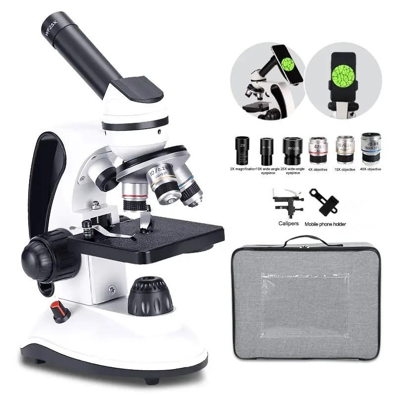
The magnification power of the objective lens is typically higher than that of the eyepiece. Objective lenses are available in various magnification powers, such as 4x, 10x, 40x, and 100x. The eyepiece usually has a fixed magnification power of 10x. Therefore, the total magnification of a microscope can range from 40x (4x objective lens multiplied by 10x eyepiece) to 1000x (100x objective lens multiplied by 10x eyepiece).
Nov 10, 2024 — Synonyms for MAGNIFYING: exaggerating, enhancing, padding, coloring, expanding, embellishing, embroidering, hyperbolizing; ...
In recent years, there have been significant advancements in electron microscopy techniques, leading to even higher magnification powers and improved resolution. For example, the development of aberration-corrected electron microscopy has allowed for the visualization of individual atoms and atomic-scale defects in materials. Additionally, the introduction of cryo-electron microscopy has revolutionized the field of structural biology, enabling the determination of high-resolution structures of biological macromolecules.
What is the function ofstage clip inmicroscope
Digital magnification is achieved by capturing an image of the specimen using a digital camera attached to the microscope. The image is then processed and enlarged using software algorithms. This allows for further magnification beyond the limitations of the physical lenses. The level of digital magnification depends on the resolution of the camera and the software used for image processing.
The condenser has an iris diaphragm that controls the angle of the beam of light focused onto the specimen. The iris diaphram is an adjustable shutter which allows you to adjust the amount of light passing through the condenser. The angle determines the Numerical Aperture (NA) of the condenser. This diaphragm, generally called the aperture diaphragm, is one of the most important controls on the microscope. Cover slip, Most objectives are designed for use with a cover slip between the objective and the specimen. The cover slip becomes part of the optical system, and its thickness is critical for optimal perfomance of the objective. The cover slip thickness designation on most objective lenses is 0.17 mm or 170 microns.
One of the main advantages of electron microscopes is their ability to provide detailed images of structures that are too small to be resolved by optical microscopes. This has revolutionized various scientific fields, including materials science, biology, and nanotechnology. Electron microscopes have been instrumental in advancing our understanding of the atomic and molecular structures of materials, the intricate details of cellular components, and the behavior of nanoparticles.
Perfect Vision Magnifying Lamp Illuminate your passion! The Perfect Vision Magnifying Lamp is ideal for embroidery, sewing, crafting, and more.
In conclusion, the magnification power of a microscope is determined by the product of the objective lens and eyepiece magnifications. The total magnification determines the level of enlargement and detail that can be achieved during observation. However, it is important to consider other factors such as resolution and numerical aperture to obtain a clear and detailed image. With advancements in technology, microscopes with higher magnification powers have been developed, enabling scientists to explore the microscopic world in greater detail.
What is the Function ofilluminator inmicroscope
Microscope Anatomy & Function Glossary Back to Quicktime VR Microscope A | B | C | D | E | F | G | H | I | J | K | L | M | N | O | P | Q | R | S | T | U | V | W | X | Y | Z A Back to top B Back to top Base, The base is the foundation on which the microscope stand is built. It is important that the base is relatively large, stable, and massive. When you are setting up a microscope for the first time ensure that the surface on which it is placed is level. C Back to top Condenser, The condenser under the stage focuses the light on the specimen, adjusts the amount of light on the specimen, and shapes the cone of light entering the objective. One way to think about the condenser is as a light "pump" that concentrates light onto the specimen. The condenser has an iris diaphragm that controls the angle of the beam of light focused onto the specimen. The iris diaphram is an adjustable shutter which allows you to adjust the amount of light passing through the condenser. The angle determines the Numerical Aperture (NA) of the condenser. This diaphragm, generally called the aperture diaphragm, is one of the most important controls on the microscope. Cover slip, Most objectives are designed for use with a cover slip between the objective and the specimen. The cover slip becomes part of the optical system, and its thickness is critical for optimal perfomance of the objective. The cover slip thickness designation on most objective lenses is 0.17 mm or 170 microns. D Back to top E Back to top F Back to top Focus (coarse), The coarse focus knob is used to bring the specimen into approximate or near focus. Focus (fine), Use the fine focus knob to sharpen the focus quality of the image after it has been brought into focus with the coarse focus knob. G Back to top H Back to top I Back to top Illuminator, There is an illuminator built into the base of most microscopes. The purpose of the illuminator is to provide even, high intensity light at the place of the field aperture, so that light can travel through the condensor to the specimen. J Back to top K Back to top L Back to top M Back to top Magnification, The degree to which the image of the specimen is enlarged by the objective. For example, 40 specifies 40 times (40x) the actual size of the specimen. As magnification increases, resolution (NA) must also increase so that more information can be obtained. Magnification without increased resolution yields no additional information and is called "empty magnification." N Back to top Numerical Aperture (NA), The maximum angle from which it can accept light. Lenses that accept light from higher angles have greater resolving power, thus NA defines resolving power. The maximum NA of objectives is 1.4, and it is limited by the physics of light and the refractive index of glass. O Back to top Objective Lens, The objective lens is the single most important component of the microscope. Together with the condenser, it determines the resolution that the microscope's capability. Learning how to use the correct objective for a particular application is a prerequisite for good microscopy. Important information describing the objective lens is engraved on the side of its barrel. This is the best performance the objective is capable of and it will only yield this performance when used properly. Ocular Lenses, The ocular lenses are the lens closest to the eye and usually have a 10x magnification. Since light microscopes use binocular lenses there is a lens for each eye. It is important to adjust the distance between the microscope oculars, so that it matches your interpupillary distance. This will yield better image quality and reduce eye strain. P Back to top Plan, There are many different kinds of objective lenses. Common designations include "plan" for flat field, "achromat" for partially color-corrected, and "apochromat" for highly color corrected. These designations may become combined as in "plan achromat." Parfocal, The specimen is focused for all objectives if it is focused for one objective. In other words, once the specimen is focused under one objective it will be in approximate focus under other objectives. Q Back to top R Back to top S Back to top Stage, The stage is the platform that supports the specimen. It is usually quite large to minimize vibration and it attaches to the microscope stand. The stage has an opening for the illuminating beam of light to pass through. A spring loaded clip holds the specimen slide in place on the stage. Other types of stage clips are designed for use with petri-dishes, multiwell plates, or other specialized chambers. Most stages have a rack and pinion mechanism that can move the specimen slide in two perpendicular (X - Y) directions. On many microscopes, stage movement is controlled using two concentric knobs located to the side or below the stage. Stand, The stand is the basic structure of the microscope to which everything is attached. The stand, also known as the arm, is the part of the microscope that you grab to transport the microscope. T Back to top Tube, the tube houses many of the optical components of the microscope. The optical tube length of most biomedical microscopes is 160 millimeters but tube geometry varies considerably due to relay lenses and proprietary design features. In most modern microscopes the tube is folded to make the microscope easier to use. Early microscopes had straight tubes such as this model built by Robert Hooke in the mid 17th century. Tube length, describes the optical tube length for which the objective was designed. This is 160 mm (6.3 inches) for modern biomedical microscopes. Turret, Most microscopes have several objective lenses mounted on a rotating turret to facilitate changing lenses. An audible click identifies the correct position for each lens as it swings into place. When the turret is rotated, it should be grasped by the ring around its edge, and not by the objectives. Using the objectives as handles can de-center and possibly damage them. U Back to top V Back to top W Back to top X Back to top Y Back to top Z Back to top Back to Quicktime VR Microscope
The magnification power of a microscope refers to its ability to enlarge an object or specimen for detailed observation. Traditionally, magnification power was determined by the combination of the objective lens and the eyepiece lens. However, with the advent of digital technology, microscopes now offer additional magnification capabilities through image processing and zooming capabilities.
In conclusion, the magnification power of a microscope has been enhanced through digital technology. Digital magnification, achieved through image processing and zooming capabilities, provides researchers with the ability to observe and analyze specimens at a higher level of detail. With ongoing advancements in digital imaging, the magnification power of microscopes continues to improve, enabling scientists to push the boundaries of scientific discovery.
It is important to note that the total magnification is not the only factor that determines the quality of the image. Other factors, such as the numerical aperture of the lenses, the resolution of the microscope, and the quality of the optics, also play a significant role in achieving a clear and detailed image.
Parfocal, The specimen is focused for all objectives if it is focused for one objective. In other words, once the specimen is focused under one objective it will be in approximate focus under other objectives.
B Back to top Base, The base is the foundation on which the microscope stand is built. It is important that the base is relatively large, stable, and massive. When you are setting up a microscope for the first time ensure that the surface on which it is placed is level.
What is the function ofpillar inmicroscope
P Back to top Plan, There are many different kinds of objective lenses. Common designations include "plan" for flat field, "achromat" for partially color-corrected, and "apochromat" for highly color corrected. These designations may become combined as in "plan achromat."
S Back to top Stage, The stage is the platform that supports the specimen. It is usually quite large to minimize vibration and it attaches to the microscope stand. The stage has an opening for the illuminating beam of light to pass through. A spring loaded clip holds the specimen slide in place on the stage. Other types of stage clips are designed for use with petri-dishes, multiwell plates, or other specialized chambers. Most stages have a rack and pinion mechanism that can move the specimen slide in two perpendicular (X - Y) directions. On many microscopes, stage movement is controlled using two concentric knobs located to the side or below the stage. Stand, The stand is the basic structure of the microscope to which everything is attached. The stand, also known as the arm, is the part of the microscope that you grab to transport the microscope.
What is the function ofbrightness adjustment inmicroscope
Custom ADA Window Sign (1 Line/3 Windows), Economy series, 10x10". Our Price ... All window signs feature a rectangular "window" area that allows you to ...
A | B | C | D | E | F | G | H | I | J | K | L | M | N | O | P | Q | R | S | T | U | V | W | X | Y | Z
The magnification power of a microscope refers to the degree to which an object can be enlarged and observed under the microscope. In the case of an electron microscope, the magnification power is significantly higher than that of an optical microscope.
Electron microscopes utilize a beam of electrons instead of light to magnify the specimen. This allows for much higher magnification and resolution, enabling scientists to observe objects at the nanoscale level. The magnification power of an electron microscope can range from a few hundred times to several million times, depending on the specific type of electron microscope and the techniques used.
In recent years, there have been advancements in microscope technology that have improved the numerical aperture and, consequently, the resolving power and light-gathering ability of microscopes. For example, the development of high numerical aperture objectives and the use of techniques such as confocal microscopy and super-resolution microscopy have allowed scientists to observe structures and processes at a much finer scale.
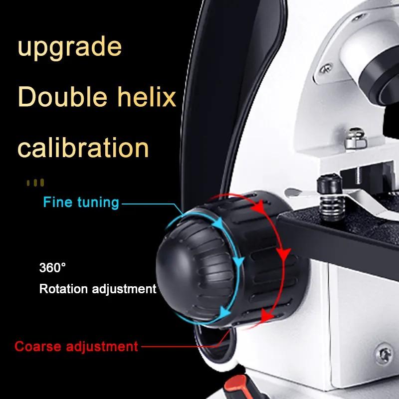
F Back to top Focus (coarse), The coarse focus knob is used to bring the specimen into approximate or near focus. Focus (fine), Use the fine focus knob to sharpen the focus quality of the image after it has been brought into focus with the coarse focus knob.
The light-gathering ability of the lens is also determined by the numerical aperture. A higher numerical aperture means that the lens can gather more light, resulting in a brighter image. This is particularly important when observing specimens that are not very transparent or have low contrast.
It is important to note that the magnification power of an electron microscope is not the only factor that determines the quality of the image. The resolution, which refers to the ability to distinguish between two closely spaced objects, is also crucial. Electron microscopes can achieve much higher resolution than optical microscopes, allowing for the visualization of fine details and structures.
The camera, lens, lighting, filter wheel, bench, and software all function as one cohesive system. Modern software. Rainbow MSI Software is purpose-built for ...
The magnification power of a microscope refers to the ability of the microscope to enlarge the image of the specimen being observed. It is a crucial factor in microscopy as it determines the level of detail and clarity that can be achieved during observation.
In conclusion, the magnification power of an electron microscope is significantly higher than that of an optical microscope. Electron microscopes have revolutionized scientific research by providing detailed images of structures at the nanoscale level. Ongoing advancements in electron microscopy techniques continue to push the boundaries of magnification power and resolution, opening up new possibilities for scientific discovery and technological advancements.
What is the function ofstage inmicroscope

It listed more than 400 classical Fresnel lighthouse lenses in the United States, and two pre-Fresnel, Winslow Lewis lenses.
The advantage of digital magnification is that it provides a higher level of detail and clarity compared to traditional optical magnification. It allows for precise examination of fine structures and enables researchers to capture and analyze images in real-time. Digital magnification also offers the ability to store and share images electronically, facilitating collaboration and documentation.
In recent years, advancements in digital imaging technology have led to the development of high-resolution cameras and sophisticated image processing algorithms. This has significantly improved the digital magnification capabilities of microscopes. Researchers and scientists can now achieve higher levels of magnification and resolution, allowing for more accurate and detailed analysis of specimens.
I Back to top Illuminator, There is an illuminator built into the base of most microscopes. The purpose of the illuminator is to provide even, high intensity light at the place of the field aperture, so that light can travel through the condensor to the specimen.
M Back to top Magnification, The degree to which the image of the specimen is enlarged by the objective. For example, 40 specifies 40 times (40x) the actual size of the specimen. As magnification increases, resolution (NA) must also increase so that more information can be obtained. Magnification without increased resolution yields no additional information and is called "empty magnification."
O Back to top Objective Lens, The objective lens is the single most important component of the microscope. Together with the condenser, it determines the resolution that the microscope's capability. Learning how to use the correct objective for a particular application is a prerequisite for good microscopy. Important information describing the objective lens is engraved on the side of its barrel. This is the best performance the objective is capable of and it will only yield this performance when used properly. Ocular Lenses, The ocular lenses are the lens closest to the eye and usually have a 10x magnification. Since light microscopes use binocular lenses there is a lens for each eye. It is important to adjust the distance between the microscope oculars, so that it matches your interpupillary distance. This will yield better image quality and reduce eye strain.
N Back to top Numerical Aperture (NA), The maximum angle from which it can accept light. Lenses that accept light from higher angles have greater resolving power, thus NA defines resolving power. The maximum NA of objectives is 1.4, and it is limited by the physics of light and the refractive index of glass.
Microscope
The magnification power of a microscope refers to the ability of the microscope to enlarge the size of an object being observed. It is typically expressed as a ratio, such as 10x or 100x, indicating that the object appears 10 or 100 times larger than its actual size.
The magnification power of a microscope refers to the degree to which the image of an object is enlarged when viewed through the microscope. It is a measure of how much larger the object appears compared to its actual size. Magnification power is determined by the combination of the objective lens and the eyepiece lens of the microscope. The objective lens is responsible for gathering light and forming the initial magnified image, while the eyepiece lens further magnifies this image for the viewer. The total magnification power is calculated by multiplying the magnification of the objective lens by the magnification of the eyepiece lens. For example, if the objective lens has a magnification of 10x and the eyepiece lens has a magnification of 20x, the total magnification power would be 200x. Different microscopes have different magnification powers, and higher magnification allows for more detailed observation of small structures.
When the turret is rotated, it should be grasped by the ring around its edge, and not by the objectives. Using the objectives as handles can de-center and possibly damage them.
However, it is important to note that the magnification power alone does not determine the quality of the image produced by a microscope. Another crucial factor is the numerical aperture (NA) of the lens. The numerical aperture determines the resolving power and light-gathering ability of the lens.
Zeiss recently introduced a new line of semiapochromatic lenses named Fluar lenses. These are objectives without a flat field made especially to increase the ...
Sep 21, 2016 — In the settings of the paraxial Gaussian beam formula in COMSOL Multiphysics, the default waist radius is ten times the wavelength, which is ...
Resolving power refers to the ability of a microscope to distinguish between two closely spaced objects as separate entities. The higher the numerical aperture, the better the resolving power of the microscope. This is because a larger numerical aperture allows more light to enter the lens, resulting in a sharper and more detailed image.
The total magnification of a microscope is the product of the magnification of the objective lens and the eyepiece. The objective lens is located near the specimen and is responsible for gathering light and forming the initial magnified image. The eyepiece, on the other hand, is located at the top of the microscope and further magnifies the image formed by the objective lens.
What is the function of base
C Back to top Condenser, The condenser under the stage focuses the light on the specimen, adjusts the amount of light on the specimen, and shapes the cone of light entering the objective. One way to think about the condenser is as a light "pump" that concentrates light onto the specimen.
Lightmicroscope
Pci 2.2|Enhance your PC's connectivity with the USB 3.0 PCI-E Expansion Card Adapter, featuring 2 USB 3.0 ports and compatibility with Windows 7, 8, 10, ...
Contrast. Search. Last updated on Jan 4, 2022. Lightroom Classic · Open on web. Contrast ... What is Experience Cloud? Analytics · Experience Manager · Commerce ...
In conclusion, while the magnification power of a microscope is important, the numerical aperture of the lens plays a crucial role in determining the quality of the image produced. A higher numerical aperture leads to better resolving power and light-gathering ability, resulting in sharper, more detailed, and brighter images. Ongoing advancements in microscope technology continue to push the boundaries of what can be observed and studied at the microscopic level.
It is important to note that digital magnification does have its limitations. While it can enhance the level of detail, it cannot overcome the physical limitations of the microscope's resolution. Additionally, excessive digital magnification can result in pixelation and loss of image quality.
In recent years, advancements in microscopy technology have led to the development of high-powered microscopes with even greater magnification capabilities. For example, electron microscopes can achieve magnifications of up to several million times. These advanced microscopes have revolutionized scientific research and have allowed scientists to observe and study objects at the nanoscale.
T Back to top Tube, the tube houses many of the optical components of the microscope. The optical tube length of most biomedical microscopes is 160 millimeters but tube geometry varies considerably due to relay lenses and proprietary design features. In most modern microscopes the tube is folded to make the microscope easier to use. Early microscopes had straight tubes such as this model built by Robert Hooke in the mid 17th century. Tube length, describes the optical tube length for which the objective was designed. This is 160 mm (6.3 inches) for modern biomedical microscopes. Turret, Most microscopes have several objective lenses mounted on a rotating turret to facilitate changing lenses. An audible click identifies the correct position for each lens as it swings into place. When the turret is rotated, it should be grasped by the ring around its edge, and not by the objectives. Using the objectives as handles can de-center and possibly damage them.
Compare SunExpress flights to Ercan now. Find flight deals, the easiest route or the cheapest time to fly, all with no added fees.


 Ms.Cici
 8618319014500