
ND Buying Guide – Breakthrough Filters - nd filter what is
oct成像
Adobe Reader is required to view the PDF file. If Adobe Reader is not installed, click the icon below to download it and then install it.
Optical coherence tomography (OCT) is a non-invasive imaging test, a lot like a photograph, which is widely used in the field of ophthalmology. It is used for the diagnosis and ongoing treatment of several ophthalmic conditions.
Cambridge University Hospitals NHS Foundation Trust Hills Road, Cambridge CB2 0QQ
A specialist allied healthcare professional will guide you through the appointment and the medical photographer or photography support will explain the scanning procedure.
We also use this clinic to see follow up patients where we have determined that a virtual clinic is suitable for their clinical needs.
optical coherencetomography是什么
oct. october
The organisations below can provide more information and support for you. Please note that Addenbrooke’s Hospital is not responsible for the quality or accuracy of any information or advice provided by these organisations:
Optical coherencetomography angiography octa
You are advised not to drive to your appointment as you may have dilating drops, which are required to obtain the best scan possible.
The scanning technique uses light waves to achieve high resolution pictures which the ophthalmologist can review at a later date.
Although you consent to this treatment, you may at any time after that, withdraw such consent. Please discuss this with your medical team.
oct检查
(Leave a message with your name, date of birth and hospital number, and a brief description of your concern, and one of the nurses will call you back.)
A clinician will review the OCT scan following the virtual OCT clinic. The consultant may want other ophthalmic tests done, to define the diagnosis, which will be performed at another appointment. The clinician will make a clinical decision for the next stage of your clinical pathway, for example treatment or a future review, and you will receive notification of this by letter or telephone.
The OCT scan provides the ophthalmologist with a high resolution, cross-sectional image of the ocular tissues at the back of the eye. This information enables the ophthalmologist to evaluate the eye in order to make management and treatment decisions.
Available at: https://www.macularsociety.org/age-related-macular-degeneration (opens in a new tab) [accessed 02.10.2020]
Smoking is not allowed anywhere on the hospital campus. For advice and support in quitting, contact your GP or the free NHS stop smoking helpline on 0800 169 0 169.
Your Optician, GP or diabetic eye screening service has seen changes or suspects changes in the retina at the back of the eye or we are following up things already seen on the retina. This diagram shows the basic structure of the eye and where the retina is in the eye.
The hospital has received a referral from your Optician, your GP or the diabetic screening program asking for further investigations of your eye. A retinal photograph and OCT (see below for information) has been requested to further investigate the conditions mentioned in the referral. The photograph and OCT scan will quickly allow the clinician reviewing your virtual clinic encounter to assess the back of your eye and decide if you need any treatment and if/when you need to be seen again.
Help accessing this information in other formats is available. You can find out more about this service on our patient information help page.
optical coherencetomography中文
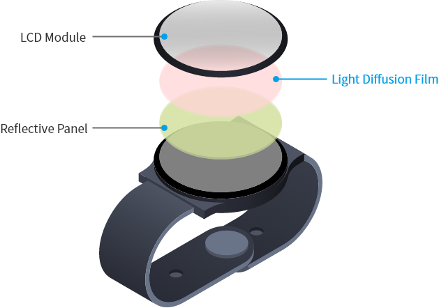
Louver structure → Anisotropic Diffusion (ADF) Columnar structure → Isotropic Diffusion (IDF)
Optical coherencetomography OCT principle and technical realization
This will depend on the outcome of the OCT scan. If the OCT scan and photos cannot fully diagnose the condition or if it shows significant abnormalities, you will normally be called for another appointment in between one and four weeks’ time. If not, you will be seen more routinely. If no abnormality is detected, you may be discharged. You will be notified of the outcome by letter most of the time. You may receive a telephone call from the booking team or the ophthalmology clinical team.
This unique light diffusion film has a refractive index distribution structure inside the film. By using light efficiently, it is able to contribute to high visibility and energy saving in display devices. The diffuse shape (anisotropic/isotropic) and diffusion angle can be adjusted depening on the purpose.
On arrival, you will have your vision checked. Please bring your current glasses, or any contact lenses you may wear to use in this test.
Optical coherencetomography
Same sex bays and bathrooms are offered in all wards except critical care and theatre recovery areas where the use of high-tech equipment and/or specialist one-to-one care is required.
This unique light diffusion film has a refractive index distribution structure inside the film. By using light efficiently, it is able to contribute to high visibility and energy saving in display devices. The diffuse shape (anisotropic/isotropic) and diffusion angle can be adjusted according to the purpose.
Blausen.com staff (2014). "Medical gallery of Blausen Medical 2014 (opens in a new tab)". WikiJournal of Medicine 1 (2). DOI (opens in a new tab):10.15347/wjm/2014.010 (opens in a new tab). ISSN (opens in a new tab) 2002-4436 (opens in a new tab).
OCT imaging can be particularly useful in detecting and monitoring multiple macula conditions such as age-related macular degeneration and diabetic macular oedema. This technology allows us to explore treatment options more efficiently and effectively.

Help accessing this information in other formats is available. To find out more about the services we provide, please visit our patient information help page (see link below) or telephone 01223 256998. www.cuh.nhs.uk/contact-us/accessible-information/


 Ms.Cici
 8618319014500