
LENGTH in inches for 10mm - length of 10mm
Future Possibilities: Fluorescence microscopy promises to open new areas of scientific exploration. From enhancing our understanding of complex biological systems to driving innovation in diagnostic tools and therapeutic interventions, the future possibilities brought about by this imaging technology are endless. Its potential to revolutionize areas such as personalized medicine and environmental protection highlights its importance as a catalyst for change.
When illuminated with light of a specific wavelength, the structures of these markers emit light at a different, longer wavelength, making them identifiable in their surroundings.
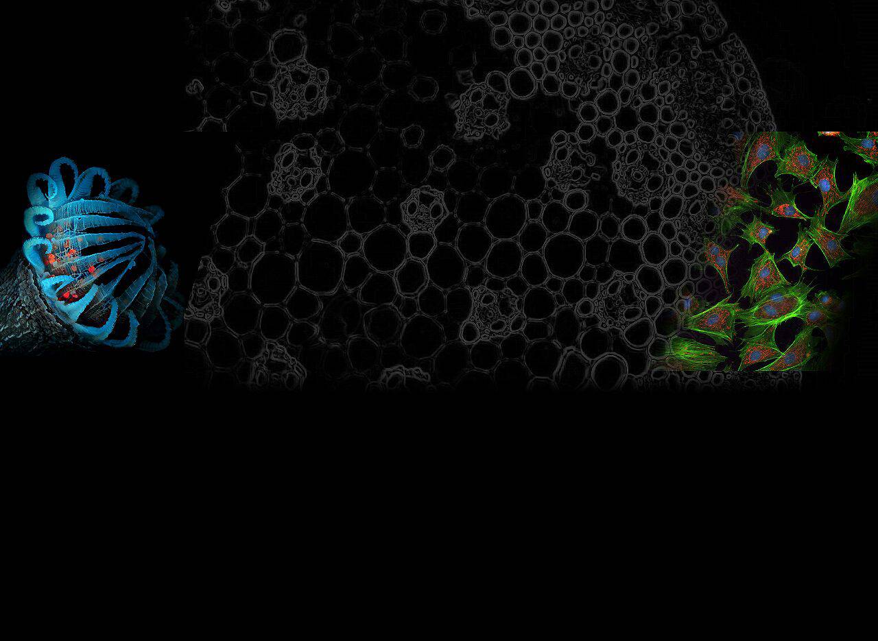
Fluorescence microscopy is an advanced imaging technique that reveals the complex world of cells and microscopic structures by making specific compounds glow.
This technology has applications across multiple scientific fields, from basic biological research to medical diagnostics to environmental monitoring, and its impact is far-reaching and diverse.
Proper use of fluorescence microscopy to effectively visualize fluorescently labeled samples. Here are a few notes on how to use fluorescence microscopy:
Emission filter: After the fluorophores in the sample are excited and emit light, this light is transmitted back through the microscope. Emission filters block the excitation light and allow only longer wavelengths of emitted light to reach the detector (eye or camera).
Fluorescence microscopy is a powerful imaging technique that allows scientists to observe and study the detailed structure and dynamic processes of cells and tissues with unparalleled clarity.
Fluorescence microscopy also plays a key role in environmental science. By analyzing the composition and behavior of microbial communities in different environments, scientists can better understand how these communities respond to environmental stresses, the degradation processes of pollutants, and their role in global ecosystems. This information is critical for ecological protection, pollution control, and environmental health assessment.
2. Many consumer products with fluorescent labels (such as laundry detergent and safety features on official documents) use fluorescent compounds for various purposes.
1. Fluorescence microscopy uses fluorescent markers in highlighters. When exposed to UV light, these markers emit bright visible light, demonstrating the principle behind fluorescence.
Dampen chassis vibrations, cabinet vibrations, and other unwanted resonances with AudioQuest Sorbothane Self-Adhesive Vibration-Damping Sheets to enjoy quieter performance from audio/video components.
This is the nicest Lighted Magnifying Glass we've found. It's made of aluminum, not plastic, by a company known for providing quality and value in optics. The ...
Its wide range of applications, from basic biological research to medical diagnosis, underscores the importance of fluorescence microscopy in science and medicine. This technology provides us with a new window to explore the nature of life and greatly advances our understanding of life sciences.
Finding Fluorescence: Adjust the focus and move the stage to find areas of interest. The fluorescence signal might be faint, so it’s often helpful to adjust the intensity of the light source and the exposure settings on the camera.
Unraveling the Mysteries of Life: Fluorescence microscopy is a powerful tool for unlocking the secrets of cell biology, allowing researchers to study dynamic processes such as cell division, protein synthesis and molecular interactions with extraordinary clarity.
Fluorescence microscopy is a special type of microscope that uses the principle of fluorescence to observe specific components in a sample. It works based on the properties of fluorescent molecules (fluorescent dyes or fluorescent proteins) that can emit light of different wavelengths when excited by light of specific wavelengths.
For advanced applications, such as live-cell imaging, additional considerations include maintaining the health of the cells with controlled temperature, CO2, and humidity. Confocal microscopy or other advanced fluorescence microscopy techniques might require additional steps and settings adjustments.
It is a powerful tool in biological and biomedical research, allowing scientists to directly observe cells and cellular components with high specificity and sensitivity by labeling them with fluorescent markers.
Alberto Pittaluga dives deep into the benefits of power conditioning, explores the relatively modest PowerQuest 3, and decides it’s staying in his system.
Reducing Exposure: Limit the exposure of your sample to the excitation light to minimize photobleaching. Only expose your sample to the light when observing or capturing images.
A. SYSTEM DESCRIPTION. Authority: Office of Management Budget (OMB) Memorandum (M) 03-22, OMB Guidance for Implementing the Privacy.
Use offluorescencemicroscope in microbiology
In addition, by observing the distribution and effects of drugs within cells, fluorescence microscopy also plays an irreplaceable role in drug development and evaluation of therapeutic effects.
Originally published in the April issue of The Absolute Sound, Derk Richardson's excellent review of Zen Widow's latest release, IV: (from one dark age to another), is now available online.
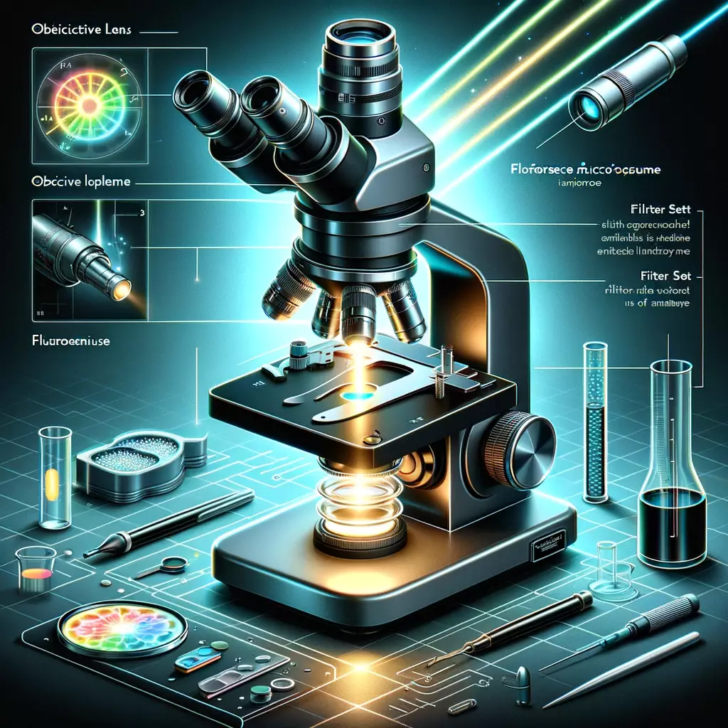
Fluorescence microscopes consist of a variety of specialized components that work together to enable the observation of fluorescently labeled samples. The following is a detailed introduction to the key components of a fluorescence microscope:
Fluorescence Filters and Cubes: A combination of excitation filters, dichroic mirrors, and emission filters are typically mounted in a single unit called a filter cube. This setup makes it easier to switch between different sets of filters and mirrors when viewing samples labeled with different fluorophores.
In fluorescence microscopy, the role of light cannot be ignored. The interaction between light and matter allows researchers to observe intricate details that would otherwise be invisible. Here are some examples from everyday life:
Vorschläge ... One suddenly finds oneself opposite a single, very small lens. Man sieht sich plötzlich einer einzigen, sehr kleinen Linse gegenüber. ... Now the ...
What is fluorescence microscopyused for
Eyepieces (Eyepieces): In traditional fluorescence microscopes, the eyepieces further magnify the image and direct it to the observer’s eyes.
Focusing: Use the microscope’s eyepiece or a camera connected to a monitor to initially focus on your sample using transmitted light (if available). Then switch to fluorescence mode.
This guide provides an overview of how technology works, from the fundamentals of fluorescence to its applications in biological research, learning how it can enhance our understanding of life at the molecular level.
What is fluorescence microscopyin microbiology
Fluorescence microscopy is a specialized optical microscope that uses fluorescence and phosphorescence phenomena instead of reflection and absorption or studies the properties of organic or inorganic substances based on reflection and absorption.
In fluorescence microscopy, a sample is first labeled with a specific dye containing fluorescent molecules and then illuminated with a light source of a specific wavelength, such as ultraviolet light. Fluorescent molecules absorb this light and emit longer wavelength light (fluorescence). Through the microscope’s specific filters, only fluorescence is detected, resulting in a vivid image.
Compared to traditional microscopy, fluorescence microscopy utilizes fluorescent dyes or proteins to label specific structures in a sample.
Combined, these components enable fluorescence microscopy to visualize and study biological samples with high specificity, sensitivity, and contrast, enabling detailed analysis of cells, tissues and molecular processes.
Labeling: Your sample should be labeled with fluorescent dyes or tagged with fluorescent proteins specific to the molecules or structures of interest.
Fluorescence microscopyprinciple
Here we typically recommend the U-Spade or banana connector. However, even though there is less weight and pressure on this side of the cable (particularly for BiWire sets), it is nevertheless important to note the binding post’s design. If a banana connector cannot seat all the way into the binding post, we recommend one of our spade connectors.
In the medical field, fluorescence microscopy is of great significance in the diagnosis and treatment of diseases. It can be used to identify abnormal structures in cells, helping to detect diseases such as cancer early.
Fluorescencemicroscope diagram
To learn more about dichroic mirrors, please read this article, which details what a dichroic mirror is and its advantages and applications.
Everyday benefits: Insights gained from fluorescence microscopy have a direct impact on daily life, impacting areas such as healthcare, technology, and environmental sustainability. This technology helps improve healthcare outcomes and quality of life by aiding early disease diagnosis and treatment monitoring.
Dichroic Mirror (Beamsplitter): A dichroic mirror is a special type of mirror that reflects certain wavelengths of light while allowing other wavelengths to pass through. It reflects the excitation light toward the sample and allows the longer wavelength emitted light to pass through the detection system.
This advanced imaging technology opens up new ways to study cellular and molecular behavior, allowing us to observe life processes with greater detail and clarity.
Scientific Advances: The impact of fluorescence microscopy extends beyond biological research, contributing to major advances in various scientific fields. From discovering new drug targets to elucidating complex disease pathways, this imaging technology drives breakthrough discoveries and advances across diverse scientific disciplines.
Light Source: Turn on the microscope’s light source, which is usually a high-intensity lamp like a mercury or xenon lamp for wide-field fluorescence microscopy, or lasers for confocal microscopy.
Advantages offluorescence microscopy
After use, turn off the light source to prolong its life, clean any oil immersion lenses with lens cleaning solution and lens paper, and cover the microscope to protect it from dust.
Stage: The stage holds the sample and allows precise movement in the X, Y (and sometimes even Z) directions to scan different areas or layers of the sample.
Mounting: Place the labeled sample on a microscope slide. If necessary, cover it with a coverslip. Use an appropriate mounting medium that preserves fluorescence and reduces photobleaching.
In this video, Walts TV hosts a discussion with Angelo, a Field Educator responsible for product training at AudioQuest. We delve into viewer comments from previous AudioQuest videos, exploring the intricacies of...
A detective obsessed with justice receives a surprising offer of help from a mysterious figure in the criminal underworld known as "Invisible."
The SnakeClamp Handheld Magnifying Glass is available with your choice of 3x, 8x, 10x or 15x glass lens magnification. Our Handheld Magnifier is available ...
Filters: Select the correct filter set (excitation filter, dichroic mirror, and emission filter) matching the excitation and emission spectra of your fluorescent dye.
Objective Lenses: Choose the appropriate objective lens based on the required magnification and numerical aperture for your sample.
What is fluorescence microscopyprocedure
by C Kim · 2023 · Cited by 1 — Highlights. •. A 3-dimensional image information provided by a light field microscope target. •. Designed to measure lateral, axial, and field of view ...
Detector: In more advanced or automated systems, a detector such as a charge-coupled device (CCD) or complementary metal-oxide semiconductor (CMOS) camera captures the emitted light to create an image. These detectors are highly sensitive and can detect the faint light emitted by fluorophores.
Objective: The objective collect the light emitted by the sample. It also plays a vital role in determining the resolution and magnification of an image. Fluorescence microscopy often uses high numerical aperture (NA) objectives to maximize light collection and resolution.
We are delighted to announce that 20 AQ products — across a wide range of categories, all representing outstanding performance and value — have received TAS’s 2024 Editors’ Choice Award.
Fluorescence microscopy is a growing imaging technology that allows scientists to observe cell interiors and microscopic structures with precise detail and contrast through the use of special light sources and fluorescent molecular markers.
Why is fluorescence microscopy important? Because it provides scientists with an understanding of the complex structures and processes inside organisms, revealing the microscopic mechanisms that control the essence of life.
AudioQuest’s founder and chief designer, William E. Low, is the featured guest in the premier episode of Stereophile’s YouTube series, “Icons of Audio.”
Often, the choice between terminating your speaker cables with spade lugs or banana plugs will come down to personal preference. However, the EU-compliant isolated binding posts used on some power amplifiers often makes it difficult if not impossible to tighten spade lugs with high torque or pressure. In these applications, the AudioQuest banana connector is the superior choice, and may be the only choice.AudioQuest has conducted a fairly extensive survey of contemporary amplifier manufacturers to determine whether spades or bananas will be most appropriate for a given brand and model of amplifier. Please contact us, provide the brand and model of the amplifier, and we’ll respond with the optimal connector type.
Taking Images: Once you have your sample in focus and properly illuminated, capture images using the microscope’s camera. Many fluorescence microscopes are equipped with software that allows you to adjust settings, and capture, and analyze images.
Excitation filter: This filter selects a specific wavelength of light that matches the excitation spectrum of the fluorophore used. It ensures that only light that excites the fluorophore reaches the sample.
Fluorescence microscopyppt
Light Source: A light source provides the illumination required for fluorescence. High-intensity light sources such as mercury or xenon lamps, LEDs, or lasers are often used because they can produce the intense light required to excite fluorophores.
In the field of cellular and molecular biology, fluorescence microscopy allows scientists to peer deeply into the dynamic processes inside cells. For example, it can be used to observe cell division, protein synthesis and molecular interactions, providing us with new ways to understand the basic mechanisms of life. By combining fluorescent markers with specific organelles, proteins, or genetic material, researchers can track the behavior of these molecules and structures with stunning clarity.
Feb 6, 2023 — l 940nm infrared LED: When invisibility is a must in some applications, such as surveillance, touch screen, proximity sensing, blood oxygen ...
DLC coating Diamond Like Carbon Coating.
Description · Infinity Plan Achromatic Objective · 4X Magnification Power · Working distance is about 19mm · RMS thread(It can set on the part (http://www.
Each fluorescence microscope might have its unique features and settings, so it’s also important to consult the manual specific to your microscope model for detailed instructions and troubleshooting tips.
KR868 Stage Micrometer 1mm with 200 divisions available with NIST certification.


 Ms.Cici
 8618319014500