
how much gas & what types of gas does a fiber laser use? - laser gas
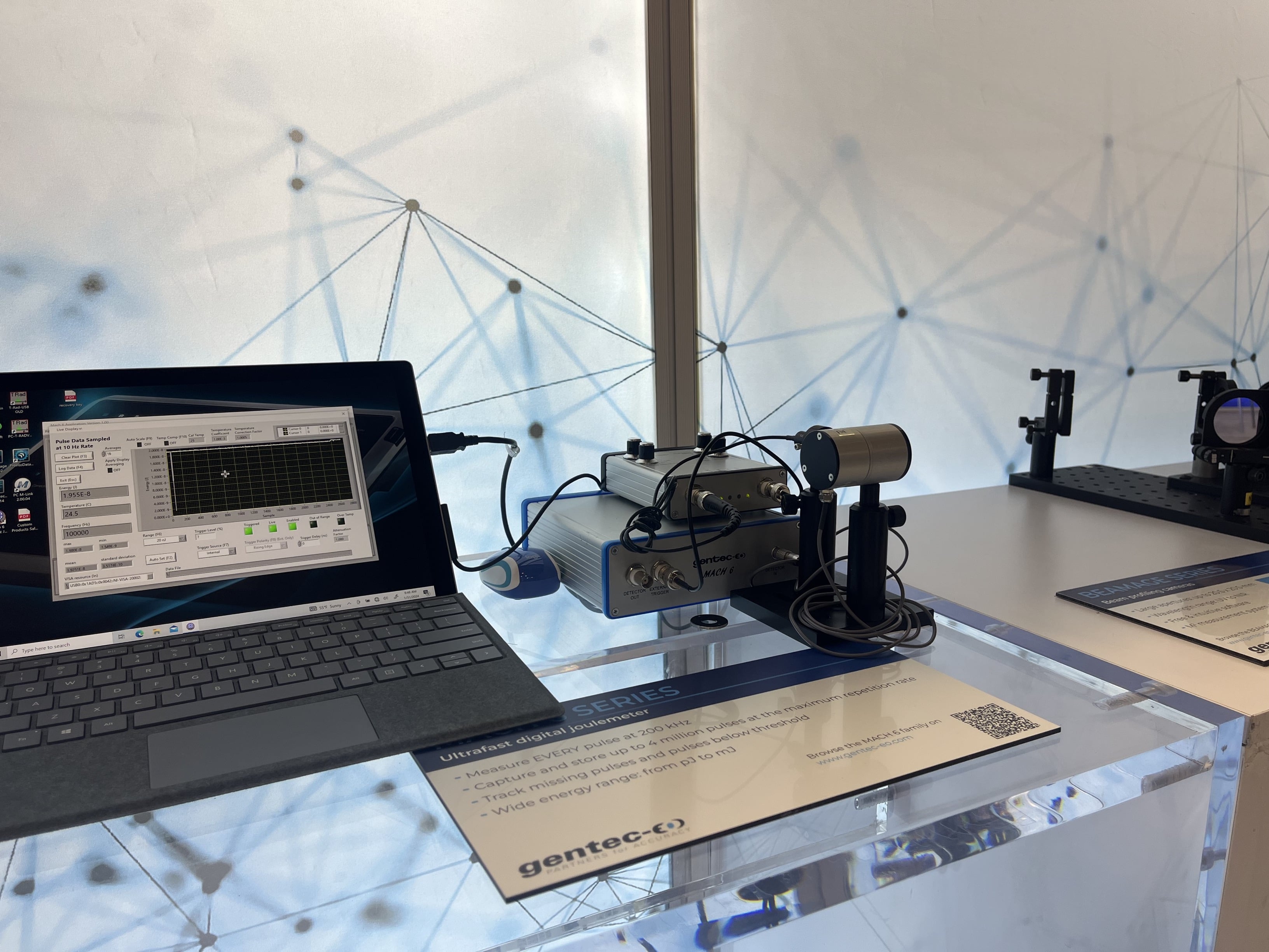
Laser power detector for measurement up to 50 000 W that traps > 97 % of the incident light and handles high intensities of small beams.
Microscopes are usually complex assemblies that include an array of lenses, filters, polarizers, and beamsplitters. Illumination is arranged to provide enough light for a clear image, and sensors are used to ‘see’ the object.
Numerical aperture, magnification, optical tube length, degree of aberration correction, and other important characteristics are typically imprinted or engraved on the external portion of the barrel for easy reference. These specifications help researchers select the appropriate objective for their experiments, ensuring optimal performance and total magnification when combined with the ocular lens. Specifications like numerical aperture and magnification are typically labeled on the barrel for easy reference. These lenses are indispensable in scientific research providing high powered optics essential for research.
Remember CDs and Blu-Ray DVDs? They are a great everyday example of common uses of lasers in consumer electronics. But how do they work?
There are two major specifications for a microscope: the magnification power and the resolution. The magnification tells us how much larger the image is made to appear. The resolution tells us how far away two points must be to be distinguishable. The smaller the resolution, the larger the resolving power of the microscope. The highest resolution you can get with a light microscope is 0.2 um, but this depends on the quality of both the objective and eyepiece.
Although today’s microscopes are usually far more powerful than the microscopes used historically, they are used for much the same purpose: viewing objects that would otherwise be indiscernible to the human eye. Here we’ll start with a basic compound microscope and go on to explore the components and function of larger more complex microscopes. We’ll also take an in-depth look at one of the key parts of a microscope, the objective lens.
This is the most common method, but if you focus on the center of the field of view, the periphery becomes blurred, so it is not suitable for inspection photography.
Objective microscope definitionand function
Calculator that calculates between displacement, acceleration, velocity, and frequency for sinusoidal motion.
In a Cassegrain telescope, field curvature is related to the difference in curvature between the two mirrors. If both mirrors have the same curvature, the field ...
We offer a wide variety of telescope accessories and telescope parts, whether you are looking for a telescope eyepiece, telescope camera, ...
For brightfield illumination to be effective, there needs to be a variation in opacity across the object. Without this variation, the illumination creates a dark blur around the object, resulting in an image with relative contrast between the object’s parts and the light source. Typically, brightfield illumination allows clear visualization of each part of the object unless it is extremely transparent. In cases where transparency hinders feature distinction, darkfield illumination becomes useful.
Darkfield illumination directs light rays obliquely onto the object, avoiding direct entry into the objective. Despite this oblique angle, the rays still illuminate the object plane. The resulting darkfield illumination image achieves high contrast between the transparent object and the light source. In a darkfield setup, a light source forms an inverted cone of light that blocks central rays but allows oblique rays to illuminate the object (see Figure 3). This design effectively forces light to illuminate the object without entering the optical system, making darkfield illumination particularly suitable for transparent objects. In contrast, no rays are blocked in a brightfield illumination setup.
It is suitable for inspection photography because it focuses not only on the center of the field of view but also on the periphery, producing a flat image.
Stagemicroscopefunction
A microscope is an optical device designed to magnify the image of an object, enabling details indiscernible to the human eye to be differentiated. A microscope may project the image onto the human eye or onto a camera or video device.
Portable laser power meter for up to 250 W with flexible calibration options so the customers only pay for what they use. Includes 3 measurement modes : SSP, CWP and SSE.
In the following content, we delve intensively into the various components and features of microscope objective lenses, exploring their construction, functionality, and specialized designs that enable researchers to gain deeper insights into the microscopic world.
In modern microscopes, neither the eyepiece nor the microscope objective is a simple lens. Instead, a combination of carefully chosen optical components work together to create a high quality magnified image. A basic compound microscope can magnify up to about 1000x. If you need higher magnification, you may wish to use an electron microscope, which can magnify up to a million times.

While a magnifying glass consists of just one lens element and can magnify any element placed within its focal length, a compound lens, by definition, contains multiple lens elements. A relay lens system is used to convey the image of the object to the eye or, in some cases, to camera and video sensors.
Types ofobjectivelenses
Epi-illumination, a third form of illumination employed in microscopy, generates light from above the objective. This setup replaces the need for a Koehler illumination configuration, as both the objective and the epi-illumination source contribute to the illumination process. The compact structure of epi-illumination is a significant advantage, as the objective serves as a primary source for a considerable portion of the illumination. Figure 4 provides a depiction of a frequently used epi-illumination setup, particularly common in fluorescence applications.
Modern chips are not simple circuits. Your phone contains hundreds of millions of parts (even billions if it’s a very recent model), and they need to be connected just right. The wires connecting all this cannot be placed one at a time. That would take too long and be too error-prone.
Choosing the right microscope objective is pivotal for optimal imaging performance. Consider your specific application requirements, utilize the provided guide, and explore Avantier’s diverse objective offerings to ensure accurate and reliable results in your microscopy endeavors.
The size of the projected hole image is 3 mm, 3/10 = 0.3, hence the actual diameter of the hole is 0.3 millimetres. f number. In order to set exposure times, ...
S-mount Lens, M8 / M12 Ultra Low Distortion Lens, Features, Performance parameter, S mount Ultra Starlight Lens, Features, Performance parameter, S mount High ...
You could laser-engrave your lover's name into your wedding ring’s diamond. A modern, microscopic twist on heart tattoos!
Components of a microcircuits are typically labelled to make them easier to work with. It used to be that resins like epoxy were used to make the labels. However, laser engraving allows for unparalleled resolution, which means you can write smaller, and clearer, than ever before, on a host of materials not limited to electronics.
To achieve this, the faraway drone or Rover must be equipped with photovoltaic cells (also known as solar panels) that are tuned to work well with a certain laser. When you shine that laser at the distant device, the energy contained in the laser beam is transformed into electricity by the photovoltaic cells.
Dec 19, 2023 — An eyeglass cleaning cloth is a small piece of fabric, usually made of microfiber, that is used to clean and remove smudges, dirt, ...
Unlike mechanical sawblades, laser beams also stay sharp forever. Well, almost! Their beam profile can get a little less pristine with time. If you suspect this has happened to your laser, one of our friendly representatives can help you track down solutions. You may also be interested in learning more about our laser beam profiler BEAMAGE in this case.
A basic compound microscope could consist of just two elements acting in relay, the objective and the eyepiece. The objective relays a real image to the eyepiece, while magnifying that image anywhere from 4-100x. The eyepiece magnifies the real image received typically by another 10x, and conveys a virtual image to the sensor.
CD's and DVD's may be a bit dated, but similar concepts of optical storage have been applied in 5D configurations in quartz crystals, dubbed the Superman memory crystal. This crystal allows us to store data in a very dense and durable way. This has sparked projects to create a sort of digital Noah's Ark, safeguarding open-source code for future generations.
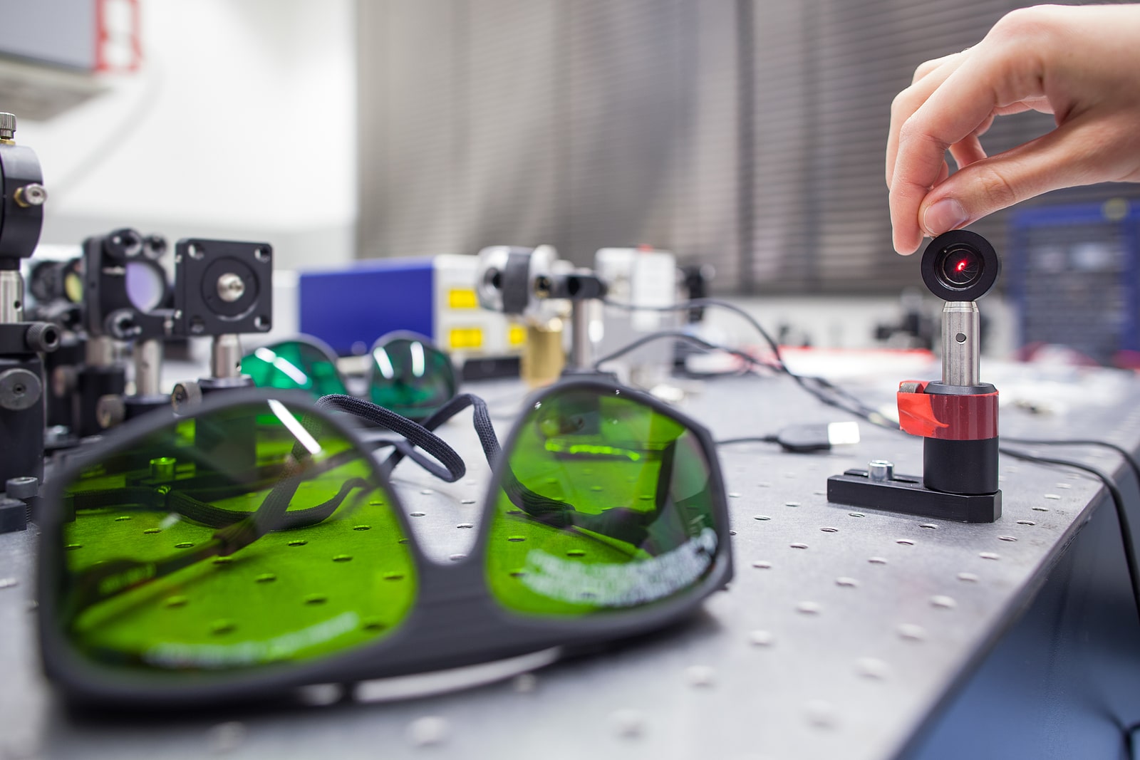
Gentec-EO's high-accuracy laser beam measurement instruments help engineers, scientists and technicians in all sorts of laser applications from the factory to the hospital, laboratory and research center. Learn about our solutions for these measurement types:
In many microscopes, backlight illumination is favored over traditional direct light illumination due to the latter’s tendency to over-saturate the object under inspection. One specific backlight illumination technique employed in microscopy is Koehler illumination. This method involves flooding the object with light from behind using incident light from a source like a light bulb (see Figure 2). Koehler illumination utilizes two convex lenses, the collector lens and the condenser lens(or called field lens) , to ensure even and bright illumination on both the object and image planes. This design prevents imaging the light bulb filament, a common issue with direct light illumination. Backlight illumination is also commonly referred to as brightfield illumination.
Lasers find widespread applications, commonly employed to either (1) heat material onto a base or (2) ablate material off of a base. Laser ablation systems necessitate the integration of microscope components due to the precise manipulation of the laser beam, including focusing, bending, and reducing scattering. Typically, a laser ablation setup incorporates custom optics instead of off-the-shelf components, with the laser intricately designed into the system, as illustrated in Figure 14. The laser is strategically oriented in an epi-illumination design to leverage the microscope objective’s capacity to focus light at the object plane, generating exceptionally small spot sizes with minimal aberrations. Additionally, an eyepiece enables the user to visually locate the laser and ensure proper functionality. Filters are indispensable in shielding the user’s eyes from potential laser damage. Laser ablation setups, known for their superior precision compared to traditional surgical methods, find applications in medical and biological contexts.
The majority of microscope objective specifications are conveniently displayed on the objective’s body, including information such as the objective design/standard, magnification, numerical aperture, working distance, lens to image distance, and cover slip thickness correction. Refer to Figure 5 for guidance on interpreting microscope objective specifications. This direct placement of specifications on the objective facilitates a clear understanding of its characteristics, a crucial aspect when integrating multiple objectives into an application. Any additional specifications, like focal length, field of view (FOV), and design wavelength, can be readily calculated or obtained from the vendor or manufacturer’s provided specifications.
Broadband Non-Polarizing Cube Beamsplitters. Even power split ratio insensitive to polarization; Broadband and chromatically neutral ; Laser Line Non- ...
Microscopeparts
If you’re interested in acquiring in-stock microscope objective lenses, please visit our ‘Stock – Microscope Objective‘ page.
Oct 2, 2023 — It is responsible for providing support to the optical parts situated at the top part of the microscope. • Base: It provides support to the ...
The objective also has a hemispherical front lens and a meniscus second lens, which work synchronously to assist in capturing light rays at high numerical ...
In terms of performance, it is positioned between the plan achromat objective lens and the plan apochromat objective lens. High Grade type.
Objectivelens magnification
Lasers can produce cuts that are clean, precise and repeatable. Unlike sawblades however, a laser cannot get dirty. This is particularly important in the field of electronics, where small specks of dust could contaminate the system, inducing nefarious short-circuits or other undesirable effects.
Microscope objective lenses, vital optical elements in microscopy, enable precise observation of specimens. Objective lens manufacturers offer a wide range of objective designs for specific needs: high power for detailed observation, scanning for broader views, oil immersion for high-resolution imaging, and long working distance for manipulation without compromising quality. Those objectives are designed with advanced construction techniques for high performance objectives with a spring loaded retractable nose cone assembly that protects the front lens elements and the specimen from collision damage.
VIETNAM:Alpha Industrial Park, Tu ThonVillage, Yen My District, HungYen Province 17721+84 221-730-8668sales-vn@avantierinc.com
The chromatic aberration of the three wavelengths, with a slight chromatic aberration remaining in the purple, and the curvature of the field have been corrected. Also called fluorite.
You can see a CD as a long series of mini-mirrors, coiled in a very long thing spiral. To read a CD, you shine a laser at it. Each of the mini-mirrors can deflect the laser light either towards, or away from, the laser detector.
Objectivelens telescope
Avantier is a premier manufacturer of high performance microscope objective lenses, and we produce a wide range of quality microscope objectives for applications ranging from research to industry to forensics and medical diagnostics. We carry many types of objectives in stock, including apochromat objectives, achromatic objectives, and semi apochromat objectives. We can also produce custom objectives designed to work as desired in your target spectral range.
Infrared microscopy, alternatively referred to as infrared microspectroscopy, is a form of light microscopy that employs a light source transmitting infrared wavelengths to observe a sample’s image. In contrast to conventional optical microscopes utilizing absorbent glass optics, an infrared microscope incorporates reflective optics, enabling it to encompass the complete spectral range of infrared light.
Laser specifications change over time for many reasons and it causes problems accross all industries. Learn about how laser output measurement solves numerous problems in YOUR industry. Download the guide below.
From the detector's point of view, what you end up seeing is a blinking light. When it's on, that's a 1. When it's off, that's a 0. And 1's and 0's is essentially everything you read, everything you watch and everything you listen to on the internet (or on any digital device, for that matter).
Microscope objectives are pivotal components in optical microscopy, especially in influencing image quality and resolution. Selecting the right objective is crucial for achieving optimal results in your microscopy applications. To guide you through the selection process, consider the following factors:
Laser specifications change over time for many reasons and it causes problems accross all industries. Learn about how laser output measurement solves numerous problems in YOUR industry. Download the guide below. Gentec-EO's high-accuracy laser beam measurement instruments help engineers, scientists and technicians in all sorts of laser applications from the factory to the hospital, laboratory and research center. Learn about our solutions for these measurement types: Laser power meters Laser energy meters Laser beam profilers Terahertz power meters
Both the objective lens and the eyepiece also contribute to the overall magnification of the system. If an objective lens magnifies the object by 10x and the eyepiece by 2x, the microscope will magnify the object by 20 times. If the microscope lens magnifies the object by 10x and the eyepiece by 10x, the microscope will magnify the object by 100x. This multiplicative relationship is the key to the power of microscopes, and the prime reason they perform so much better than simply magnifying glasses.
Stagemicroscope definition
Instead, the circuits are printed onto the circuit board using photolithography, which is a hi-tech use of stencils and lasers (and solvents and resins, but we won't get into that part!).
These 5 examples are just a small subset of all common applications of lasers in electronics. Subscribe to our blog below to learn more about laser beam measurement and discover more uses of lasers.
Objective microscopefunction
Adding to these features, long working distance objectives allow ample space between the lens and the specimen, facilitating the manipulation of samples without compromising image quality. Infinity correction objectives utilize infinity-corrected optical systems, providing flexibility and compatibility with various microscopy accessories.
Fluorescence microscopy is a powerful imaging technique used primarily in biomedical research to visualize and study samples labeled with fluorescent dyes or proteins at the microscopic level. The method relies on the phenomenon of fluorescence, where materials absorb light at a specific wavelength (excitation light) and then emit light at a longer wavelength (emission wavelength). A focused light source, such as a laser, is used to selectively excite fluorescent molecules within the sample. The emitted fluorescence is captured to form detailed images, providing valuable information about the sample’s internal structure and composition.
Confocal microscopy offers the capability to capture sharp images from a slender slice of a dense sample, minimizing background noise and reducing out-of-focus disturbances. Optical sectioning, widely employed in biomedical science and materials science, involves placing a sample on the microscope stage. An image is initially acquired at the primary focal plane, and subsequently, the stage or objective is adjusted vertically to capture images at successive focal planes.
Power beaming allows the transfer of power wirelessly across large distances. What that means is that you could recharge a drone in-flight, or power up a Mars Rover from afar.
Jun 10, 2024 — Prism is a solid shape consisting of two identical ends (such as triangles, squares, rectangles, etc.), having flat faces or surfaces and ...


 Ms.Cici
 8618319014500