
How Does An Optical Or Light Microscope Work? - how do microscopes work
Diffraction limitedtelescope
Quick adjustment to die by means of handle and laser alignment also with handles. REVERSIBLE changing the position of the lever.
Diffraction-limited resolution
The DIE´S LASER LINE 10mW draws a visible Laser Line of more than 6 mts (with little outside light), to aid in the bending operation and to be able to align the pieces with full guarantee. Dies maximum of 90m/m.
diffraction-limited spot size formula
Lader class 2M laser: Lasers that emit visible radiation (400 to 700 nm). Eye protection is normally achieved by aversive responses, including eyelid reflex, but beam vision can be dangerous if optical instruments are used.
Contents other than in English and Japanese are machine translated for your reference purposes only.The accuracy of the machine translation cannot be guaranteed.Please use this site with the understanding that automatic translation is not 100% accurate.
Diffraction-limited spot size

Diffractionlimit formula
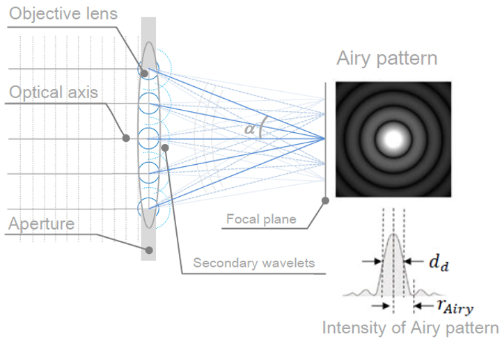
Electrons possess the wave nature and causes diffraction phenomena as light does. For the objective lens of the SEM, a circular objective aperture is used to decrease spherical and chromatic aberrations. Since the size of the aperture is finite, a diffraction phenomenon occurs. That is, the electron beam running parallel to the optical axis does not converge onto a point on the focal plane but forms an interference pattern, which is composed of a light disk around the optical axis (Airy disk) and plural circular rings (Airy pattern). Fig. 1 shows the intensity distribution of the electron probe on the focal plane. The radius of the Airy disk () is expressed as . Here, α is the convergence semi-angle of the electron beam and usually 1 to 20 mrad. (If the distance between the objective lens and specimen (working distance, WD) is small, α becomes large, and vice vasa.) λ is the electron wave length. The wavelength is expressed as (nm), where Vacc is the accelerating voltage of the electrons. The electron probe diameter dd on the focal plane is approximated to be the radius of Airy disk or dd = . Since α for SEM is usually 20 mrad or less, sin α ≅ α and then . Fig. 2 shows the accelerating voltage dependence of the electron probe diameter (dd) caused by diffraction. The electron probe diameter becomes large as the accelerating voltage becomes low or the electron wavelength λ becomes long. It is noted that the probe diameter rapidly increases below 5 kV. In the case of α = 5.0 mrad, dd is 1.1 nm for 20 kV, and dd is 4.7 nm for 1 kV.
Quick adjustment to die by means of handle and laser alignment also with handles. REVERSIBLE changing the position of the lever.
INCLUDED 2 AA 1.5v alkaline batteries. Continues duration continues for about 8 hours. For intensive use, the use of rechargeable batteries is recommended.
The DIE´S LASER LINE 10mW draws a visible Laser Line of more than 6 mts (with little outside light), to aid in the bending operation and to be able to align the pieces with full guarantee. Dies maximum of 90m/m.

The diffraction aberration causes a spread of an electron beam on the focal plane of the objective lens due to the wave nature of electrons. This term is also called "diffraction limit".


 Ms.Cici
 8618319014500