
Diffraction Grating Monochromators - monochromator
microscope: definition biology
A simple rug with a contemporary feel. Andrew Martin Aurum has a luxurious velvet texture underfoot and in a cool steel grey tone, with a soft metallic ...
Photography: What, How, Why Copyright © 2023 by Maria Politarhos and Randy Matusow is licensed under a Creative Commons Attribution 4.0 International License, except where otherwise noted.
Whatis microscope in science
Microscopic examination confirms the laboratory tests that may be positive for the disease. Technicians count the number of red blood cells infected with the virus or parasite to give the doctors an idea of how advanced the disease is in a patient.
Electron microscopes help prepare the small surfaces for sectioning into small slices. Microscopes enlarge the images of silicon chips to help the engineers create more efficient electronic devices. When more circuits are fitted onto a small chip, the computational power of silicon microchips increases.
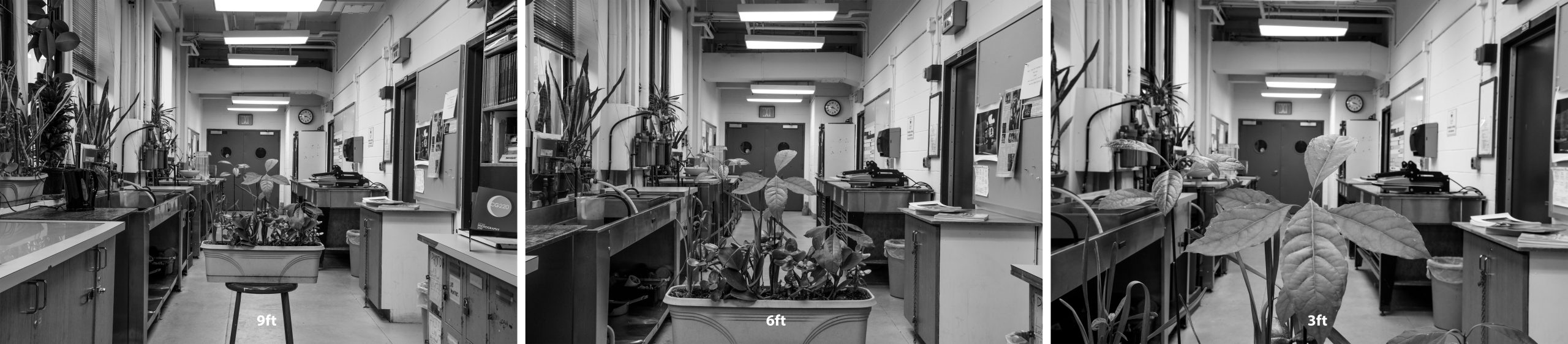
Notice how the size of the leaves in the planter gets much bigger while the size of the round windows on the door remains about the same. The depth seems to increase because the camera was brought closer to the subject.
Jun 13, 2014 — In general, given similar cost and materials, the fixed focus lens should have a bit better IQ particularly with regard to the image corners.
Microscopes are not just used to observe cells and their structure but are also used in many industries. For example, electron microscopes help create and observe extremely tiny electrical circuits found on Silicon microchips. Scanning microscopes are much more sophisticated and they have higher magnifications than light-refracting microscopes.
Scanning electron microscopes have magnifications up to several million times to view the molecules, the viruses and the nano-particles. They use the corrective software to increase the magnification and the resolution of images. The computers help the nano-technologists use high-powered electron microscopes to view the objects.
The larger the opening of the lens (small F Stop number) the less the Depth of Field. Only the person is sharp when using F 1.4 and the background appears blurred/out of focus.
Microscopes use the simple visible light refracting lenses. Electrons, x-rays and infrared rays Scanning electron microscopes are able to resolve the viruses which are far smaller than any cell. They enlarge the view of tiny viruses, which allows scientists to develop the vaccines and cures for infectious diseases in the humans and the animals.
Types of microscope
GraphPad Prism Software | Axes Format | X-Axis | Y-Axis | Titles and Fonts | Colour. MechTech with SK · 3:37 · How to make Colour graph using GraphPad Prism ...
Microscopes are also used to diagnose illness in hospitals and clinics all over the world. Microscopes magnify the blood samples, so the doctors or the pathologist can see the viruses and the parasites attacking the red blood cells and take the necessary steps to cure it.
Light microscope
When you focus your lens on a subject (the lamp post in our example) the depth of field will change depending on the F stop you are using.

Whatis a microscope used for
About the Laboratory. Mass spectrometry is defined as a technique that involves analyzing ions according to their mass to charge ratio (M/Z). This technique can ...
Depth of Field (DoF) refers to the distance between the closest and farthest objects that appears acceptably sharp in a photograph.
Palintest photometers are available in single and multiparameter formats; suitable across the range of applications, covering all major water quality tests.
When it comes to biology, Microscopes are important because biology mainly deals with the study of cells (and their contents), genes and all organisms. Some organisms are so small that they can only be seen by using magnifications of 40x-1000x, which can only be achieved with the use of a microscope. Cells are too small to be seen with the naked eye.
Microscopes have opened up many doors in science. By using Microscopes scientists, researchers and students were able to discover the existence of microorganisms, study the structure of cells and see the smallest parts of plants, animals and fungi.
10 uses of microscope
Microscope diagram
Next Gen Walking War Robots is here! (Armor Attack Review + Interview). 3.8K views ; [3.25] Forbidden Rite is Back! (Tanky + All content) - Path of Exile Best ...
Mastermind Toys Mini Fiber Optic Light 9'' Assorted ... Turn on the mini fiber optic light in a dark room and watch as the light travels up through the fibers and ...
Microscope parts and functions
All branches of biology use Microscopes especially in Molecular Biology and Histology (the study of cells). Microscopes are the backbone of studying biology. The biologists use them to view the details that cannot be seen by the naked eye such as the small parasites and small organisms which is important for the disease control research.
Just as in the previous example, the closer the subject is to the camera, the shallower the Depth of Field. These three photographs are all focused on the same subject (planter-box in the middle of the hallway) and were taken with the same F stop and shutter speed, the only difference is the distance of the camera from the subject being photographed. The photographer kept walking closer to the subject (planter box).
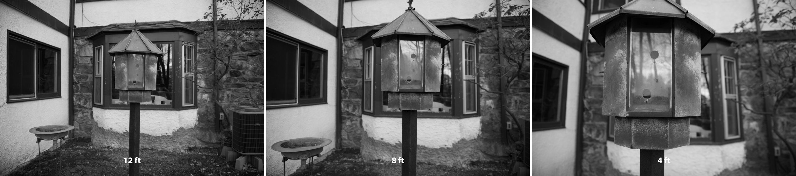
These three photographs above are all focused on the same subject (bird feeder) and were taken with the same F stop and shutter speed, the only difference is the distance of the camera from the subject being photographed. The photographer kept walking closer to the subject (bird feeder).
Notice how the size of the leaves in the bird feeder gets much bigger while the size of the windows directly behind the feeder remains about the same. The depth seems to increase because the camera was brought closer to the subject.
Fits common size (DIN/JIS international standard) Compound Biological Microscopes. The infinite plan or infinity-corrected objective is the most advanced ...
Apart from biological research use and industrial use, Microscopes are also used in the field of genetics. Genetics is the study of variations in an organism generation after generation. Genetic engineering requires the mixing of genes. Genes are even smaller than cells, which is why microscopes are essential in this field.
Ball lenses are perfectly round optical spheres that consist of just one transparent substrate such as UV grade fused silica, BK7, or sapphire. Shaped like a ...
The microscope objective is a key component for reaching high performance of a microscope. It is the part which is placed next to the observed object.
The smallest opening (large F stop number) the greater the Depth of Field. The person and the background are sharp when using F 22.


 Ms.Cici
 8618319014500