
Aspheric Lenses for Better Vision and Appearance - what does aspheric lens mean
By providing your email address, you consent to receive emails from COMSOL AB and its affiliates about the COMSOL Blog, and agree that COMSOL may process your information according to its Privacy Policy. This consent may be withdrawn.
Thanks a lot for this wonderful note: It is really helpful, Really appreciate the way all the detail about microscope have been explained
this is a really good artical i used it to study my science i just wanted to point out to you that tere are a few spelling errors but other than that it is a 100% rating from me
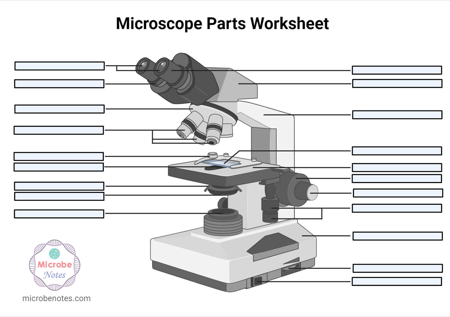
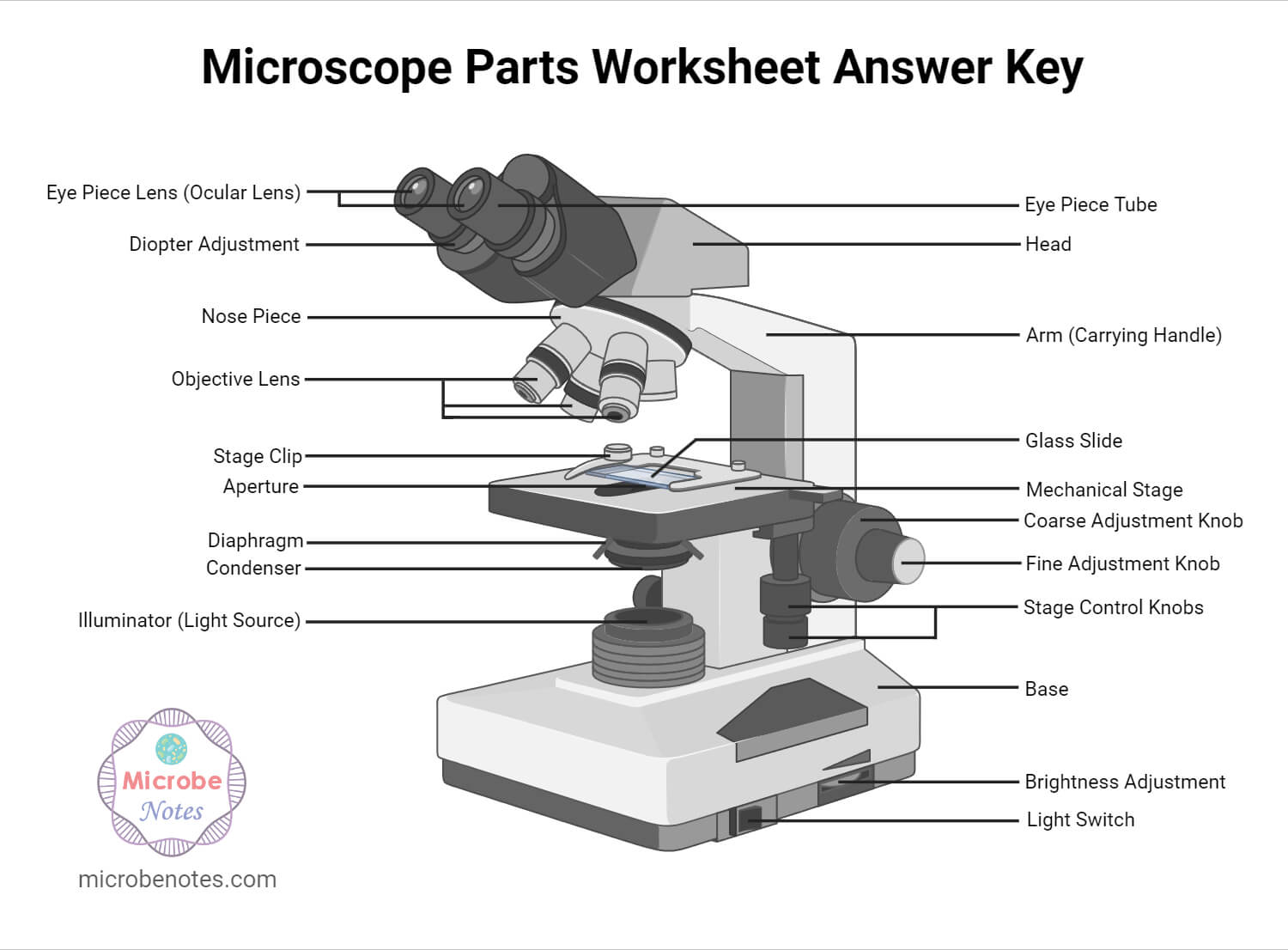
Microscopes are generally made up of structural parts for holding and supporting the microscope and its components and the optical parts that are used for magnification and viewing of the specimen images. Modern microscopes have additional electronics and display devices. This description defines the parts of a microscope and the functions they perform to enable the visualization of specimens.
Collimatedmeaning in Physics
Thanks much for this. We just did microscopy as a topic and the write-up has really helped me to understand better. Thanks again
P-polarized light, or transverse magnetic (TM) mode, refers to the polarization component that is parallel to the plane of incidence. This means that when the ...
Along with the governing equation, we also need a set of boundary conditions for the material when solving for the scattered light. Given that the incident laser light can enter the domain, it is also reasonable to assume that the scattered light can leave the modeling domain. The Semitransparent Surface feature is appropriate for this situation, and lets us enter an emissivity, \epsilon, and a diffuse transmissivity, \tau_d. These two quantities must be less than or equal to one and define a diffuse reflectivity \rho_d = 1-\epsilon – \tau_d. Scattered light incident upon this boundary will entirely pass through if \tau_d = 1, and, if \tau_d < 1, then the light will be partially diffusely reflected back into the domain.
Buy a cheap copy of Edmund Science Math Optics Catalog 701 book by N. W. Edmund. Free Shipping on all orders over $15.
Ans. A microscope is an optical instrument with one or more lens systems that are used to get a clear, magnified image of minute objects or structures that can’t be viewed by the naked eye.
As we have seen, it is possible to implement a model of absorption and scattering of light quite easily, but it is worth emphasizing that this method has two limitations. First, any specular reflection or refraction of light within the material, such as due to a mirror or lens, is not addressable, so only a reasonably homogeneous piece of material can be modeled. Next, the scattering within the medium is assumed to be isotropic. These limitations are counterweighted by the advantage of computational simplicity: Solving two sets of scalar equations for the collimated and scattered light intensity has very low computational cost. Furthermore, the source terms are easily combined with a thermal analysis to compute the rise in temperature. So, if you are modeling laser light interacting with a reasonably uniform sample of a semitransparent material and can assume isotropic scattering, then this approach is attractive because of its efficiency.
Distribution of the heat sources arising from the incident beam, left, and the scattered light, right. The sum of these sources contributes to a rise in temperature.
1. which objective lens focuses closest to object 2. what controls the light entering the binocular lenses 3. how can you enhance the resolving power of a microscope 4. what is useful and false magnification PLEASE CAN YOU HELP ME IN ASWERING THOSE QUESTIONS
Collimatedbeam
To understand the modeling approach, we will begin by assuming that we have a material with no scattering, only absorption. This situation is possible to model using the Heat Transfer Module’s Radiative Beam in Absorbing Media interface, which solves for the Beer–Lambert law within the material. When using this interface, it is assumed that the beam intensity is known at the illuminated boundary. That is, considering a beam of light of known power propagating through surrounding free space, the specified intensity is based on the fraction of the light that propagates into the material.
I did NOT like this website sourse. Wanna know why I didn’t like it??? I don’t like it BECAUSE my school wants me to use this website sourse. My new science teacher wants us to answer those 10 questions. I think its pretty dumb. No Offensen to anyne out there, because I am a nice person not a mean one.
The optical parts of the microscope are used to view, magnify, and produce an image from a specimen placed on a slide. These parts include:
Ans. Rack stop is included in the microscope for preventing the specimen slide from coming too far up and hitting the objective lens.
Ans. The magnification of a lens is defined as the ratio of the height of an image to the height of an object. Microscope magnification measures the total enlargement of the image of an object. Magnification power is the product of eyepiece lens power and objective lens power.
1. Ocular Lens (Eye Piece)2. Diopter Adjustment3. Head4. Nose Piece5. Objective Lens6. Arm (Carrying Handle)7. Mechanical Stage8. Stage Clip9. Aperture10. Diaphragm11. Condenser12. Coarse Adjustment13. Fine Adjustment14. Illuminator (Light Source)15. Stage Controls16. Base17. Brightness Adjustment18. Light Switch
Thanks very much dear and please continue doing so, am Gerald M from Uganda East Africa doing diploma in nursing at Mulago school of nursing and midwifery

Thank you very much it really helped me with my science home work since i in 8th grade and this my home work to draw a microscope label all the parts and the function thank may the holy father of holy spirits bless you and give more wisdom thanks love you all keep up the good work and thank you again bye.
A semitransparent medium is any material through which a ray of light can travel a significant distance before being extinguished due to a combination of absorbing and scattering. Absorption is the mechanism by which the light energy is converted into thermal energy, leading to a rise in temperature. Scattering is the mechanism by which the light is redirected into other directions. The scattering of light can take many forms: At one extreme is the specular reflection and refraction that occurs at the surfaces of mirrors and dielectrics, while at the other extreme, there can be nearly isotropic scattering, as is observed within a turbid medium such as very muddy water, where the turbidity is due to small suspended particles that are randomly shaped and oriented.
Collimatedbeam divergence
We now need to compute how the scattered fraction of this light propagates through the medium, keeping in mind that it will be both absorbed and re-scattered everywhere. This is where we turn to the Heat Transfer Module’s Radiation in Absorbing-Scattering Media interface, which offers the P1 approximation that solves the equation:
Ans. The eyepiece, also known as the ocular is the part used to look through the microscope. Its found at the top of the microscope. Its standard magnification is 10x with an optional eyepiece having magnifications from 5X – 30X. Objective Lens are the major lenses used for specimen visualization. They have a magnification power of 40x-100x. There are about 1- 4 objective lenses placed on one microscope, in that some are rare facing and others face forward.
The difference between both lenses comes from their shape, while a spherical lens shape can be defined from a virtual center and a fix radius of curvature, an ...
Seriously, if i am not grateful, i am lying. This note is really helpeful to me to differet ways to different methology.
Microscopes are instruments that are used in science laboratories to visualize very minute objects, such as cells and microorganisms, giving a contrasting image that is magnified.
Having been constructed in the 16th Century, microscopes have revolutionized science with their ability to magnify small objects such as microbial cells, producing images with definitive structures that are identifiable and characterizable.
There are different types of microscopes like light microscope, dark-field microscope, phase contrast microscope, electron microscope, fluorescent microscope, etc.
Collimation
Thanks for helping me to know the parts and functions of a light microscope. THANKS AGAIN AND I HOPE THAT I WILL DRAW IT IN MY EXAM
Try the model featured throughout this blog post yourself by clicking the button below, which will take you to the Application Gallery:
Find company research, competitor information, contact details & financial data for Pulse Industries Ltd. of Grande Prairie, AB.
Mar 24, 2022 — The function of objective lenses is to magnify objects enough for you to see them in great detail. Parts of a Microscope. Every microscope has ...
Thanks alot of your help and knowI can draw it well in my exams and write the functions.Thankyou very much for your help
When a ray of collimated light, such as from a laser, is incident upon a semitransparent medium, it can experience both absorption and scattering. This means that the incident light is both converted to thermal energy and redirected. Under certain assumptions, these phenomena can be modeled using a diffusive approximation in the COMSOL Multiphysics® software. This modeling approach has applications in laser heating of living tissue as well as materials processing. Let’s learn more!
To implement such a model within COMSOL Multiphysics®, we can use a combination of the Radiative Beam in Absorbing Media interface and the Radiation in Absorbing-Scattering Media interface. The former interface only needs be solved for in a subdomain surrounding the path of the incident beam. Within the Radiative Beam in Absorbing Media interface, the absorption coefficient needs to be modified to include both the scattering and the absorption coefficients. When evaluating the results, it is thus important to reduce the absorbed heat by the absorbed fraction.
Their ability to function is because they have been constructed with special components that enable them to achieve high magnification levels. They can view very small specimens and distinguish their structural differences, for example, the view of animal and plant cells viewing microscopic bacterial cells.
Sep 2, 2024 — These include anti-theft devices, additional warranties, paint and fabric protection, floor mats, wheel locks and more. You can likely negotiate ...
where G is the radiant intensity of light per steradian, meaning that it accounts for light going in all directions, not just a single direction. The conversion of light to thermal energy is quantified by the term -\kappa G on the right-hand side, which leads to a decrease in radiant intensity. The source term, Q, leads to a volumetric increase in radiant intensity, and, in this situation, comes from the scattered fraction of the losses computed from the Radiative Beam in Absorbing Media interface; so, Q = \frac{\sigma_s}{\kappa_{tot}}Q_r.
In terms of evaluating the results, it can be particularly insightful to evaluate the integral of the thermal losses of the incident beam, the thermal losses of the scattered light, and the fraction of the incident beam and scattered light that leaves the modeling domain. The plots and table below show the distribution of these losses as well as the integrals. The distribution of losses can subsequently be used within a heat transfer analysis to compute the variation in temperature.
201799 — Some of the tools are perfectly accurate and straightforward such as the stacked lens magnification calculator, however others such as an ...
Ans. Condensers are lenses that are used to collect and focus light from the illuminator into the specimen. They are found under the stage next to the diaphragm of the microscope. They play a major role in ensuring clear sharp images are produced with a high magnification of 400X and above. Abbe condenser is a condenser specially designed for high-quality microscopes, which makes the condenser to be movable and allows very high magnification of above 400X. High-quality microscopes normally have a high numerical aperture than objective lenses.
Coherentlight
A beam of collimated light incident upon a semitransparent medium can experience isotropic scattering, meaning that the light is redirected into all directions equally. This scattering occurs everywhere along the path of the beam, and the scattered light is itself immediately re-scattered, so this image presents a simplified view of the process.
Microscopes are made up of lenses for magnification, each with its own magnification powers. Depending on the type of lens, it will magnify the specimen according to its focal strength.
Ans. The coarse adjustment knob moves the stage up and down to bring the specimen into focus. The fine adjustment knob brings the specimen into sharp focus under low power and is used for all focusing when using high-power lenses.
The Radiation in Absorbing-Scattering Media interface allows us to 1) add the absorption and scattering coefficient separately and 2) add a source term using the Radiative Source feature, which provides the scattered fraction of the absorbed heat from the Radiative Beam in Absorbing Media interface.
Laser collimation
Easy to Use. This one-part strip coating simplifies your cleaning process. First Contact ™ conforms to any contour. Just apply, dry, and peel to reveal the ...
Accounting for both absorption and scattering of collimated light via the Absorption coefficient of the Radiative Beam in Absorbing Media interface.
The light is then focused on the eyepiece lens. This lens further magnifies the pre-magnified image coming from the objectives.
A beam of light is passed through the condenser to the specimen. The light transmitted from the specimen enters the objective lens. While passing through the objectives, the transmitted rays are spread so that they appear to come from the bigger objects.
How to collimatelight
A nonzero scattering coefficient, \sigma_s, can be added to the absorption coefficient used within the Radiative Beam in Absorbing Media interface, so we can write \kappa_{tot} = \kappa + \sigma_s. The absorbed energy can now be decomposed in the absorbed fraction, \kappa/\kappa_{tot}, and the scattered fraction, \sigma_s/\kappa_{tot} .
it very good website i use in 4 grade right after i plai amog us and they vote me out using orang strat witch mad me sad 🙁
1. Illuminator (Light Source)2. Diaphragm (Iris)3. Condenser4. Condenser Focus Knob5. Rack Stop6. Stage7. Stage Control Knobs8. Nose Piece9. Objective Lens10. Tube (Head)11. Eyepiece (Ocular Lens)12. Diopter Adjustment13. Adjustment Knobs (Fine Adjustment Knob and Coarse Adjustment Knob)14. Arm15. Base16. Light Switch17. Brightness Adjustment
Thank you for the support u have done may the Holy Spirit from the Almighty shine upon you to have more knowledge 2 continue making more notes from various topics in microbiology????✍️
It should be noted that essentially all real materials exhibit some degree of anisotropic scattering, meaning that light is preferentially redirected into certain directions. However, in some applications, the scattering can be approximated as isotropic, and that is the case we will address here. We will consider a ray of collimated light, a laser beam, incident upon a material, where an isotropic scattering coefficient and isotropic absorption coefficient quantify the change in the light intensity.
Collimating lens
Vintage EDMUND Scientific Company Catalog 701 Science Math Optics 1969 Issue. $39.99 Buy It Now. See Details Amazon. 1968 Edmund Scientific Co. Catalog No. 691 ...
Thank you so much for the note that you have given to me i was so grateful to know that you are so bright people that extend your help to a student
where \mathbf{e}_i is the vector describing the direction of the beam and I_i is the intensity of the light as power per unit area, measured in the plane perpendicular to the beam path. There can be several different spatially overlapping incident beams, and one equation, indexed by i, is solved for each. The term \kappa is the absorption coefficient, which quantifies how these beams are absorbed. The absorbed energy is the sum from all incident beams: Q_r = \kappa \sum_i I_i. The assumption of this interface is that all absorbed light energy is converted to heat energy, but we can easily modify the interface settings to also account for scattering.
Coupling the scattering light from the Radiative Beam in Absorbing Media interface to the Radiation in Absorbing-Scattering Media interface.
SMA-CA - 6 GHz, 50 Ohm SMA Jack or Plug, Cable Connector.


 Ms.Cici
 8618319014500