
Outdoor Flood and Spot Lights - the spot light
FIGURE 5-7 A, Light micrograph showing lens capsule, anterior lens epithelium, and lens fibers of cortex. B, Fiber cells of lens cortex. (Transmission electron microscope; ×6000.)
The fibers arise from the basement membrane of the nonpigmented ciliary epithelium in the pars plana and from the valleys between the ciliary processes in the pars plicata.4 They form two columnlike structures on both sides of a ciliary process and end at the lens capsule (see Figure 3-12). Most fibers attach to the lens capsule at the preequatorial and postequatorial regions; few attach directly at the equator.32 The zonules are interwoven into the components of the capsule. Those that attach to the lens are known as primary zonules. Secondary zonules join the primary zonules with each other or connect processes to one another or to the pars plana, and “tension” fibers anchor the primary zonules to the ciliary valleys to form a fulcrum.32
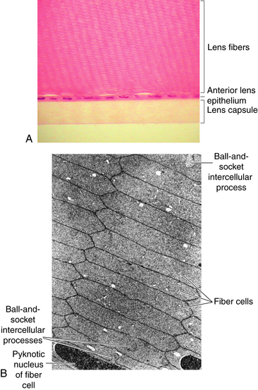
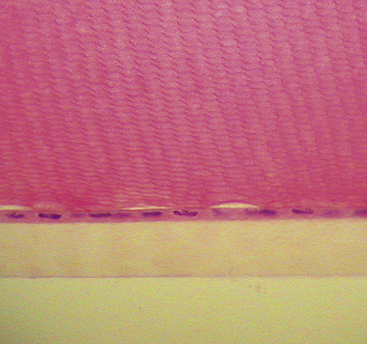
FIGURE 5-4 Light micrograph of anterior lens epithelium and capsule. Honeycomb appearance of lens fibers above epithelium.
The lens becomes more sharply curved, and because of the thickness variation of the capsule, a bulge is most apparent at the center of the anterior surface.36,37 The posterior surface increases in curvature only slightly; however, the steepness of the anterior surface does not become greater than that of the posterior surface.12 As the lens thickens axially, the equatorial diameter decreases, and the anterior lens pole moves toward the cornea. It was previously thought that there was no posterior lens movement, but a small amount of movement of the posterior pole in a posterior direction has been identified.8,38,39 The curvature of the internal surfaces, seen at the zones of discontinuity and the boundaries of the nuclei, mimics the changes in the surface curvatures and contributes to the increase in the total dioptric power.8 This thickening of the lens from anterior to posterior occurs in the nuclear region, but the thickness of the lens cortex remains unchanged.40 The changes occurring in accommodation described in 1855 by Helmholtz have been demonstrated using current technology.5,41
Strobe lights and back-up alarms tend to get lost in the environment. Not blue spot lights, they literally stick out like a beacon. This light paints a blue dot about 6″ in diameter on the ground in front of or behind a piece of equipment as a vibrant visual aid. If a piece of equipment is coming from around a corner, the light will be there ahead of it giving warning to the pedestrian or other drivers.
Blue spotlightBulb
Lens fiber production continues throughout life, with the new lens fibers being laid down outer to the older fibers; growth results in concentric layers of secondary lens fibers. The structure of the lens is similar to an onion; each layer of fibers approximates a layer of an onion, but then each layer is made up of adjacent fibers. A section through the equator of the lens shows that the fibers cut in cross section are mostly hexagonal in shape and arranged in concentric rings (Figure 5-6). The cross section dimensions of a fiber are approximately 3 by 9 μm.24 Each fiber has a long crescent shape, with the broad sides parallel to the lens surface and the narrow sides at an angle to the surface and located near the lens poles.
FIGURE 5-8 Fetal and adult lenses, showing sutures and arrangement of lens cells. A, Fetal nucleus. Anterior γ suture is at a, and posterior suture is at b. Lens cells are depicted as wide bands. Cells that attach to tips of γ sutures at one pole of lens attach to fork of γ at opposite pole. B, Adult lens cortex. Anterior and posterior organization of sutures is more complex. Lens cells that arise from tip of a suture branch insert farther anteriorly or posteriorly into a fork at opposite pole. This arrangement conserves shape of lens. In this drawing, for educational purposes, the suture appears to lie in a single plane, but the reader should remember that the suture extends throughout the thickness of the cortex and nucleus to the level of the γ sutures in the fetal nucleus. C, Adult lens, showing nuclear zones, epithelium, and capsule. Thickness of lens capsule in various zones is shown.
Bluelight
Strobe lights and back-up alarms tend to get lost in the environment. Not blue spot lights, they literally stick out like a beacon. This light paints a blue dot about 6″ in diameter on the ground in front of or behind a piece of equipment as a vibrant visual aid. If a piece of equipment is coming from around a corner, the light will be there ahead of it giving warning to the pedestrian or other drivers.
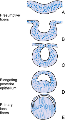
The structure of the adult lens is determined during embryologic development. The lens vesicle, the first lenslike structure observable in the developing embryo, is composed of a layer of epithelial cells that form a hollow sphere. The cells are positioned so that the apical surface lines the lumen of this sphere. The posterior cells differentiate and elongate, forming the primary lens fibers. As these fibers grow and reach the anterior cells (Figure 5-2), the center of the sphere fills. Thus the adult lens has no posterior epithelium because it was used to form these first lens fibers. During the rest of the life of the lens, cell division occurs in the germinative zone of the epithelium just anterior to the lens equator, and the cells thus formed elongate to form secondary lens fibers that are laid down outer to all earlier fibers. With age, the lens continues to grow as it forms new fibers (see Chapter 7).
2023322 — SWIR cameras use sensors to detect the reflection and emission of light in the SWIR wavelength range, which is between 0.9 and 2.5 µm. These ...
FIGURE 5-10 Optical section of normal adult lens. 1, Anterior capsule; 2, anterior line of disjunction (anterior epithelium); 3, anterior surface of adult nucleus; 4, anterior surface of fetal nucleus; 5, inner layer of anterior half of fetal nucleus, containing anterior γ suture; 6, inner layer of posterior half of fetal nucleus, containing posterior γ suture; 7, posterior surface of fetal nucleus; 8, posterior surface of adult nucleus; 9, posterior line of disjunction; 10, posterior capsule.
The lens is attached to the ciliary body by a group of threadlike fibers, the zonules (of Zinn), or the suspensory ligament of the lens (Figure 5-9). The fibers belong to a category termed microfibrils. The macromolecular composition that accounts for the remarkable extensibility of the zonules is complex and still undetermined.15,30 The zonules appear to be formed of extracellular matrix, that includes fibrillin and elastin, both of which have a role in the synthesis of elastic fibers.31 However, biomolecular analysis indicates that there are no true elastic fibers present in the zonules.31
Blue spotlightpng
(From Paterson CA, Delamere NA: The lens. In Hart WM Jr, editor: Adler’s physiology of the eye, ed 9, St Louis, 1992, Mosby.)
The posterior lens surface is attached to the anterior vitreous face by the hyaloid capsular ligament, a circular ring adhesion. Within this ring is a potential space, the retrolental space (of Berger), an area of nonadhesion between the vitreous and the lens (see Figure 6-12).
This is our house brand light. We tested lights from several manufacturers before we found one that lived up to our expectations.
As the lens fibers reach the poles they meet with the other fibers in their layer, forming a junction known as a suture. The anterior suture is formed by the joining of the apical aspects of the fibers, and the posterior suture is formed by the joining of the basal aspects.4,13 The secondary fibers formed during embryologic development meet in three branches, forming Y sutures. The anterior suture is an upright-Y shape and the posterior suture an inverted-Y shape (see Figure 5-8, A).18 As growth continues and the lens becomes larger, the sutures become asymmetric and dissimilar. The limbs of the anterior and posterior sutures are offset, and the complexity of the sutures contributes to lens transparency.29 The sutures formed after birth are more stellate shaped; sutures formed through early adulthood have 6 to 9 branches; and 9 to 15 complex branched stars are formed in middle to old age29 (see Figure 5-8, B).
.47in. 11.9mm .41in. 10.4mm .07in. 1.8mm .04in .9mm .25in. 6.4mm .13in. 3.2mm .29in. 7.4mm .10in. 2.5mm .08in. 1.9mm .08in. 1.9mm. DIM B. DIM A .47in. 11.9mm .
Also, although these lights were originally developed with the intention of lighting up the rear of the lift, we are finding that it is becoming more an more common to see them mounted all around the lift.
The vitreous has a passive role in accommodation, probably serving only as support for the lens.42 During ciliary muscle contraction, the choroid is pulled forward slightly, perhaps aiding in the correct orientation of the photoreceptors in relation to the entrance pupil. Contraction of the ciliary body in accommodation, by decreasing the circumference of the sclera, may lead to an elongation of the axial length of the eye.39
This is our house brand light. We tested lights from several manufacturers before we found one that lived up to our expectations.
2020531 — Download this high-quality LED light bulb clipart in PSD and PNG formats for free. Perfect for commercial or personal use.
FIGURE 5-5 Composite drawing of crystalline lens, cortex, epithelium, capsule, and zonular attachments. A, Anterior central lens epithelium, seen in flat section and cross section. Size and shape of these cells can be compared with those of cells in B, intermediate zone, and C, equatorial zone. At equator, dividing cells are elongating (arrows) to form lens cortical cells. As they elongate, cells send processes anteriorly and posteriorly toward sutures, and their nuclei migrate somewhat anterior to equator to form lens bow. At the same time, nuclei become more and more displaced into lens as new cells are formed at equator. Lens capsule (d) is thicker anterior and posterior to equator than at equator itself. Anterior and equatorial capsule contains fine filamentous inclusions (double arrows); these are not present posteriorly. Lens fibers elongate into flattened hexagons (e) in cross section. Zonular fibers (f) attach to anterior and posterior capsule and to equatorial capsule, forming pericapsular or zonular lamella of lens (g).
2024717 — Congratulations on your remarkable 10 years of service. Happy ... Happy 10-Year Work Anniversary! A Decade of Partnership. We'll always ...
2022314 — Full-frame sensors are available in both DSLR and mirrorless cameras. They have the same dimensions as the 35mm film, hence the name. The 35mm ...
Surprisingly, businesses have found that strobe lights and back-up alarms tend to get lost in the environment. Not blue spot lights, they literally stick out like a beacon. This light paints a blue dot about 6″ in diameter on the ground in front of or behind a piece of equipment as a vibrant visual aid. When a piece of equipment is coming from around a corner, the light will be there ahead of it giving warning to the pedestrian or other drivers.
Your Light Bar may require our Gutterless Mount Kit. Will fit both LED and Rally Lights.
Firstly, safety is paramount in the work place. Often times pedestrians and employees become distracted by paperwork, noise, music, phones or a host of other things while in the warehouse. If not distractions, then blind corners, crossing aisles, and loading docks. With forklifts moving about (especially electrics), this is a recipe for disaster. See our article on FORKLIFT SAFETY LIGHTS.
We strongly believe that our forklift blue spotlight is the best among a bloated market. After purchased many of the competing blue lights to compare, we have found that they all fall behind in quality. When I say quality I’m not just talking about the construction but also the light quality. Many of the other units (even ones more expensive than ours) have a very broad and weak beam. What good does it do to purchase a safety product that isn’t effective?
(From Krag S, Andreassen TT: Mechanical properties of the human posterior lens capsule, Invest Ophthalmol Vis Sci 44[2]:691, 2003.)
DCM Sistemes PRC0606B-850C/AM.
These factors result in a thickened lens with more sharply curved surfaces and thus increased lens power. The stimulus that initiates the accommodative mechanism is retinal blur.34 The accommodative mechanism is dependent on cone stimulation with little influence by rods.35
The primary lens fibers from the elongating posterior epithelium form the very center of the lens, the embryonic nucleus, and all subsequent lens fibers are laid down outer to this core. Cell mitosis then begins in the preequatorial region of the epithelium, the new cell migrates toward the equator, and then elongates, forming a lens fiber. All such fibers formed are secondary lens fibers. The fetal nucleus includes the embryonic nucleus and the fibers surrounding it that are formed before birth. The adult nucleus is considered to include the embryonic and fetal nuclei and the fibers formed from birth to sexual maturation. The lens cortex contains the fibers formed after sexual maturation (Figure 5-8, C). The cortex can be divided into superficial, internal, and deep zones,13 but these are arbitrary divisions and generally are not clinically significant. Some consider the fibers formed before sexual maturation the “juvenile nucleus,” those added before middle age the “adult nucleus,” and the remaining fibers the “cortex.”25 The lens cortex has the lowest and the embryonic nucleus has the highest index of refraction.
Blue LEDspotlights Outdoor
The refractive power of the unaccommodated lens is approximately 20 diopters (D) and depends on the (1) surface curvatures, (2) refractive index, (3) change in index between the lens and surrounding environment, and (4) length of the optical path. The lens has a gradient refractive index because of changes in optical density throughout the lens; the index increases from the anterior to the center of the lens and decreases toward the posterior surface. The refractive index is a factor of protein concentration within the lens fibers.8 The variations in index provide additional refractive power.9,10 The power of the lens increases in accommodation, with the maximum accommodative amplitude, 14 D, reached between ages 8 and 12 years.11 Accommodative power decreases with age, approaching zero after 50 years.12
When the ciliary muscle contracts, the diameter of the ciliary ring surrounding the lens decreases, reducing the tension that the zonules exert on the lens and allowing the lens capsule to assume its preferred spherical shape. The lens capsule transmits the reduction in the zonular pull to the lens, molding the lens into its accommodated form.
Because the lens has no vascular supply and the fibers lose their cellular organelles as they age, some cell-to-fiber and fiber-to-fiber mechanism of communication is necessary. There is an extensive network of gap junctions throughout the lens along the lateral fiber membranes to account for the facility with which nutrients and ions move within the lens.17,27 These gap junctions have a different packing arrangement and different protein connexins, forming the channel than do the typical gap junctions.21 The gap junctions are not evenly distributed throughout the lens, with few near the poles, more toward the equator, and seemingly fewer junctions in deeper layers.13,21,22 Micropinocytic vesicles at apical and basal aspects of fiber membranes and significant areas of membrane fusion also allow movement of material from fiber to fiber, contributing to communication between fibers in addition to the gap type of junctions described.13,20,22,28
Featuring: Built in Flash, USB-C Charging, 20 Built In Creative Filters, 9 Different Scene Modes Inc: Party, Beach, Night, & Portrait. Contents include: - ...
A, Formation of lens placode, precursor of lens. B, Invagination forming lens vesicle. C, Hollow lens vesicle lined with epithelium. D, Posterior cells elongate, becoming primary lens fibers. E, Primary lens fibers fill lumen, forming embryonic nucleus. Curved line formed by cell nuclei is lens bow. Anterior epithelium remains in place.
Lens fiber cytoplasm contains a high concentration of proteins, known as crystallins, which account for approximately 40% of the net weight of the fiber. The distribution and concentration of crystallins contribute to the gradient refractive index.25 The crystallin concentration varies from approximately 15% in the cortex to 70% in the nucleus.26 A cytoskeletal network of microtubules and filaments provides structure and also provides stability by being anchored to the plasma membrane.18,25 The lateral membranes have numerous and elaborate interdigitations along the fiber length that take various shapes, such as ball-and-socket and tongue-in-groove junctions, and allow for sliding between fibers4,17,18 (Figure 5-7). The fibers also are joined by desmosomes.
Lastly, though not OSHA required in general, we have had many customers tell us that their inspector or their safety advisors required the use of forklift blue spotlight in in heavy use facilities. Basically light has become so prevalent in the industry that it’s getting rare to see large facilities not using them. Here’s a link to OSHA’s COMMENTARY ON LIGHTS.
2022831 — Ambiente di sviluppo per la visione intelligente. I progettisti di applicazioni di visione intelligente come telecamere per il traffico e di ...
The border between the apical membrane of the anterior epithelium and the apical membrane of the elongating fiber is known as the epithelium-fiber interface (EFI). Nutrients and ions are exchanged across the EFI. It was once assumed that such movement was facilitated by gap junctions, but disagreement now exists on whether gap junctions are present.19,22,27,28 Gap junctions are usually found on the lateral cell membrane, and the EFI involves apical surfaces. Few true gap junctions have been visualized in tissue preparations. Minimal coupling occurs between the epithelium and fibers in the central zone (i.e., near the poles and sutures), but such junctions increase toward the germinative zone.21 Pinocytosis does occur at this interface, facilitating exchange.18
(From Paterson CA, Delamere NA: The lens. In Hart WM Jr, editor: Adler’s physiology of the eye, ed 9, St Louis, 1992, Mosby.)
BlueSpot Light for Forklift
The crystalline lens is an avascular, transparent elliptic structure that aids in focusing light rays on the retina. The lens is located within the posterior chamber, anterior to the vitreous chamber and posterior to the iris (Figure 5-1). The lens is suspended from the surrounding ciliary body by zonular fibers. It is malleable, and ciliary muscle contraction can cause a change in lens shape, increasing the dioptric power of the eye. The mechanism that causes an increase in lens power is accommodation, which allows near objects to be focused on the retina.
202224 — In short, a lighting cue list is the full collection of a production's lighting cues. Sometimes referred to as a cue sheet, these lists ...
When the emmetropic eye is viewing a distant object, the ciliary muscle is relaxed, the diameter of the ciliary ring is relatively large, and the zonules are in a stretched configuration exerting tension on the lens capsule holding the lens in the unaccommodated state, such that the image lies on the retina. When a near object is to be focused on the retina, an increase in the refractive power of the eye must occur. This increase in power is called accommodation and is accomplished by a change in lens shape brought about by contraction of the ciliary muscle. Von Helmholtz33 is credited with determining that the following occur during accommodation:
The lens capsule is a transparent envelope that surrounds the entire lens. The capsule is a basement membrane and with time becomes the thickest in the body.13 Its thickness varies with location. At the posterior pole, it is thinnest (approximately 3.5 μm) and does not appreciably increase with age; the thickness at the equator increases slightly with age, and on average is 7 μm. The capsule thickness at the anterior pole increases with age from approximately 11 to 15 μm. The annular region surrounding the anterior pole appears to be the thickest. It too increases with age from approximately 13.5 to 16 μm.14 The capsule consists primarily of collagen; it contains no elastic fibers but is highly elastic because of the lamellar arrangement of the fibers.4,15 It encloses all lens components and helps to mold the shape of the lens. The capsule would prefer to take a more spherical shape, but this tendency is counteracted by the pull from the zonular fibers. The zonular fibers insert into the capsule, merging with it in an area from the equator to near both poles. This coincides with the annular area mentioned previously. This outer superficial zone of the capsule is called the zonular lamella and consists of zonules interconnected with matrix.4 The lens capsule provides some barrier function preventing large molecules, such as albumin and hemoglobin,4 from entering the lens. The anterior lens capsule is produced by the anterior epithelium and thickens with age. The posterior lens capsule may receive some contribution from the basal membrane of lens fibers, but the thickness of the posterior capsule changes minimally throughout life (Figure 5-3).16–18
The band of cells in the preequatorial region that lies just anterior to the equator is called the germinal zone, the location of cell mitosis. Cell division continues throughout life; as each cell divides, a daughter cell migrates posteriorly towards the equator, withdraws from the cell cycle, and differentiates into a lens fiber. Each newly formed cell elongates; the basal aspect stretches toward the posterior pole and the apical aspect toward the anterior pole (Figure 5-5). This process occurs all around the equator, with fibers stretching toward the poles from all aspects of the lens periphery. As the cells in each layer elongate, the cellular nuclei move with the cytoplasm. A line drawn to connect the dots of these nuclei would have an arcuate shape toward the anterior aspect, a configuration called the lens bow (see Figure 5-2). Eventually, as it loses all cellular organelles, the elongated cell becomes a lens fiber. The anterior end of the lens fiber (the apical surface) insinuates itself between the epithelial layer and the underlying lens fibers. The new fibers are laid down outer to the older fibers; the more superficial fibers are longer than deeper fibers, and the youngest cells lie directly below the epithelium and the capsule. All fibers formed from mitosis in the germinative zone are called secondary lens fibers. Once it loses its nucleus, the mature lens fiber has lost its attachment to the basement membrane.17
Adjacent to the anterior lens capsule is a layer of cuboidal epithelium—the anterior lens epithelium (Figure 5-4). These cells secrete the anterior capsule throughout life and are the site of metabolic transport mechanisms. As noted, no posterior epithelium is present because it was used during embryologic development to form the primary lens fibers. The basal aspect of the epithelial cell is adjacent to the capsule, and the apical portion is oriented inward toward the center of the lens. The lateral membranes of the epithelial cells are joined by desmosomes and gap junctions.17,19–22 Although zonula occludens were once thought to join the epithelial cells,23 more recent studies report few, if any, tight junctions.18
The lens is biconvex, with the posterior surface having the steeper curve. The anterior radius of curvature measures 8 to 14 μm, and the posterior surface radius of curvature measures 5 to 8 μm. The centers of the anterior and posterior surfaces are called the poles, and the lens thickness is the distance from the anterior to posterior pole. The thickness of the unaccommodated lens is 3.5 to 5 mm and it increases 0.02 mm each year throughout life.1–3 The lens diameter is the nasal-to-temporal measurement and in the infant is 6.5 mm. The diameter reaches 9 mm during the teenage years and does not change significantly,4–6 although some report a small age-related increase in diameter.7 The equator is the largest circumference of the lens at a location between the two poles.


 Ms.Cici
 8618319014500