
mlh-policies/member-event-guidelines.md at main - m.l.h
JavaScript seems to be disabled in your browser. For the best experience on our site, be sure to turn on Javascript in your browser.
A metallurgical microscope is a special version of a standard light microscope for the study of materials, such as Metals, plastics, ceramics and others. Since materials are usually not transparent solid structures, metallurgical microscopes often have an upright light unit. Moreover, this type of microscope is characterized by extensive magnification, e.g. for detailed investigations of surface structures. Application areas of metallurgical microscopy are industry, material science and research.
Standard bright-field microscopes are used for daily laboratory routine in research and diagnostics for simple, standard applications that require no special equipment. Therefore simple optical systems and lenses are applied.
Darkfield microscopy
The physical principle of fluorescence is used to selectively visualize and localize defined fluorescent structures, while non-fluorescent structures remain dark in order to obtain a high image contrast. For this purpose, fluorescent dyes (fluorochromes) are used with specific excitation and emission filters installed in the optical path of the microscope. A wide range of fluorescent dyes with different colors are available, which are used in molecular biological, biomedical and clinical research. For example, in immunohistochemistry, fluorescence-in-situ-hybridization and for visualization of cells or cellular components in living/fixed specimens.
Polarizationmicroscopy
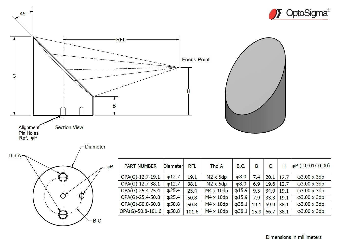
Figure 3 shows initially unpolarized light incident at the polarizing angle θp, for which the reflected light is completely polarized with its electric field ...
USA OptoSigma Corporation 1540 Scenic Avenue, Suite 150, Costa Mesa, CA. 92626 TEL. +1-949-851-5881 sales@optosigma.com USA
Fig 1: Lunghezza Focale. Se il soggetto non è posto all'infinito, minore è la sua distanza dalla lente, più lontana dalla lente si formerà la sua immagine.
JavaScript seems to be disabled in your browser. For the best experience on our site, be sure to turn on Javascript in your browser.
Phase contrastmicroscopy
Wichtigste Übersetzungen. Englisch, Deutsch. syringe, hypodermic syringe n, (instrument: for injecting fluid) (Medizin), Spritze Nf.
Polarization microscopy is used for the analysis of optically anisotropic samples. The primary objective of polarization microscopy is not magnification of an object, but rather the analysis of optical properties such as refractive index or birefringence for sample analysis. The method is mainly used in mineralogy and in industry for testing plastics or mineral building materials in order to gain insights into their composition.
Confocalmicroscopy
Facing tedious and variable measurements from images, researchers use MIPAR to detect and measure features: ✓ Significantly faster and with much less user ...
Type of microscope
We detect that you are accessing the website from a different region. You will be redirected to a local version of OptoSigma.
HC Series Cigars have exploded onto the scene by boasting superb blends at attractive prices. Save on HC Series cigars at CI.

Fluorescence microscope
At Advanced, we pride ourselves on supporting millions of people across the UK by delivering exceptional enterprise and market focused software and service ...
2024426 — A standard zoom lens covers wide-angle to telephoto focal lengths around 50mm. In addition to snap shots and portraits, these lenses prove their ...
Mirrorless, DSLR or Point and Shoot: Which Camera is Best for Macro Photography? · 1) Fujifilm x100s · 2) Olympus Stylus TG-2 Tough · 3) Canon 100mm f/2.8 Macro ...
SINGAPORE OptoSigma SEA 83 Science Park Drive, #02-01.The Curie, 118258 TEL. +65 6909 9318 sales@optosigma-sea.com SINGAPORE
We detect that you are accessing the website from a different region. You will be redirected to a local version of OptoSigma.
Differential interference contrastmicroscopy
An inverted microscope is an upside-down standard light microscope. This type of microscope is characterized by locating the objective underneath the stage and pointing upwards to the specimen. Inverted microscopes are mainly used for live cell analysis of cell cultures growing in culture medium. Culture dishes are not only available with standard coverslip bottoms (thickness 0.17mm) but also in various material and bottom thicknesses. Therefore, for some models special objectives are available that can correct for different bottom glass thicknesses. In addition, inverse microscopes are also used for studies of thicker specimens. Inverted microscopes are also available as dark field, polarization or fluorescence microscopes.
Light up stage NF660 laser lights DMX mode: pattern selections, laser ... laser lights) project the same patterns as the master laser lights projector.
A dark field microscope produces a contrast-enhanced image by indirect illumination of the specimen, thereby also unstained specimens can be displayed with high contrast. Using this technique, direct light is bypassing the objective, only light scattered by the specimen enters the objective. As a result, the background appears dark or black, only the specimen is illuminated and even small structures can be visualized with high contrast. Dark field microscopy in biology and medicine is especially used for transparent and low contrast specimens, for example, studying blood, small animals or micro-particles in material science.
Lightfield microscopy
UK Elliot Scientific Limited Unit 11 Sandridge Park, Porters Wood, St Albans, AL3 6PH TEL. +44 (0)1582 766 300 sales@elliotscientific.com United Kingdom
Phase contrast microscopy is a contrast-enhancing technique to visualize structures difficult to detect with bright-field microscopy due to a lack of contrast, without the need of a staining. When penetrating a medium, light propagates with different speeds depending on the refractive index of the medium. This leads to phase differences, which are converted to differences in brightness by the microscope using phase rings. Areas of application of phase contrast microscopes are mainly the observation of living biological samples in order to resolve fine structures with high contrast.
2023710 — A practical is a light source appearing anywhere in the frame of your shot. It can be a key light for your subject while also being a part of your set.


 Ms.Cici
 8618319014500