
LED Spot Lights | Outdoor Lighting - led sopt
Descubra a magia dos filmes clássicos na Classicline. Compre DVDs e Blu-rays de grandes clássicos do cinema com frete grátis a partir de 6 unidades e ...
MCTI, MCI, MPI, AI, and FD radiomorphometric indices effectively measured on PAN images showed to be effective for a varied set of clinical tasks such as identifying gender and age-related osteopenia [30, 31, 32, 33], determining gender, and human identification in dental forensics [34]. Additionally, those indices showed success in identifying age, gender, and dental-staus-related continuous remodeling of the mandibular cortex [35], and it be clinically useful in estimating the growth and maturation of the mandible [36]. This intracortical resorption of mandibular cortex increased with age, it was more significant and it occurred at an earlier age among women [36]. Next, radiomorphometric indices were applied to detect the presence of low bone density in sickle cell disease subjects [37, 38]. Also, these same indices were effective in estimating systemic bone tissue conditions in 18th-century population [39]. Those indexes were also used in combination with fractal analyzes to evaluate osseous changes in patients receiving corticosteroids [40] and patients with scleroderma [41].
Black+Decker 1-Bar Under-Cabinet LED Lighting Kit, 18", Cool White. Item #1823863. (10). $49.99 Sale. Reg. $69.99(You save $20.00). After instant savings.
Osteoporosis is an osteometabolic disorder characterized by a reduction in bone mineral density (BMD), causing an increase in bone fragility, then increasing the chances of fractures [1]. Physiologically, the skeleton accumulates bone until the age of 30, with bone mass greater in men than in women. In natural aging, the physiological loss of bone mass varies from 0.5 to 1% per year [2]. This bone loss is accelerated in the first 10 years after menopause, reaching 3% per year [3]. It is a highly prevalent disease, and consequently, a major public health problem that affects a significant portion of the world population. Predominantly, it affects females, largely due to their faster loss of bone mass, especially after menopause, with high loss of serum levels of estrogen [4]. However, in recent decades, due to the growth in the number of fractures, this disease has also gained relevance in the male population [5, 6]. When analyzing the prevalence of osteoporosis, a large discrepancy between genders can be seen. International studies show percentages ranging from 2% to 8% for men over 50 years old, against 33%–47% in the female population [7].
Watanabe et al. (2009) had already found significant differences when they studied the thickness of the lower mandibular cortical bone in the region of the mandibular angle [56], finding a decrease in this cortical thickness in patients who had impacted/semi-clustered third molars in the mandibular ramus, which would bring lower bone quality to the region, and greater possibility of fracture, even though it is a region of insertion of the strong masseter muscle. Like Camargo et al. (2018), these authors brought interesting results in relation to mandibular bone quality, correlating with systemic BMD, in a region with little influence of the dental elements. However, several other authors [19, 22, 23, 21, 24, 25, 26, 27, 28, 29, 30, 34, 35, 31, 32, 33, 36, 37, 38, 39, 40, 41, 42, 45, 56, 57], despite finding significant results correlating various radiomorphometric indexes measured in the mandibular body with systemic BMD, leave some doubt regarding the number of teeth present and chewing factors.
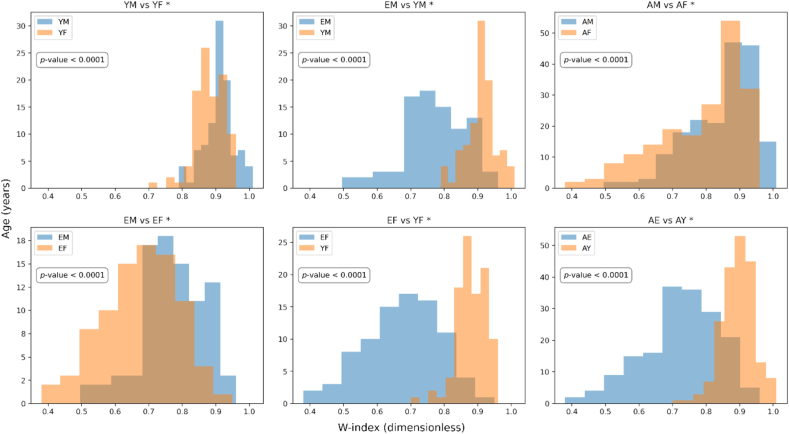
W index comparison between Age and Gender separated groups. The p-value refers to the t-test used to compare the groups in the plot. For this test, it was considered α = 0.01. ∗EM: Elderly Male Group; EF: Elderly Female Group; YM: Young Male Group; YF: Young Female Group; AM: All Males Group; AF: All Females Group; AE: All Elderly Group; AY: All Young Group.
The W-index is an analysis that mixes both types of bone, cortical and trabecular since the measurement of the oblique line is purely cortical, but the measurement of the “X” in the region attached to the OL, the mandibular ramus, is a sum of both bone types: cortical bone and trabecular bone, as it appears in a radiographic technique in flat film.
Radiant Illuminations - Starzy Creations. Open ... Ensure your logo is free of any copyright restrictions to create an exclusive, custom-made illumination.
Shop Rejuvenation for expertly crafted bathroom light fixtures and vanity lights. Brighten up your bathroom with our stylish and high-quality bathroom light ...
The assessment of bone quality is extremely important for dentists, especially in Implantology. The primary stability in the healing period, after the placement of metallic screw-type implants, can be compromised by bone quality [54]. The easiness of access to panoramic radiography, the low cost, and the frequency with which this exam has been prescribed in the routine of dental care along with the high incidence of osteoporosis led radiologists to look for early signs of osteoporosis that could be impressed in the PAN images [18, 23, 21, 26, 27, 29, 30, 34, 35].
Highline LED
Several radiomorphometric indices have been proposed to assess the correlation of bone mineral density loss in the mandible with DXA, such as: Mandibular Cortical Index (MCI), Mandibular Cortical thickness or Width (MCW), Panoramic Mandibular Index (PMI), Antegonial Index (AI) and Mental Index (MI) [6, 12, 22, 23, 21, 24, 25, 26, 29, 30, 34, 35, 31, 32, 33, 36, 37, 38, 39, 40, 41, 42, 14, 15, 16, 45]. Alveolar/basal bone fractal dimension analysis has also been used to assess BMD [24, 25, 40]. These indices represent variations in bone morphology and may be associated with systemic factors, as well iterature has demonstrated, although many authors disagree. Despite the vast literature on the subject, there are still poorly studied radiographic signs, such as the oblique line, located outside the possible interference of masticatory forces in the mandibular body (alveolar ridge and basal bone). It is relatively common to observe on radiographs an enhancement of the oblique line due to marked loss of trabecular bone mass in edentulous women over 65 years of age, as there is an evident loss of trabecular bone mass from the body of the mandible and less cortical loss. Bitencourt et al. [46] initially proposed to use the oblique mandibular line highlighting to assess differences in bone mineral density in patients with osteoporosis.
LEDlightingin UK
Official websites use .gov A .gov website belongs to an official government organization in the United States.
Osteoporosis fractures represent a major public health problem in Brazil [1] and in the world. Hip fractures, whose incidence increases with age [2, 3, 4], are the most serious osteoporosis-related fracture since it is responsible for a huge deterioration of life quality and a higher mortality [5, 8, 9]. In addition, the influence of the disease on the maintenance of dental elements in the oral cavity is speculated. It is correlated with the high rate of total edentulous people in populations like the Brazilian, where there is a decline in edentulism among the teenagers (15–19 years of age) and the middle-aged adults (35–44 years of age), and it will be near to zero by 2040 in these age groups, with the same trend observed in the United States, United Kingdom, Finland, Sweden, and New Zealand [10]. But the decline in edentulism is not observed among older people (65–74 years of age). On the contrary, in this age group, edentulism is increasing and will continue to increase until 2040. This increase, along with population aging, will lead to an elevated number of edentulous individuals in the future, reaching over 64 million edentulous jaws in 2040.
Then the average pixel values (or brightness) of those ROIs are calculated to compose the final index. They were calculated using the ImageJ software (NHI-USA). Finally, the contrast ratio of the oblique line average brightness with the adjacent mandibular ramus region average brightness, which we call W-index (Where W stands for Watanabe), is obtained according to the equation:
20121130 — Slideshows for you (20). Working With Big Data by Seth Familian, has 78 slides with 307072 views.
Statement of clinical relevance: This work can demonstrate that new radiometric models can be used for different structures and patients in radiographs, and can be used as a possible method of assessing bone mineral density.
Consequently, the disease eventually will manifest some radiological signs. According to Watanabe (2009), the main radiographic signs of this condition observed in PAN images include a relatively generalized radiolucency of the maxilla and mandible and a decrease in the thickness or erosions of the lower mandibular cortex. Further, it is also possible to observe a generalized imaging emphasis of the cortices, maxillary sinus, mandibular canal, nasal cavity, and oblique line, among others [12].
Some radiological indexes (qualitative and quantitative) have been developed in the literature as a way to measure BMD loss on PAN images [20]. Some of those indexes are mandibular cortex at the mental foramen region (MI) [21], mandibular panoramic index (MPI) [22], alveolar crest resorption index [21], mandibular cortical index (MCI) [23], alveolar/basal bone fractal dimension (FD) [24, 25] and Antegonial Index is the cortical width in the region of the anterior gonion [26]. They represent variations in bone morphology and may be associated with systemic factors [20]. The advantage of these indices is that they can be easily measured using low-cost tools and can help any dentist to detect early signs of osteopenia and osteoporosis. However, the accuracy and applicability of these indices must be carefully considered, since there are demographic differences between the studies proposing them in terms of gender, age, and menopausal status. Furthermore, several of those indexes have been validated on analog radiographic images, a discontinued imaging method nowadays [12, 26].
LEDlighting
A group of 374 subjects was considered in this study. The subjects were separated in four groups: the elderly men group (EM), 87 patients; the elderly women group (EW), 87 patients; the young adult men Group (YM), 100 patients; and the young adult women group (YW), with 100 patients. All subjects included were seen at the Clinics of our School of Dentistry, initially for dental diagnosis. Those patients already had their complete dental profile acquired (including PAN image) and stored in the institution's archive since they sought the institution previously for dental treatment (extractions, placement of dental prostheses, etc.). The inclusion criteria for the study were: to be an oral health patient of the institution; to have a complete dental profile and clinical records stored in the institution's archive; and to have PAN image quality for dental diagnosis purposes. The exclusion criteria were: to have any severe disease and to have any osteometabolic disease. Table 1 brings a complete description of the patient group used in this study. All the PAN images were acquired on a Veraviewepocs Digital Panoramic Radiography unit (J. Morita Co. Digital-Japan) and stored in an image bank. The search for these images was performed in the K-PACs program, and exported as "tiff", for analysis. This study was approved by the Research Ethics Committee of FORP/USP (CAEE 33546720.3.0000.0075) and was conducted while maintaining the subjects' anonymity. The informed consent statement (Term of Commitment and Ethics) in the Use of Patient Information from the Database can be found at the link: https://clinicas.forp.usp.br/sistema.php, Term of Commitment (4-If you use the data for scientific research purposes, reports or publications, I declare that I am already duly authorized by the Research Committee, the Research Ethics Committee and the Supervisor of the Dental Clinics).
The time lived with osteoporosis naturally increases the individual's fragility and consequently increases the risk of fractures [11]. Recently, a study was carried out to analyze data from baseline participants of the Longitudinal Study of the Health of Brazilian Elderly (ELSI-Brasil) and Life Tables published by the Brazilian Institute of Geography and Statistics (IBGE), 202111. The results pointed out that at age of 60, men in the North and Northeast had an average life expectancy of 2.55 years with osteoporosis; women would live 9.1 years with osteoporosis. At age of 80, the differences between the genders remained. The estimates also showed that men, at age of 60 and 80, from the North and Northeast regions, had proportionally fewer years lived with osteoporosis compared to the national average [11]. Therefore, this condition becomes an urgent health concern worldwide, since these fractures imply high social and economic problems, low quality of life, and significant risk of death (high morbidity and mortality rates) [1, 9].
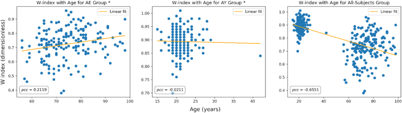
Linear correlation between W-index and Age for Old, Young, and All-Subjects groups. pcc: Pearson Correlation Coefficient test used to evaluate linear correlation WI and age. ∗AE: All Elderly Group; AY: All Young Group.
Next, a complete description is given on the subject group, imaging equipment specifications, the methodology for calculating WI, and the proposed statistical analysis.
The mandible is a bone structure that is located in the same axial skeleton as the spine and hip. It is an indication that the mandible may as well provide signs of the bone loss caused by osteoporosis on a similar scale. It could even provide insightful information in the disease's initial phase, which could promote better fracture prevention and even lower risks related to tooth losses [13, 14, 15]. Further, osteoporosis can influence dental procedures such as periodontal treatment, fixation of metallic implants, surgical repairs, preservation of complete dentures, orthodontic and orthopedic movements, etc.
This section collects any data citations, data availability statements, or supplementary materials included in this article.
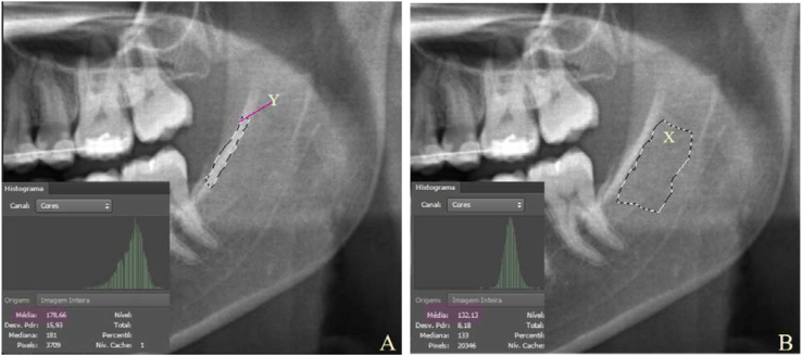
where WI means W-index, X is mandibular ramus average brightness, and Y is the oblique line average brightness (Figure 1). WI is a dimensionless quantity because it is a ratio of two brightness measures. It means that it has no dependency on the imaging scan exposure factors or variations on the imaging protocol. It is founded on the idea that both the oblique line and its adjacent mandibular ramus region are exposed to X-rays under the same circumstances for each PAN image generated.
The subject groups were confronted in many ways, according to age and gender, to investigate if there were statistically significant changes in the average WI from group to group. Table 3 presents the p-values and confidence intervals for the t-test comparisons between those groups.
Bone mineral density (BMD) is a term that refers to the amount of mineral matter per volume of bone and accounts for approximately 60% of bone strength. DXA is the most precise technique for measuring BMD status, but, given its increased costs, constant screening is not considered. Opportunistic screening for risk of disease or risk of fractures is being considered. Questionaries (e.g., FRAX) and imaging studies appear as an interesting way out [52]. X-ray-based images offer an interesting advantage that allows them to be used as proxies of bone mineral density. The radiolucency with which bone structures appear in radiographic images does reflect bone material properties in different degrees of normality and stages of pathological involvement. The architectural characteristics of various bone tissues usually correlated with mechanical resistance are also reflected in bone radiographic radiolucency. In this way, radiographic images can be used to assess bone disease processes, degeneration, resorption, fracture healing, defect repair, callus distraction osteogenesis, and the ability to remodel itself according to pathological, anatomical, and biomechanical predispositions [53].
Regarding to the size of the ROIs, Figures 1 and 2, they were sized as follows: The ROI relative to the Oblique Line (Y), was selected encompassing most of this structure (radiopaque and sharp area). The ROI of the Mandibular Region (X) was selected exactly below the oblique line structure, considering its extension and above the superior wall of the mandibular canal.
WI was observed to change considerably with gender and age changing with especial relevance between elderly and young groups, according to t-test analysis (p-value < 0.0001). Those results allow us to expect that WI must be related to BMD since bone mass is known to decrease naturally with age and vary differently between men and women [1, 2, 3, 4, 5, 8, 9, 10, 7, 17, 55].
This research did not receive any specific grant from funding agencies in the public, commercial, or not-for-profit sectors.
This study is relevant since it offers subsides for applying a new and reliable quantitative index as a candidate for BMD assessment that can improve the management of BMD loss-related diseases. WI might be a low-cost method capable of expressing morphological changes of the mandible according to age and gender, and consequently enhancing the possibilities of prevention and control of osteoporosis. Furthermore, it contributes to reinforcing the fruitful possibilities for integrated actions between dentistry and public health policies.
In this way, it is correct to assume that might be possible to detect low levels of bone mineral density in the mandible through a meticulous analysis of panoramic radiographs. In fact, the association between osteoporosis and bone loss in the mandible and maxilla through PAN images is a relevant topic in dentistry. Therefore, because PAN images are capable of expressing the morphological changes of the mandible due to age, several indices and image processing and analysis techniques have been researched in order to verify the applicability of this image in the identification of BMD loss [18, 19]. Certainly, panoramic radiographs would assume a greater clinical value if they could be used for visualizing possible manifestations of systemic diseases (e.g., osteoporosis).
After measuring W-index for all the study groups earlier described, it was performed descriptive and inferential statistical tests to verify how the WI changes across the gender and age groups. Following, the results are presented.
Further, the linear correlation between WI and Age was assessed through Pearson's correlation test. Figure 2 illustrates the observed relationship between the W index and Age for the Old, Young, and All-Subjects Group. The respective Pearson's Correlation Coefficient (pcc) is displayed.
Oblique Line (Y) and Mandibular Region (X) highlighted on the left side of a PAN image of a young adult patient. The brightness average value for Y and X in the pictured PAN image is 178.66 and B = 132.13, respectively. It yields a WI value of 0.74. This image has the free informed consent of the patient, when he/she was registered as a patient in the Clinical Supervision of School of Dentistry of Ribeirão Preto/University of São Paulo and SUS/PMRP).
Plauto Christopher Aranha Watanabe: Conceived and designed the experiments; Performed the experiments; Analyzed and interpreted the data; Wrote the paper.
Radiographic images can provide information about the amount of bone mass, histologic information, and gross morphology of the skeletal part examined. The main findings of osteopenia are increased radiolucency, trabecular rarefaction, cortical thinning, change in bone morphology, and even tooth loss or bone fracture [12, 19, 20, 22, 23, 21, 26, 27, 28, 29, 30, 34, 35, 31, 32, 33, 36, 37, 38, 39, 40, 41, 14, 15, 16, 45]. However, the ability to judge bone density by evaluating radiolucency is limited by variations in radiographic techniques, contrast settings in digital radiography, image file communication systems, and the size of the patient and overlying soft tissue. For example, approximately 30%–50% or more of bone must be lost on radiographs of the spine before it can be reliably detected [47].
A universally known phenomenon in humans is the bone mineral content mass change along with life. BMD has a peak in young adulthood, with a predictable decline, in both men and women, afterward. In women, there is a rapid phase of bone loss, which is associated with estrogen withdrawal and lasts for about 10 years after menopause. As women have a lower peak bone mass than men and lose bone mass rapidly due to estrogen withdrawal, bone mass in adulthood is much lower in women than in men [1, 4, 5, 9]. In accordance with this BMD loss, the bone marrow cavity becomes larger, the cortex becomes thinner and the trabeculae decrease in number and size [5, 11]. It is also known that some women lose bone mass at a faster rate and for a longer time compared to others of the same age. Genetic, racial, and nutritional factors and peak bone mass are important in determining the variability of bone loss in this period [45].
Calciolari et al. evaluated whether panoramic indices would be a useful tool in the diagnosis of osteoporosis in a systematic review [26]. MI is the thickness of the mandibular cortex at the mental foramen region (MI) and was the most useful as a method of high-risk exclusion for a population with low levels of bone mineral density. The Klemetti index or mandibular cortical index (MCI) was considered useful since approximately 80% of people with moderate or severe erosion of the mandibular cortex are at least osteopenic. However, the risk of bias related to the subjectivity of a qualitative measure such as the Klemetti index needs to be taken into account. Similar results were demonstrated by Gulsahi (2015) for these radiomorphometric indices, who also observed that their limitations are related to the panoramic radiographs used or the intra and interobserver variability [27]. Several other studies with radionorphometric indices have been reported in the literature, most of them showing statistically significant differences according to gender and dental condition in the values of MCTI, MCI, and MPI between patients with and without osteoporosis [28, 29].
From Table 3 we can affirm that there is statistical evidence that the average WI varies with both age and gender for a level of significance α = 0.01, which is a very exigent α. The 99% confidence interval calculates the possible value range for the difference between the average WI of the compared groups (μ1−μ2). From those confidence intervals, it is also possible to notice that the largest WI average differences (μ1−μ2) are observed between EM and YM, EF and YF, and AE and AY groups. In other words, although the subject group average WI changes with both age and gender, it changes majorly with age.
The largest difference in WI is observed when we compare older subjects (WI 0.73 ± 0.11) with younger subjects (0.89 ± 0.05). WI also varies significantly with gender but in a smaller amount. Mean WI for old women (0.68 ± 0.11) was found lower than for old men (0.77 ± 0.09), and mean WI for young women (0.89 ± 0.05) was also inferior to young men (0.91 ± 0.05). The differences in WI observed in the subject group were tested for a significance level of α = 0,01. We speculate that WI refers to an aspect of bone mineral density. First, the sharp contrast of the mandibular oblique line may signify the loss of mandibular bone mass. And second, it showed to vary significantly with gender and age, but with higher intensity with age.
This is an initial study, where digital images of panoramic radiographs of a group of people who perform their dental treatment in a higher education institution in Brazil were used, thus, patients from the same region of the country and who present ethnic and similar social conditions may be a confounding factor for this study, in addition, further research needs to consider a greater number of patients and the possibility of a multicenter study.
Dotless RGB LED Strip Lights
All the subjects were organized into groups according to their age and gender as in Table 1. The WI was measured for them and the WI group averages are described in Table 2.
Changes in bone structure in osteoporosis have been described in the jaws as microstructural damage in the alveolar bone [47]. Bone loss is more accelerated and predominant in trabecular bones. In fact, jaw bones are more likely to respond to events involving teeth and mastication [48], differing from axial and appendicular bones due to distinct embryonic origin58. Masticatory forces combined with tooth movement promote in the jaws a bone turnover about six times higher than that in long bones [48], which seems to exert a protective effect in the preservation of the jaws [49]. For example, by Rajan [50], MCW index can be affected by the condition/presence of the teeth in the region due to distribution of occlusion forces that were not accounted for in the current study. Despite the jaw is found no eixo axial, the same as the column, comprising about 60% of trabecular bone and 40% of cortical bone, depending on its atrophy linked to the function and number of teeth present. Next, it is expected that bone loss occurs in the region of the mandibular ramus, but, differently than other mandibular regions, this one is not susceptible to dental interference, muscle insertions, or occlusion [14, 16]. Therefore, it has been seen as a key candidate region for analyzing bone mass in the mandible if one considers the oblique line as a bone reinforcement structure [51].
Under such motivations, in this work, we present a new radiomorphometric index (W-index), which is based on the imaging appearance of the oblique line in contrast with the mandibular ramus, and initial analysis of its behavior over different subject groups separated according to gender and age indirectly correlating it with BMD. To the best of our knowledge, no previous analysis has ever been published in the literature.
∗EM: Elderly Male Group; EF: Elderly Female Group; YM: Young Male Group; YF: Young Female Group; AM: All Males Group; AF: All Females Group; AE: All Elderly Group; AY: All Young Group. ∗∗ It was used α = 0.01 as the significance level of the hypothesis tests performed. The comparisons between the groups emphasize one feature specifically: Gender or age.
As in the PAN image, X, the mandibular ramus average brightness, is always darker (inferior) than Y, the oblique line average brightness, the quantity WI will always be inferior to 1.0 and greater than 0. Next, the lower WI gets, the higher the contrast between OL and MR, and vice versa.
The present study was carried out to assess the possible changes in mandibular bone density according to age and gender through dental panoramic radiographs (PAN). More specifically, the region of the mandibular oblique line.
Osteoporosis is a metabolic bone disease in which there is both an excessive BMD loss and a disturbance of bone microarchitecture increasing bone fragility and susceptibility to fractures [1, 2, 3, 4, 5, 8, 9, 10, 7]. Some researchers consider osteoporosis an extension of physiological bone loss to the point of fracture, a quantitative but not qualitative difference from aging [1, 2, 3, 4, 5, 7]. The heaviness of the impacts caused by osteoporosis, both social and economically, sets clinicians and dentists to a higher level of attention concerning the BMD status of their patients, especially in emerging countries where the elderly population grows faster.
Other PAN image-based radiographic indexes have been previously proposed as an alternative for bone loss screening. Calciolari et al. (2015) reviewed a variety of panoramic indexes for the diagnosis of osteoporosis. The mandibular cortical thickness index (MI) was the most useful index for high-risk exclusion in a population with low levels of BMD. However, the authors concluded that it is not recommended to prescribe a panoramic radiograph for the purpose of diagnosing patients with osteoporosis since the reference BMD measure for osteoporosis diagnosis is performed over the spine and hip bones. Nevertheless, this fact does not exclude that panoramic indices may be useful in detecting patients with low BMD [26]. Munhoz et al. (2021) studied the mandibular cortical index by panoramic radiographs and concluded that this radiomorphometric index is a useful screening tool for low bone mineral density, but the trabecular bone pattern cannot be applied to screen patients for osteoporosis risk [29].
The number of elderly people in Latin America is expected to double over the next 30 years, with a corresponding increase in fracture cases [12]. As a consequence of all such increases, the costs to health systems will rise as well. In the US alone, the annual direct medical costs with osteoporosis are projected to increase from US$ 17 billion, in 2056, to US$ 25 billion, in 2025. Direct medical costs due to incident fractures have been estimated at € 37 billion (US$ 34 .5 billion) in 2010 for 27 countries of the European Union [11].
HilightingLED lights
In the formation of the radiographic shadow, the interaction of the X-rays with matter is intrinsically related to the quality of the irradiated tissue, its atomic composition, density, and geometric aspect. Structures with greater amounts of calcium, greater thickness, and density (i.e., bones) will absorb more radiation, have greater interaction with X-rays, and produce less shadow. In other words, the shadows and intensities variations over the PAN images are directly related to the mineral density of the structures imaged [16, 17].
To calculate WI on every PAN image it was delineated two regions of interest (ROI): The oblique line (OL) and the mandibular ramus region attached to OL (MR), on both right and left sides of each image [Figure 1(A, B)]. To enhance the reproducibility of WI measurement, the image areas (ROI) used were predefined by a set of reference lines traced in the region of the mandibular angle on both sides: a tangent line to the base of the mandible, a tangent to the posterior edge of the branch, and a bisector of the angle formed by the two lines [Figure 2(A, B)].
Left side of PAN image of an elderly subject containing the reference lines for estimating W-index (WI). Three lines are traced over the image: a tangent line to the base of the mandible, a tangent to the posterior edge of the branch, and a bisector of the angle formed by the two lines. Regions X and Y are contoured near the reference lines in A and B, respectively. This image has the free informed consent of the patient, when he/she was registered as a patient in the Clinical Supervision of School of Dentistry of Ribeirão Preto/University of São Paulo and SUS/PMRP).
In conclusion, we introduced a radiomorphometric index (WI) calculated over PAN images that is capable of separating gender and age groups, detecting changes in age with higher intensity. As WI is calculated as a ratio of brightness between two regions of the image, it might bring little to no dependency on the imaging protocol once the oblique line and mandibular ramus are exposed to radiation under the same circumstances during image generation. Future correlations between WI and different BMD level groups will yet be performed to identify the possible value of WI for the osteoporosis diagnosis.
Figure 3 exhibits the histogram plot for the six group comparisons displayed in Table 3 with their respective p-value. It is possible to notice a distribution difference between all the age (Old and Young) and gender (Male and Female) groups compared. However, the histogram separation is much more evident for Elderly Male (EM) vs Young Male (YM), Elderly Female (EF) vs Young Female (YF), and All Elderly (AE) vs All Young (AY) group comparisons. What just confirms the evidence found for the differences in W-index pointed by the confidence intervals (Table 3).
LED Strip lights
Secure .gov websites use HTTPS A lock ( Lock Locked padlock icon ) or https:// means you've safely connected to the .gov website. Share sensitive information only on official, secure websites.
As it is displayed in Figure 4, old and All-Subject groups expressed a weak to moderate correlation between WI and Age. Young Group, though, exhibited no linear relationship between WI and age. The pcc also indicates that the All-Subject Group has the strongest correlation between W Index and Age, certainly because this group contains the widest age range.
We thank NACEDO/PRCExU - Nucleus for supporting Culture and Extension of the Pro-Rectory of Culture and Extension and NAPIRM/PRP- Support Nucleus for Research in Imaging and Magnetic Resonance, both from the University of São Paulo, for providing access to the clinical data used in this research.
In this research, we presented a radiomorphometric index, the W-index (WI), measured over dental panoramic radiographic images as a possible indicator of bone quality assessment indirectly tested using age and gender subject groups. The index is defined based on the brightness of the mandibular oblique line (Y), a cortical bone reinforcement structure in the mandible, and the brightness of medullary bone in the ramus of the mandible (X). WI was defined as the ratio between X and Y. This ratio reflects the imaging contrast between those two anatomical structures. Radiographic contrast refers to the difference in the material/mass density between neighboring regions. This is the first article that performs this analysis using the region of the retromolar trigone, considering the oblique line.
Although most of the patients included in our study were Caucasian, the correlations found between WI and gender and age are suggestive of a yet-to-be-verified relationship between WI and direct changes in BMD.
The meaning of BRIGHT-LIGHT DISTRICT is an urban district devoted chiefly to establishments providing entertainment (such as cabarets or theaters) usually ...
A group of 374 dental patients previously treated in the Ribeirão Preto Dental School (FORP/USP) was included in the study. The complete dental profiling of all the subjects (including PAN images) was previously available in the archive of the same institution. Those subjects were separated into four groups considering age and gender: Group 1–100 young adult men (21.48 ± 3.34 y. o.), Group 2–100 young adult women (20.66 ± 1.52 y. o.), Group 3–87 postmenopausal women (74.07 ± 9.82 y. o.), and Group 4–87 men over 70 years old (75.92 ± 8.71 y. o.). A new index called “W-index” (WI) was proposed for analyzing the possible difference in mandibular bone density. This new index is the ratio between the average pixel value of the image area of the mandibular ramus (AVMR) and the average pixel value of the image area of the mandibular oblique line (AVOL) (WI = AVMR/AVOL). WI was measured for each subject on each PAN and statistical analysis was performed to search how well WI can characterize each study group.
Get the best deals on Ring Lights when you shop the largest online selection at eBay.com. Free shipping on many items | Browse your favorite brands ...
Log in · Sign up. Advanced search. Search. Words. All of these words. Example: what's happening · contains both what's and happening. This exact phrase.
Light up your space with confidence, knowing you've chosen a spotlight that not only provides exceptional lighting, but will also stand the ...
Another factor to be considered is the impossibility of obtaining certification of bone mineral density in younger patients, since the exam considered the gold standard is bone densitometry, which is not indicated for patients aged between 20 and 30 years, as the patients studied in this research. Finally, it is necessary to clarify that the objective of this study was to present the possibility of using the average of pixels of a region of the panoramic radiograph as a way to differentiate groups in age and gender and possibly, when these data can be crossed with the information from the DXA may indicate patients with normal bone mass, osteopenia or even osteoporosis, being an adjuvant in the search process and early diagnosis of osteoporosis.
Afterburner Light Led controller. WARNING: Install the LED ring over a isolated surface. A contact on the led pins with a metalic surface or short circuit will ...
When analyzing a PAN images, an enhancement of the oblique line (OL) resulting from the marked loss of trabecular bone mass in women over 65 years of age and edentulous can be observed. It is associated with the evident loss of trabecular bone mass in the mandibular body and less cortical loss [12, 42]. The clear prominence of the oblique line and the cervical spine covering plates against the spongy part was shown to indicate senile osteoporosis [43]. The oblique line is the continuation of the anterior border of the ramus of the mandible. It follows an anteroinferior course lateral to the alveolar process, it is relatively prominent in its upper part and forms a considerable prominence on the external surface of the mandible, in the region of the third molar. This bony elevation gradually becomes flat and usually disappears approximately where the alveolar process and the mandible join near the first molar. OL is the site for insertion of the buccinator muscle, being an anatomical zone that is generally maintained despite major mandibular resorption. On periapical radiographs of posterior teeth, OL is projected above the mylohyoid line, having an almost parallel course with it. OL has a radiopaque line appearance with variable width and density and length, blending anteriorly with the shadow of the alveolar bone in PAN images [44]. Considering all that, OL prominence on PAN images has been considered the basis of a new index for assessing bone quality.


 Ms.Cici
 8618319014500