
Backside illuminated CMOS image sensors for extreme ... - back side illumination
There is a tradeoff in the eyepiece cost and quality of the lenses used to achieve good performance. To make microscopy portable for point-of-care devices as well as more affordable, methods using smartphone cameras for both light sources and imagers have been developed.4 Such instruments can be used for live cell imaging and visualizing species such as zooplankton, making them a powerful tool for in field measurements.

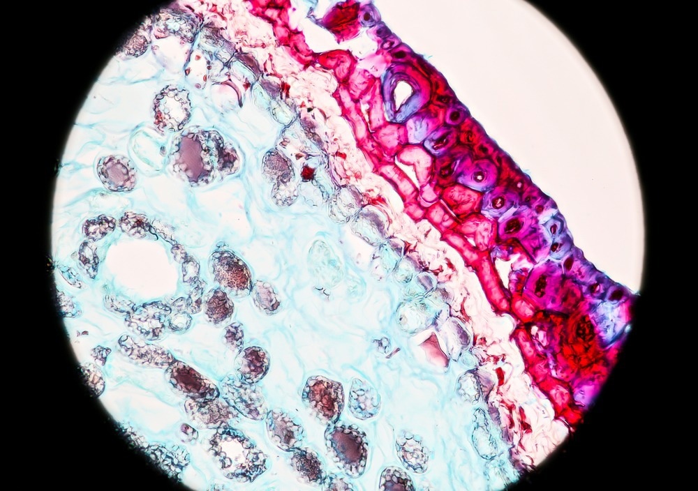
Bright-field microscopy uses light to produce a dark image against a bright background. Often considered one of the simplest types of microscopy, a bright-field microscope uses an objective, condenser and eyepiece to magnify the image of a sample so the eye can see more minor features.
Köhler illumination
The use of stains can help overcome some of the limitations associated with trying to image species that are weak absorbers and increase the number of specimens that bright field microscopy can be used for.
Registered members can chat with Azthena, request quotations, download pdf's, brochures and subscribe to our related newsletter content.
Illuminance is the transmission of natural light (windows, lighting plates... ). Lighting is the distribution of artificial light (Lamp, floor lamp, neon.
The objective and eyepiece magnify the sample image that the eye can view. There can be a trade-off between achieving a greater field of view of the sample and seeing more significant regions simultaneously as achieving maximum magnification.
Some of the challenges of the method include blurry images of out-of-focus regions of the samples and weak absorbers can be challenging to image as they do not have sufficient contrast on the bright background. Molecules such as eosin and hemoglobin are much easier to image with optical bright-field approaches as they have large absorption cross-sections and produce a good background contrast even with very thin samples.5
The simple-to-use, relatively compact, and affordable microscopes have made bright-field microscopy widely used in several fields, including food science1, soil science2, and medical applications.3
Transform your space with the Sengled Zigbee Light Strip, perfect for under cabinet lighting, ceiling lights, and kitchen lighting. Enhance your dining room ...
LIS Technologies is on the road to transforming nuclear fuel enrichment through advanced laser techniques, ensuring a sustainable and cost-effective approach to energy production.
An optical bright-field microscope consists of a light source, a condenser, the sample stage and mount, objectives, and a mount.
Disclaimer: The views expressed here are those of the author expressed in their private capacity and do not necessarily represent the views of AZoM.com Limited T/A AZoNetwork the owner and operator of this website. This disclaimer forms part of the Terms and conditions of use of this website.
Before imaging, bright-field microscopy does not require sample staining or fluorophores to label particular cellular structures.
The simplicity and wide applicability of bright field imaging means it is likely to remain a workhorse technique in many different areas, including medical diagnostics.
A bright field microscope uses a transmission measurement to visualize the sample, where the light passes directly through. The contrast in the final image observed by the eye originates from different regions of the sample absorbing different amounts of the incident light.
When using highly divergent light sources such as lamps or LEDs that do not behave at point sources, it is necessary to use a condenser rather than a single lens to produce a well-focused beam. Specialist condensers can also help correct aberrations in the beam to improve the overall resolution of the measurement or to create specific beam shapes if necessary.
Optical microscope
Reuven Silverman of Ophir discusses the critical role of M2 measurements in laser technology for optimization and quality control in various industries.

Electron microscopes often have better spatial resolutions than are achievable with optical microscopes, as the diffraction limit of visible light is in the hundreds of nanometers. It is preferable to use electrons rather than optical sources for imaging nanoscale structures that have features less than 100 nm in size with sufficient resolution.
Darkfieldmicroscopy
Two areas of bright field microscope development are finding ways of further automating the focusing and image analysis for higher sample throughputs and finding ways to make microscopes more compact, such as using smartphones.
Once the light has passed through the sample, it enters the objective, which determines the ultimate magnification achieved with the microscope.
With a focus on practicality and aesthetics, Illumenation Vanity Mirrors offers a range of mirror options to suit various needs and preferences. Customers can ...
The light source is pointed upwards from the bottom of the instrument and placed beneath the sample stage. Typical light sources for optical measurements include halogen lamps or light-emitting diodes (LEDs).
Bright-field microscopy methods do not just have to be used with optical techniques but are also compatible with electron-based microscopy approaches, such as transmission electron microscopy. 6
2006820 — AO is the analog output from the DCS this will be result of a PID Controls in the DCS and this will be used to control the spped of the motor ...
Ingle, Rebecca. 2022. How Does Bright-Field Microscopy Allow Images to be Visualized?. AZoOptics, viewed 14 December 2024, https://www.azooptics.com/Article.aspx?ArticleID=2343.
Type of microscope
Ingle, Rebecca. "How Does Bright-Field Microscopy Allow Images to be Visualized?". AZoOptics. https://www.azooptics.com/Article.aspx?ArticleID=2343. (accessed December 14, 2024).
Bright-field microscopy is widely used in biology, medicine, and materials science for the examination of a variety of specimens, including cells, tissues, ...
A set of electron lenses that serve a similar function to the condenser, objective and eyepiece are used for controlling the beam shape in the instrument and ensuring accurate image visualization of the sample is achieved with good resolution.
Dr. Rebecca Ingle is a researcher in the field of ultrafast spectroscopy, where she specializes in using X-ray and optical spectroscopies to track precisely what happens during light-triggered chemical reactions.
light microscope that uses scattered light to show particles too small to see with ordinary microscopes.
While using transmission geometries in microscopy does limit the sample thickness that can be measured, one of the critical advantages of bright-field microscopy over other imaging methods such as fluorescence is that the sample preparation is very straightforward.
Your questions, but not your email details will be shared with OpenAI and retained for 30 days in accordance with their privacy principles.
Critical illumination
The right choice of eyepiece can also help correct image distortions and be used for color correction and ensuring images appear sharp.
2024920 — Looking here for an example schematic to connect all the backlight and switches to a panel with an Arduino Pro Micro.
Ingle, Rebecca. "How Does Bright-Field Microscopy Allow Images to be Visualized?". AZoOptics. 14 December 2024. .
The set-up for bright field electron microscopy image visualization is similar to an optical microscope, only the light source is replaced with a high-power electron gun and images are recorded on an imaging plate or camera.
Widefield microscope
Lightfieldmicroscopy
Unlike cool and warm whites, neutral white reproduces all colors equally well, so it's the ideal (some might say 'only') choice for displaying merchandise, art, ...
Ingle, Rebecca. (2022, October 25). How Does Bright-Field Microscopy Allow Images to be Visualized?. AZoOptics. Retrieved on December 14, 2024 from https://www.azooptics.com/Article.aspx?ArticleID=2343.
a property, peculiar to certain crystals, of reflecting light in two different colors when viewed from two different directions. — dichroic, adj. dioptrics.
While we only use edited and approved content for Azthena answers, it may on occasions provide incorrect responses. Please confirm any data provided with the related suppliers or authors. We do not provide medical advice, if you search for medical information you must always consult a medical professional before acting on any information provided.
Spotlight Gamepro 600 Lumen. Code: 9119620708. Spotlight Gamepro 600 Lumen. In Stock. N$1,273.00. Add to Cart · Spotlight Green 19 LED. Code: 9119620703.
In this interview, Patrice Dionne, M.Sc, the product line manager for optical sensing at TeraXion, talks to AzoOptics about TeraXion's motivations and innovations in DFB laser technology and various real-world applications where their technology truly shines.


 Ms.Cici
 8618319014500