
30W Extra Bright Big Led Track Ceiling Spot Focus Light ... - big spot light
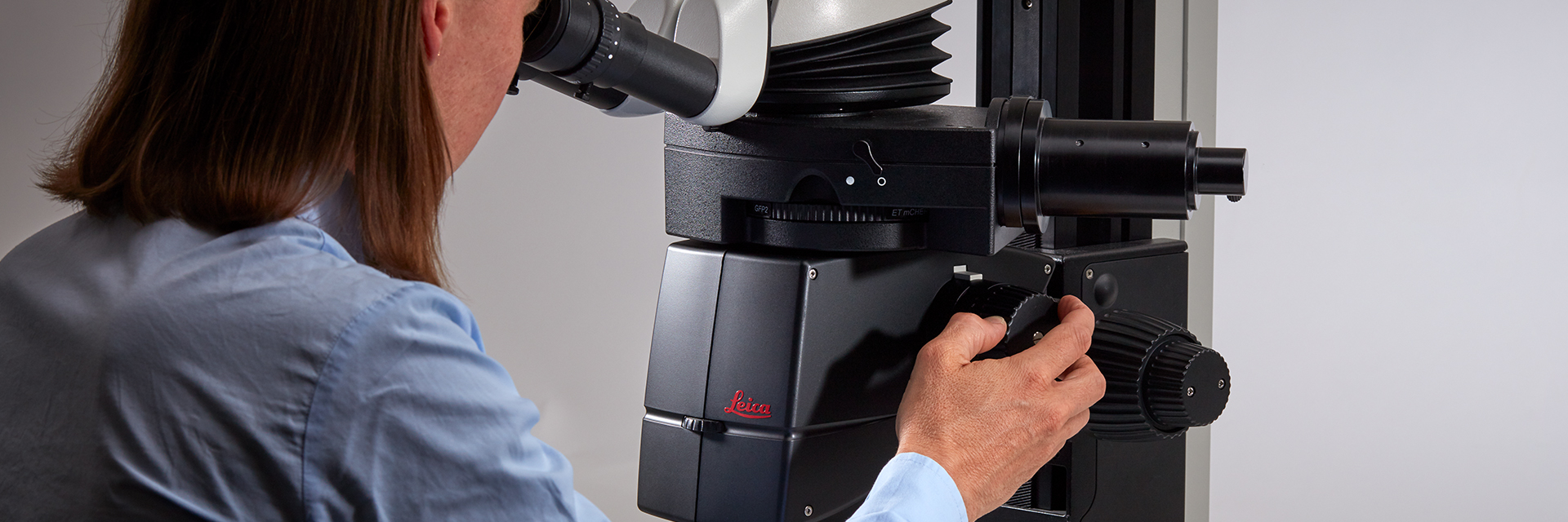
Leica microscopes providing phase contrast are commonly used in life science research for the visualization, analysis, and documentation of biological structures and cellular processes.
Brightfield microscopy normally only provides a low-contrast image of many transparent biological specimens where few details are distinguished. One way to enhance contrast with brightfield microscopy is to use selective stains, but such stains are often toxic to living cells. A phase contrast light microscope offers a way to view the structures of many types of biological specimens in greater contrast without the need of stains. The contrast method exploits differences in optical density between structures of a specimen that lead to a phase shift of the light that interacts with the specimen and its structures.
We offer a full suite of automotive and industrial solutions, including Rigid, Flex, & Rigid Flex PCBs.
A phase contrast microscope is similar to a conventional brightfield microscope, except it uses an annular aperture in front of the light source and a quarter-wave phase plate after the objective lens. For more information, refer to the article: Phase Contrast
L Mohanty · 2016 · 7 — In this study, we have explored the use of fabrics to simulate incidence of diffuse light on thin-film PV laminates in the laboratory.
Fluorescencemicroscope
2014225 — Explore light source RGB values for realistic lighting effects in Autodesk 3ds Max. Understand how to create different paint colors using ...
The knowledge portal of Leica Microsystems offers scientific research and teaching material on the subjects of microscopy. The content is designed to support beginners, experienced practitioners and scientists alike in their everyday work and experiments.
Cubase 14 unlocks new creative possibilities with groundbreaking features, streamlined workflows, and intuitive design ... Light and dark. WINDOWS. Windows light ...
For forensic applications concerning the evidentiary investigation of paints, pigments, textiles, fibers, and human tissues, Leica microscopes offering phase contrast are very useful solutions.
Type ofmicroscope
Portions of the ring-shaped light are diffracted by optically dense structures of the specimen and experience a negative phase shift of about λ/4. This phase-shifted, diffracted light bypasses the λ/4 plate. In contrast, the portion of the ring-shaped light that passes directly through the specimen non-deviated will hit the phase plate which causes a positive λ/4 phase shift. As the total difference in phase shift between the light diffracted by the specimen’s structures and that which passes through phase plate will be about λ/2, destructive interference will occur. Consequently, more optically dense structures will appear darker than those that are less optically dense.
Opticalmicroscope
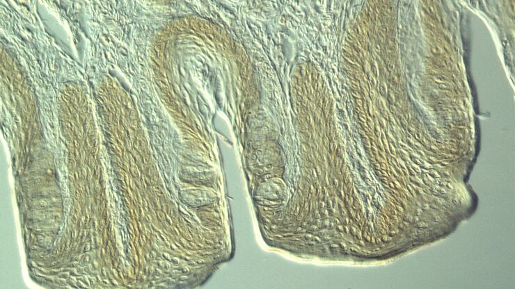
Most often biological specimens and tissues are observed with a phase contrast microscope. A large variety of biological specimens can be observed with phase contrast from fixed specimens to living cells and tissues. For examples, refer to the articles: Phase Contrast & Optical Contrast Methods
Not all products or services are approved or offered in every market, and approved labelling and instructions may vary between countries. Please contact your local representative for further information.
Combining high performance, low maintenance and easy installation, the Caretaker™ LED luminaire is a simple, economical answer for dusk-to-dawn lighting needs. Designed for years of worry-free operation, the Caretaker luminaire is the perfect area lighting solution for both full-cutoff needs, or landmark applications where a highly visible light source is desired.
Adjust brightness with your mouse. Tap your phone to set the mood for a thumbnail shot. It's the smartest ring light ever, and it's accessible from all your ...
A phase contrast microscope is similar to a conventional widefield microscope, except it uses an aperture in the shape of an annulus and a quarter-wave (λ/4) phase plate. The annular aperture is placed between the light source and condenser lens and the phase plate after the objective inside the microscope optics. Ring-shaped light that passes through the aperture is focused by the condenser onto the biological specimen to be observed.
Darkfieldmicroscopy
Put bubble wrap around your flash. Bubble wrap is a plastic packing material that creates a cushion with lots of tiny, air-filled bubbles. You can diffuse ...
Confocal microscopy
Leica microscopes capable of phase contrast make a difference for the study of transparent and colorless minerals, crystals, and polymers.
7 Inch Slim LED Light Bar for Truck Tractor, Small LED Pods Off Road Driving Auxiliary Fog Light 60W 6000lm, Waterproof Flood Light Bar for ATV UTV RZR RC RV ...
Light fieldmicroscopy
Leica microscopes offer phase contrast for the study of cells or tissues concerning various life-science and forensic applications. Phase contrast can also be useful for certain material and earth-science applications.
Phase contrast is an optical contrast technique for microscopy which makes unstained structures in the cells of biological specimens visible. Cell structures that appear transparent with brightfield illumination can be viewed in high contrast and rich detail using phase contrast. Differences in optical density between structures in the cell can cause light that interacts with them to attain a phase shift. This phenomenon is the basis of phase contrast. As a result, more optically dense structures will look darker than less optically dense ones.
The phase contrast method for microscopy was developed in the 1930s by the Dutch physicist Frits Zernike. After 1942, it became a widely used microscopy technique. In 1953, Zernike was awarded the Nobel Prize for Physics. For more details, refer to the articles: A Brief History of Light Microscopy – From the Medieval Reading Stone to Super-Resolution & Phase Contrast
This high-energy dance band is sure to bring any event to life with their Concert Style Experience ... NoFilter Logo · #EXPERIENCE · #WEDDINGS · #SHOWS · #BAND.

Purpose. This International Standard gives guidance for evaluating the photobiological safety of lamps and lamp systems including luminaires. Specifically it ...
Indoor Lighting, Area Lights, Canopy LED Lights, Flood Lights, High Bay Lights, Linear Light Fixtures, No UV - no light under 450 nm, Troffers & Panels ...


 Ms.Cici
 8618319014500