
Image Sensors - what is image sensor
Yaesu repeater controller
Darkfield microscopy has many advantages. Its dark background offers a high degree of contrast, making it easy to see samples on difficult backgrounds. This technique is easily accessible since many brightfield lab microscopes can be configured for darkfield illumination. Besides the microscopy benefits, darkfield images can even look like works of art with the sample beautifully illuminated.
Right: silkworm larva spiracle and trachea. Tiny pieces of fragmented wood take on an unusually beautiful appearance when illuminated under darkfield conditions with a transmitted light microscope. Left: butterfly. Butterflies, because of the wide spectrum of wing scale patterns exhibited by members of this order, are one of the most colorful members of the insect world. The digital image shows the many miniature scales that decorate most of a butterfly wing’s surface. The wing scales were illuminated with a darkfield substage condenser and captured at low magnification (50x).
RLCcontrollers
Darkfield illumination requires blocking most of the light that ordinarily passes through and around the specimen, allowing only oblique rays to interact with the specimen.
Unfortunately, darkfield illumination is less useful for revealing internal details. There are also other conditions to consider if you want to make the most out of darkfield illumination.
Repeater controller kit
Darkfield microscopy is a technique that takes advantage of oblique illumination to enhance contrast in specimens that are not imaged well under normal illumination conditions.
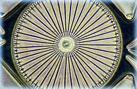
Left: this spectacular darkfield photomicrograph of the diatom Arachnoidiscus ehrenbergi was captured on an Olympus microscope by Mortimer Abramowitz. The specimen was illuminated with a high numerical aperture darkfield condenser with immersion oil placed between the microscope slide and the objective and condenser front lenses.
MTR2000 controller
Non-biological specimens include mineral and chemical crystals, colloidal particles, dust-count specimens, and thin sections of polymers and ceramics containing small inclusions, porosity differences, or refractive index gradients.
Care must be taken when preparing specimens for darkfield microscopy because the features that lie above and below the plane of focus can scatter light and contribute to image degradation. The cleanliness of the slide is an important factor when imaging, but it is even more important in darkfield since every piece of debris will be illuminated and can take away from what you’re trying to see.
Darkfield illumination is best for revealing outlines, edges, boundaries, and refractive index gradients. Ideal candidates for darkfield illumination include minute living aquatic organisms, diatoms, small insects, bone, fibers, hair, unstained bacteria, yeast, cells in tissue culture, and protozoa.
Simple repeater controller
These condensers are relatively simple and offer the high numerical aperture (NA) required to create the cone of illumination needed for darkfield. Yet, switching condensers based on the illumination type is impractical for everyday microscope use. Luckily, there’s a workaround.
After the direct light has been blocked by an opaque stop in the condenser, light passing through the specimen from oblique angles is diffracted, refracted, and reflected into the microscope objective to form a bright image of the specimen superimposed on a dark background.
Alec De Grand is a product manager for virtual slide scanning and upright microscopes for life science at Evident. He has been with Evident for over 10 years, during which he has managed clinical products, marketing initiatives, imaging courses, and trade shows.
Raspberry Pi repeater controller
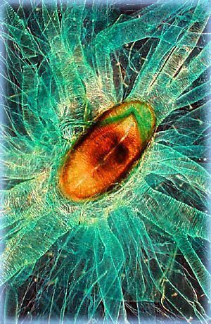
Right: liquid crystalline DNA. This highly concentrated DNA solution has undergone a series of liquid crystalline phase transitions to form a densely packed hexagonal phase. This photomicrograph was taken using a compound optical microscope with a 10x objective in darkfield illumination.
Many condensers can accommodate inserts that can create a cone of illumination. These inserts don’t offer an NA as high as a dedicated darkfield condenser, so not all objectives can be used. However, the inserts give you the flexibility to have multiple observation methods on the same condenser.
Arcom controller
Middle: basswood. Stained specimens are often excellent candidates for darkfield microscopy, yielding beautiful images that are rendered in color on a dark background. This photomicrograph illustrates a stained thin section of a basswood tree under darkfield illumination.
When a specimen (especially an unstained, non-light absorbing one) is placed on a slide, the oblique rays interact with the specimen and are diffracted, reflected, and/or refracted by elements in the sample, such as the cell membrane, nucleus, and internal organelles. This allows these faint rays to enter the objective. The result is a bright specimen on a black background.
ICScontroller asthma
In general, objects imaged under the proper conditions of darkfield illumination are spectacular to see. Often, specimens containing very low inherent contrast in brightfield microscopy shine brilliantly in darkfield.
The top lens of a simple Abbe darkfield condenser is spherically concave, allowing light rays emerging from the surface of the top lens to form an inverted hollow cone of light with the focus centered on the specimen plane. In places where there is no sample and the condenser’s numerical aperture is greater than the objective’s, the oblique rays cross each other and miss the objective, leaving those areas dark.
This dark background provides a high degree of contrast and can make samples with difficult backgrounds stand out with relatively little effort. Check out some examples in the images below.
Almost any brightfield laboratory microscope can be easily converted to perform darkfield illumination. The easiest way to do this is to switch out your current condenser with one that is designed for darkfield illumination (Figure 1).


 Ms.Cici
 8618319014500