
Zenni Optical: Glasses Online & Contacts | Affordable ... - new start optics
Other transgenic mouse models have been successfully used for in vivo platelet imaging. For instance platelets of the floxed confetti reporter mouse crossed with a megakaryocyte-specific Cre mouse (Pf4-Cre mouse) are individually color-labeled due to the stochastic expression of one or several of three different transgenes, cytoplasmic YFP (yellow fluorescent protein), cytoplasmic RFP (red fluorescent protein) and/or plasma membrane CFP (cyan fluorescent protein) [8]. Such platelets, labeled individually by different color combinations, can be easily tracked in a complex 3D environment like a blood clot. Other examples are transgenic mice expressing a FRET (Förster resonance energy transfer)-biosensor to follow the activation of kinases like ERK and PAK during in vivo thrombus formation [9]. A FRET-biosensor for kinase activities is typically composed of a phosphorylatable peptide substrate fused to a donor chromophore and combined through a linker sequence with a phosphopeptide-binding domain fused to an acceptor chromophore. The ratiometric FRET quantification using stoichiometric Donor-Acceptor probes is probably the most popular quantification method both in vitro and in vivo. However, it often faces limits related to spectral bleed-through, sequential acquisition, incomplete fluorescent protein maturation and in-depth light scattering. Moreover, deciphering signaling networks often requires several biosensors for a simultaneous read-out. Recent technological developments using dark acceptor FRET couples (consisting of a fluorescent Donor and an absorbing but non-emitting or “dark” Acceptor) and fast FLIM imaging allow such multiplexing and an intensity-independent read-out of FRET efficiency [10]. Furthermore, super-resolution optical microscopy such as STED takes benefit of such probes by a resolution increase to nm scale due to non-linear effects [11]. So far, however, no reported applications in the field of platelet research can be found.
Laseroptics procedure
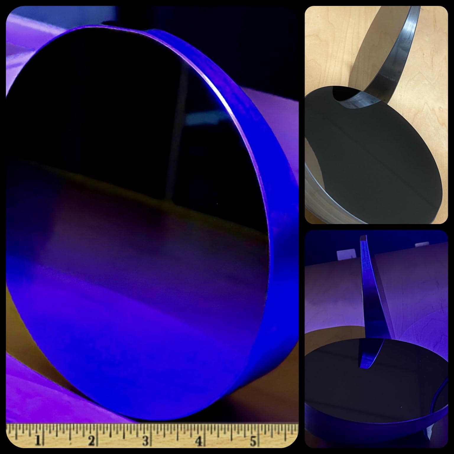
Aside from standard size glass wafers such as 100mm, 150mm, 200mm and 300mm, Sydor Optics can provide custom wafers with diameters up to 450mm and thin wafers with thicknesses down to 0.1mm.
Laseroptics course
Flat optics are made from materials determined by the wavelength of use. Sydor can provide laser optics for deep-ultraviolet (DUV), extreme-ultraviolet (EUV) and longer-wave UV laser applications with various materials such as calcium fluoride, magnesium fluoride, fused silica, ULE, Zerodur and Borofloat.

Many considerations go into the design, fabrication and use of optics for laser applications. Rather than modifying the geometry of a laser beam, flat optics can modify other properties such as direction or spectral content. These optics are used for mirrors and debris shield windows among other applications.
A consequence of these innovative developments has led to their application for in vivo functional imaging of tumor-ECM interactions. The feasibility of imaging the motility and metastatic differences between carcinoma cells of differing metastatic potential in the same animal at subcellular resolutions was elegantly demonstrated in a recent study by Sahai et al. (16). In this study, to compare directly the behavior of cells with different metastatic potentials in the same tumor microenvironment in vivo, tumors were generated by injecting a mixed population of poorly metastatic cancer cells labeled with green fluorescent protein (GFP) and highly metastatic cancer cells labeled with cyan fluorescent protein (CFP). This was followed by simultaneously imaging GFP, CFP fluorescence, and collagen second harmonic imaging in vivo with multiphoton laser scanning microscopy. SHG imaging in tumors has also been applied to dynamically visualize the structure of fibrillar collagen, estimate relative diffusive hindrance, and the dynamics of enzymatic modification of tumor collagen with relaxin in vivo (17). In other studies, quantum dots, which are fluorescent semiconductor nanocrystals, were used to image and differentiate tumor vessels from the ECM and also track the recruitment of quantum dot-labeled bone marrow-derived precursor cells to the tumor vasculature (18).
Bestoptical laser
Laser optics have a limited use expectancy based on the damage created by the laser source. Through our unique process capabilities for both flat and wedged optics, this life is extended by leveraging our superior polishing expertise. Volume production can be accommodated up to 25,000 components per month.
Laseroptics Physics
The hypothesis that RF fields specifically induce RAS or ERK activity or impact chemically-induced RAS or ERK activity in live cells was tested using a BRET based assay. In that protocol, we took advantage of previously described RAS and ERK FRET probes (Komatsu et al. 2011) that we modified into BRET probes by replacing the cyan fluorescent protein-energy donor with Renilla Luciferase (see material and methods). These new RAS and ERK BRET probes comprise, therefore, a sensor domain and a ligand domain connected by a flexible linker, a yellow fluorescent protein (YFP) serving as energy acceptor and Renilla Luciferase serving as energy donor in place of the Cyan Fluorescent protein in the original FRET probes (see material and methods). The sensor domain changes its conformation upon action of either RAS or ERK endogenous protein. This sensitized sensor domain interacts with the ligand domain, thereby inducing a global change of the biosensor conformation and a concomitant increase of the BRET efficiency from the donor to the acceptor (Figure 2(A)).
Sydor Optics manufactures IR windows utilizing a range of infrared materials suited for the NIR, SWIR, MWIR and LWIR spectrums ranging from 0.75µm to 15µm for applications including thermal imaging, mineral & gas detection and monitoring and other forward-looking infrared (FLIR) imaging applications.
Optical laserfor sale
Our robust inspection processes and extensive equipment list for quality assurance provides our customers a stunning 99.94% level of perfection on delivery with nearly zero returns.
Improved imaging technolog y and exploitation of genetically encoded fluorescence technolog y have also been combined to measure subcellular microdomains of second messengers. Transfected proteins contain two different fluorescent molecules that come into close proximity to each other on binding of a second messenger. This produces a transfer of fluorescence, or Forster resonance energy transfer (FRET) between donor and acceptor chromophores such that the emission ratio between their two wavelengths changes as demonstrated in Figure 19.3. For example, cyclic adenosine monophosphate (cAMP) FRET sensors are structurally based on exchange protein directly activated by cAMP (Epac), protein kinase A, or cyclic nucleotide-gated channels and often use a cyan fluorescent protein donor and a yellow fluorescent protein acceptor to tag the regulatory and catalytic subunits, respectively. The FRET signal can be pH-sensitive; important control experiments involve demonstrating that the FRET signal is within the dynamic range and not saturated, and also transfecting a dead sensor control that contains the chromophores but has lost the ability to bind the second messenger.
Optical laserlevel
Reducing surface roughness plays a significant role in lowering scatter and increasing laser performance. We can precisely control a wide array of characteristics – reflected wavefront (flatness), transmitted wavefront, parallelism, TTV, Bow & Warp, among others, and have a sound understanding of the extreme importance in controlling surface scratch and dig cosmetic defects. We can minimize shipment to shipment variability with “Copy Exactly!” process controls upon customer request so that every optic delivered will be the same to the last, no matter how much time has passed between orders.
Optical LaserLight
Some of the most-popular wavelengths for flat laser optics in the UV to the near-infrared (near-IR) spectral region are 266, 355, 405, 488, 532, 461, 633, 785, 980, 1064, and 1550 nm.
LaserOptics pdf
Emmanuelle Poque, Delia Arnaud-Cormos, Lorenza Patrignoni, Hermanus J. Ruigrok, Florence Poulletier De Gannes, Annabelle Hurtier, Rémy Renom, André Garenne, Isabelle Lagroye, Philippe Lévêque, Yann Percherancier
Published in Martha Skinner, John L. Berk, Lawreen H. Connors, David C. Seldin, XIth International Symposium on Amyloidosis, 2007
Optical wedges with ± 10 arc seconds of wedge angle tolerance. Superior craftsmanship made to your specifications in a variety of shapes, sizes, and materials.
Reflect and transmit any combinations of wavelengths with precision plate beamsplitters. UV, VIS, or IR, Polarized or Unpolarized, 10:90 – 90:10 Reflection:Transmission(R:T).
Published in Martin G. Pomper, Juri G. Gelovani, Benjamin Tsui, Kathleen Gabrielson, Richard Wahl, S. Sam Gambhir, Jeff Bulte, Raymond Gibson, William C. Eckelman, Molecular Imaging in Oncology, 2008
Best practices suggest that when coating or setting up an optical component, it’s best to use a witness sample, also called a “test piece,” to ensure that the actual part will match your specific coating specifications and your application.
Cells were cultured to 80% confluency in Petri dishes and medium was changed to serum free medium one hour prior to transfection. A solution of 20μg pB.RIP2.DEVD12 and 20μg pcDNA3 vector with 10nM polyethyleneimine and 5% sucrose in a total volume of 175μl was mixed and let to precipitate for 10 minutes and then added to 9ml of medium. Cells were incubated for 6 hours and FBS was thereafter added to a final concentration of 10%, and selection medium containing 0.4mg/ml G-418 antibiotics was added to the cells 24 hours after transfection. A stable clone of pB.RIP2.DEVD12 expressing cells were isolated and cultured into black 96 well plates. Sample volume was set to 100μl and Krebs-Ringer with Hepes and Glucose (KRHG) (120mM NaCl, 4.7mM KCl, 2.5mM CaCl2, 1.2mM MgSO4, 0.5mM KH2PO4, pH 7.4 with 2mM D-glucose, 20mM HEPES and 200nM adenosine) was used as assay buffer. The apoptotic effect of 50μM synthetic IAPP in 1% DMSO was studied and controls included in the assay were the apoptotic agent staurosporine (2μM), in 1% DMSO and 1% DMSO in KRHG buffer. Cells that express pB.RIP2.DEVD12 will produce a fusion protein consisting of Enhanced Cyan Fluorescent Protein (ECFP) and Enhanced Yellow Fluorescent Protein (EYFP) connected by the amino acid residues DEVD and FRET will occur at 440nm excitation wavelength (Figure 1A) (5). The DEVD sequence is a specific substrate for caspase-3 protease, and if the transfected cells undergo apoptosis, the DEVD is cleaved and the two fluorophores will disperse, and a loss of FRET can be monitored as a decrease of the 535nm/480nm ratio (Figure 1B). Loss of FRET was measured in a Wallac 1420 multilabel counter (Perkin


 Ms.Cici
 8618319014500