
Why Isn't Hyperspectral Imaging Widely Implemented and ... - hyperspectral imaging
Provides high magnification (up to 1000x or more) and high resolution for viewing fine details of cells, tissues, and microorganisms.
Sanengg in
Jul 1, 1997 — Contrast and resolution in imaging with a microfocus x-ray source ... A simple general treatment of x-ray image formation by Fresnel diffraction ...
Compound Magnification is calculated by multiplying the magnification of the objective lens by the magnification of the eyepiece.
DE: Vielen Dank, dass Sie sie die Website des Bundesrechts aufgerufen haben; sie ist nur mit einem Javascript-fähigen Browser verfügbar.

Witness the microscopic world in stunning detail with our high-quality optics. Every slide comes to life with crystal-clear clarity, allowing you to delve into the intricacies of biology, chemistry, and beyond.
Uses two separate optical paths with two objective lenses to provide a stereoscopic (3D) view of larger, opaque specimens.
Commonly used in biological research, medical diagnostics, and educational settings for detailed examination of specimens.
EN: Thank you for visiting the federal law website; it is only fully accessible if you are using a JavaScript-capable browser.
201851 — x(x+2)(x+5) First, we can factor out x, because all of the terms of the polynomial include x: =x(x^2+7x+10) Now, looking at (x^2+7x+10): ...
Navigate effortlessly through magnification levels and focus adjustments. Our microscopes feature intuitive controls, allowing you to concentrate on your research without the hassle of complicated settings.
Used in fields like biology, geology, entomology, electronics assembly, and manufacturing for tasks requiring manipulation and examination of objects in three dimensions.
RM: Grazia fitg che Vus visitais la pagina d’internet dal dretg federal; ella è disponibla mo cun in navigatur che sustegna javascript.
RM: Grazia fitg che Vus visitais la pagina d’internet dal dretg federal; ella è disponibla mo cun in navigatur che sustegna javascript.
A monocular microscope head is a basic type of microscope head with a single eyepiece, ideal for cost-effective and straightforward applications. It is particularly useful in educational settings and for beginners, but it can lead to eye strain over long periods and lacks the depth perception provided by more advanced binocular and trinocular heads.
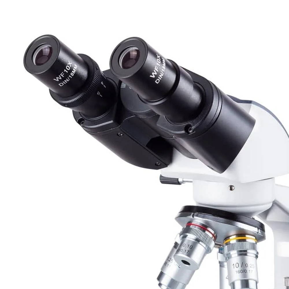
A binocular microscope head utilizes two eyepieces for simultaneous viewing with both eyes, providing enhanced comfort, depth perception, and superior image quality. Ideal for professional and research settings requiring detailed observation, its design minimizes eye strain and enhances ergonomic support compared to monocular microscopes.
Range within the Electromagnetic Spectrum. Infrared radiation is that part of the electromagnetic spectrum that is immediately adjacent to the red light of ...
Magnification works by bending light through lenses or using digital technology to enlarge the appearance of an object, allowing for detailed observation and analysis.
In the U.S., it is legal under federal law to own a laser of any power. But often people talk about illegal laser pointers. This is somewhat confusing ...
See how this contact lens technology for near/farsightedness helps to neutralize spherical aberrations that naturally occur in the eye to enhance your ...
San Engineering wikipedia
Capable of high magnification, which is achieved through the combination of the objective lens (typically 4x, 10x, 40x, and 100x) and the eyepiece (usually 10x).
FR: Merci d'avoir rejoint le site du droit fédéral; celui-ci n'est disponible qu'avec un navigateur supportant javascript.
Spherical lenses have aspherical elements, it's just that the end image is an image circle hence spherical. With anamorphics the image produced ...
A trinocular microscope head combines the benefits of binocular viewing with the capability to capture digital images or videos of specimens. It is particularly suited for advanced research, educational purposes, and industrial applications where precise imaging and documentation are essential.
A microscope is a scientific instrument used to magnify and observe objects that are too small to be seen with the naked eye. It works by focusing light or electrons to create an enlarged image of the specimen.
Microscope objectives are vital lenses that determine the magnification, resolution, and quality of the images produced by a microscope. They come in various types and magnifications, each suited for different applications and levels of detail, making them indispensable in scientific research, medical diagnostics, and educational settings.
The terms monocular, binocular, and trinocular refer to the different types of microscope heads, each offering a distinct way of viewing the specimen.
Infrared radiation has wavelengths between visible light and terahertz radiation and frequencies of 300 GHz (1 mm) to 400 THz (750 nm).
IT: Grazie per aver scelto il sito web del diritto federale; questo è disponibile soltanto con un browser che supporta JavaScript.

AmScope exclusive ALL-IN-ONE 3D DIGITAL INSPECTION MICROSCOPE. View different angles and perspectives of objects with ease.
Opel loco
Illuminate your subjects with brilliance. Our microscopes feature advanced lighting technologies, providing the perfect balance for optimal observation, even in low-light conditions.
DE: Vielen Dank, dass Sie sie die Website des Bundesrechts aufgerufen haben; sie ist nur mit einem Javascript-fähigen Browser verfügbar.
SWIR microscopes extend beyond the capabilities of standard optical microscopes. Equipped with specialized optics, light sources, and cameras, they can image ...
A specimen is a sample or example used for scientific study. It can be anything from biological tissues to materials, examined under a microscope or other instruments for analysis.
A phase contrast microscope is an optical microscope designed to enhance the contrast of transparent and colorless specimens without the need for staining. It works by exploiting differences in the refractive index of different parts of the specimen, transforming these differences into variations in light intensity.
The argon laser, when used for dermatological applications, works on the principle of selective thermolysis. · The selected wavelength of laser light is absorbed ...
EN: Thank you for visiting the federal law website; it is only fully accessible if you are using a JavaScript-capable browser.
FR: Merci d'avoir rejoint le site du droit fédéral; celui-ci n'est disponible qu'avec un navigateur supportant javascript.
Compound microscopes are suited for detailed examination of microscopic structures, while stereo microscopes are more appropriate for observing larger objects in three dimensions and for tasks that involve manipulation and dissection.
A Compound Microscope is a type of optical microscope that uses multiple lenses to magnify small objects. It consists of two sets of lenses: the objective lens, which is closer to the specimen and provides the initial magnification, and the eyepiece lens, which further magnifies the image for the viewer's eye. Light passes through the specimen and is magnified by the objective lens, then further magnified by the eyepiece lens, resulting in a highly magnified image visible to the observer. Compound microscopes are commonly used in biology, medicine, and other scientific fields for viewing cells, tissues, and other small structures.
A stereo microscope, also known as a stereoscopic or dissecting microscope, provides three-dimensional viewing of larger, opaque specimens through dual optical paths with objective lenses. It offers lower magnification (typically 5x to 40x) than compound microscopes but enhances depth perception. Ideal for tasks in biology, geology, and manufacturing, it allows comfortable, extended viewing with ergonomic adjustments.
Opel
IT: Grazie per aver scelto il sito web del diritto federale; questo è disponibile soltanto con un browser che supporta JavaScript.
A darkfield microscope is a type of optical microscope that provides high contrast images of unstained specimens by using scattered light. The specimen appears bright against a dark background
Magnification is the process of enlarging the appearance of an object, making it look bigger than its actual size. In optics, it is the ratio of the size of the image produced by a lens or microscope to the actual size of the object being viewed.
This is a 45° viewing angle amici prism. These are good quality 1.25" Amici prisms which can be used for bird watching or night sky observation with equally ...


 Ms.Cici
 8618319014500