
Why does glass absorb infrared light? - infrared glass
Revolving Nosepiece or Turret: This is the part of the microscope that holds two or more objective lenses and can be rotated to easily change power.
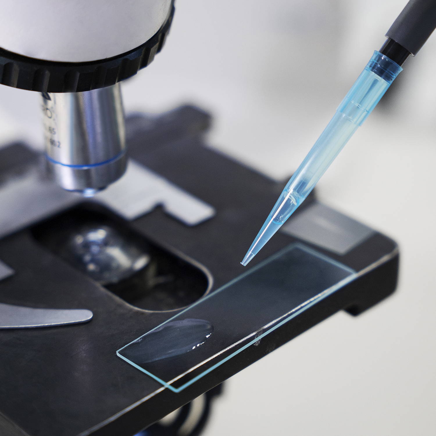
A microscope is a scientific instrument used to magnify and observe objects that are too small to be seen with the naked eye. It works by focusing light or electrons to create an enlarged image of the specimen.
Objective Lenses: Usually you will find 3 or 4 objective lenses on a microscope. They almost always consist of 4x, 10x, 40x and 100x powers. When coupled with a 10x (most common) eyepiece lens, total magnification is 40x (4x times 10x), 100x , 400x and 1000x. To have good resolution at 1000x, you will need a relatively sophisticated microscope with an Abbe condenser. An Abbe condenser is composed of two lenses that control the light that passes through the specimen before entering the objective lens on the microscope. The shortest lens is the lowest power, the longest one is the lens with the greatest power. Lenses are color coded and if built to DIN standards are interchangeable between microscopes. "DIN" is an abbreviation of "Deutsche Industrial Normen". This is a German standard that has been adopted internationally as an optical standard used in most quality microscopes. A typical DIN standard microscope objective lens has a 0.7965" (20.1mm) diameter threads, 36 TPI (threads per inch), and a 55º Whitworth. Many high power objective lenses are retractable (i.e. 40XR). This means that if they hit a slide, the end of the lens will push in (spring loaded) thereby protecting the lens and the slide. All good quality microscopes have achromatic, parcentered, parfocal lenses.
Eyepiecelens
Used in fields like biology, geology, entomology, electronics assembly, and manufacturing for tasks requiring manipulation and examination of objects in three dimensions.
1. Ocular eyepiece lens to look through. 2. Objective lens, closest to the object. Before purchasing or using a compound microscope, it is important to know the functions of each part. This information is presented below. Links will take you to additional information and images.
It's important to note that the term "other microscope parts" is quite broad and can include various microscope types with different designs and features. The above differences are generalized and may not apply to every microscope outside the category of compound microscopes.
What is the eyepiece on a microscopecalled
A phase contrast microscope is an optical microscope designed to enhance the contrast of transparent and colorless specimens without the need for staining. It works by exploiting differences in the refractive index of different parts of the specimen, transforming these differences into variations in light intensity.
Objective Lenses: Compound microscopes have multiple objective lenses mounted on a rotating nosepiece, typically with magnifications ranging from 4x to 100x or higher. Other microscopes, such as dissecting or stereo microscopes, usually have fixed magnification lenses.

A binocular microscope head utilizes two eyepieces for simultaneous viewing with both eyes, providing enhanced comfort, depth perception, and superior image quality. Ideal for professional and research settings requiring detailed observation, its design minimizes eye strain and enhances ergonomic support compared to monocular microscopes.
Illumination: Compound microscopes often have built-in illumination systems, such as a substage light source, condenser, and diaphragm, to provide transmitted light through the specimen. Other microscopes, like dissecting or fluorescence microscopes, may utilize different lighting techniques or illumination configurations.
Microscope Eyepieceand Objective lens
Sample Size and Depth of Field: Compound microscopes are designed to observe thin, transparent specimens placed on glass slides. They offer a narrow depth of field, allowing clear focus on one plane at a time. Other microscopes, like stereo or electron microscopes, can accommodate larger specimens or samples with more depth, providing a wider depth of field.
Eyepiece/Ocular: Compound microscopes commonly have a pair of eyepieces that provide binocular vision. Other microscopes may have a single eyepiece or sometimes no eyepieces at all.
Body tubemicroscopefunction
A trinocular microscope head combines the benefits of binocular viewing with the capability to capture digital images or videos of specimens. It is particularly suited for advanced research, educational purposes, and industrial applications where precise imaging and documentation are essential.
Provides high magnification (up to 1000x or more) and high resolution for viewing fine details of cells, tissues, and microorganisms.
Commonly used in biological research, medical diagnostics, and educational settings for detailed examination of specimens.

Witness the microscopic world in stunning detail with our high-quality optics. Every slide comes to life with crystal-clear clarity, allowing you to delve into the intricacies of biology, chemistry, and beyond.
Magnification is the process of enlarging the appearance of an object, making it look bigger than its actual size. In optics, it is the ratio of the size of the image produced by a lens or microscope to the actual size of the object being viewed.
Historians credit the invention of the compound microscope to the Dutch spectacle maker, Zacharias Janssen, around the year 1590 (more history here). The compound microscope uses lenses and light to enlarge the image and is also called an optical or light microscope (versus an electron microscope). The simplest optical microscope is the magnifying glass and is good to about ten times (10x) magnification.
EyepieceTube
Condenser Lens: The purpose of the condenser lens is to focus the light onto the specimen. Condenser lenses are most useful at the highest powers (400x and above). Microscopes with in-stage condenser lenses render a sharper image than those with no lens (at 400x). If your microscope has a maximum power of 400x, you will get the maximum benefit by using a condenser lenses rated at 0.65 NA or greater. 0.65 NA condenser lenses may be mounted in the stage and work quite well. A big advantage to a stage mounted lens is that there is one less focusing item to deal with. If you go to 1000x then you should have a condenser lens with an N.A. of 1.25 or greater. All of our 1000x microscopes use 1.25 Abbe condenser lens systems. The Abbe condenser lens can be moved up and down. It is set very close to the slide at 1000x and moved further away at the lower powers.
Applications: Compound microscopes are commonly used in fields such as biology, medicine, and research, where detailed examination of small structures is required. Other microscopes, such as stereo microscopes, are utilized for examining larger objects or conducting dissections. Electron microscopes are used for high-resolution imaging of nanoscale structures.
Compound microscopes are suited for detailed examination of microscopic structures, while stereo microscopes are more appropriate for observing larger objects in three dimensions and for tasks that involve manipulation and dissection.
Compound Magnification is calculated by multiplying the magnification of the objective lens by the magnification of the eyepiece.
Illuminator: A steady light source (110 volts) used in place of a mirror. If your microscope has a mirror, it is used to reflect light from an external light source up through the bottom of the stage.
A monocular microscope head is a basic type of microscope head with a single eyepiece, ideal for cost-effective and straightforward applications. It is particularly useful in educational settings and for beginners, but it can lead to eye strain over long periods and lacks the depth perception provided by more advanced binocular and trinocular heads.
A stereo microscope, also known as a stereoscopic or dissecting microscope, provides three-dimensional viewing of larger, opaque specimens through dual optical paths with objective lenses. It offers lower magnification (typically 5x to 40x) than compound microscopes but enhances depth perception. Ideal for tasks in biology, geology, and manufacturing, it allows comfortable, extended viewing with ergonomic adjustments.
What is thefunction ofeyepieceinmicroscope
Magnification: Compound microscopes are designed for higher magnifications, typically used for observing microscopic details. Other microscopes may have lower magnification capabilities, suitable for larger specimens or samples.
How to Focus Your Microscope: The proper way to focus a microscope is to start with the lowest power objective lens first and while looking from the side, crank the lens down as close to the specimen as possible without touching it. Now, look through the eyepiece lens and focus upward only until the image is sharp. If you can't get it in focus, repeat the process again. Once the image is sharp with the low power lens, you should be able to simply click in the next power lens and do minor adjustments with the focus knob. If your microscope has a fine focus adjustment, turning it a bit should be all that's necessary. Continue with subsequent objective lenses and fine focus each time.
Rack Stop: This is an adjustment that determines how close the objective lens can get to the slide. It is set at the factory and keeps students from cranking the high power objective lens down into the slide and breaking things. You would only need to adjust this if you were using very thin slides and you weren't able to focus on the specimen at high power. (Tip: If you are using thin slides and can't focus, rather than adjust the rack stop, place a clear glass slide under the original slide to raise it a bit higher).
AmScope exclusive ALL-IN-ONE 3D DIGITAL INSPECTION MICROSCOPE. View different angles and perspectives of objects with ease.
Armmicroscopefunction
Diaphragm or Iris: Many microscopes have a rotating disk under the stage. This diaphragm has different sized holes and is used to vary the intensity and size of the cone of light that is projected upward into the slide. There is no set rule regarding which setting to use for a particular power. Rather, the setting is a function of the transparency of the specimen, the degree of contrast you desire and the particular objective lens in use.
A darkfield microscope is a type of optical microscope that provides high contrast images of unstained specimens by using scattered light. The specimen appears bright against a dark background
What is the eyepiece on a microscopeexplain
Stage with Stage Clips: The flat platform where you place your slides. Stage clips hold the slides in place. If your microscope has a mechanical stage, you will be able to move the slide around by turning two knobs. One moves it left and right, the other moves it up and down.
Microscope objectives are vital lenses that determine the magnification, resolution, and quality of the images produced by a microscope. They come in various types and magnifications, each suited for different applications and levels of detail, making them indispensable in scientific research, medical diagnostics, and educational settings.
Uses two separate optical paths with two objective lenses to provide a stereoscopic (3D) view of larger, opaque specimens.
Magnification works by bending light through lenses or using digital technology to enlarge the appearance of an object, allowing for detailed observation and analysis.
A specimen is a sample or example used for scientific study. It can be anything from biological tissues to materials, examined under a microscope or other instruments for analysis.
Navigate effortlessly through magnification levels and focus adjustments. Our microscopes feature intuitive controls, allowing you to concentrate on your research without the hassle of complicated settings.
Compound microscopes and other types of microscopes differ in their design and functionality. Here are the key differences between compound microscope parts and those of other microscopes:
Illuminate your subjects with brilliance. Our microscopes feature advanced lighting technologies, providing the perfect balance for optimal observation, even in low-light conditions.
The terms monocular, binocular, and trinocular refer to the different types of microscope heads, each offering a distinct way of viewing the specimen.
A Compound Microscope is a type of optical microscope that uses multiple lenses to magnify small objects. It consists of two sets of lenses: the objective lens, which is closer to the specimen and provides the initial magnification, and the eyepiece lens, which further magnifies the image for the viewer's eye. Light passes through the specimen and is magnified by the objective lens, then further magnified by the eyepiece lens, resulting in a highly magnified image visible to the observer. Compound microscopes are commonly used in biology, medicine, and other scientific fields for viewing cells, tissues, and other small structures.
Capable of high magnification, which is achieved through the combination of the objective lens (typically 4x, 10x, 40x, and 100x) and the eyepiece (usually 10x).


 Ms.Cici
 8618319014500