
Uniform Shop | St Edmund's College - edmund sec
Their ability to function is because they have been constructed with special components that enable them to achieve high magnification levels. They can view very small specimens and distinguish their structural differences, for example, the view of animal and plant cells viewing microscopic bacterial cells.
Ans. Condensers are lenses that are used to collect and focus light from the illuminator into the specimen. They are found under the stage next to the diaphragm of the microscope. They play a major role in ensuring clear sharp images are produced with a high magnification of 400X and above. Abbe condenser is a condenser specially designed for high-quality microscopes, which makes the condenser to be movable and allows very high magnification of above 400X. High-quality microscopes normally have a high numerical aperture than objective lenses.
Function ofnosepiecein microscope
Microscopes are instruments that are used in science laboratories to visualize very minute objects, such as cells and microorganisms, giving a contrasting image that is magnified.
Ans. The magnification of a lens is defined as the ratio of the height of an image to the height of an object. Microscope magnification measures the total enlargement of the image of an object. Magnification power is the product of eyepiece lens power and objective lens power.
Function ofbody tubein microscope
Ans. The coarse adjustment knob moves the stage up and down to bring the specimen into focus. The fine adjustment knob brings the specimen into sharp focus under low power and is used for all focusing when using high-power lenses.
In shallow depth of field, the distance between the nearest and the furthest elements in focus is very short. This puts only a small portion of your image in focus. You can achieve a shallow DoF by using a large aperture.
Diopter adjustmentmicroscope function
Using the proper depth of field can go a long way in creating the perfect image for that ad or website icon you're designing. But, to get more fulfilling results, you'll need to combine your expertise with an efficient graphics design software like CorelDRAW.
Thanks much for this. We just did microscopy as a topic and the write-up has really helped me to understand better. Thanks again
What is the function of eyepiece in microscopepdf
There are two common types of depth of field; shallow depth of field and deep depth of field. Both types bring out different aspects in your image and, therefore, have different benefits.
Thank you very much it really helped me with my science home work since i in 8th grade and this my home work to draw a microscope label all the parts and the function thank may the holy father of holy spirits bless you and give more wisdom thanks love you all keep up the good work and thank you again bye.
Thanks a lot for this wonderful note: It is really helpful, Really appreciate the way all the detail about microscope have been explained
What is eyepiece in microscope
Microscopes are generally made up of structural parts for holding and supporting the microscope and its components and the optical parts that are used for magnification and viewing of the specimen images. Modern microscopes have additional electronics and display devices. This description defines the parts of a microscope and the functions they perform to enable the visualization of specimens.
Oct 24, 2022 — The microscope lens functions like a magnifying glass, bending light to make the object appear wider to get the desired magnification effect.
this is a really good artical i used it to study my science i just wanted to point out to you that tere are a few spelling errors but other than that it is a 100% rating from me
The world leader in infrared conversions, modifications & DIY IR conversion tutorials. Scratched sensor replacement, UV & Full spectrum conversions. The world ...
What is the function ofarmin microscope
Ans. Rack stop is included in the microscope for preventing the specimen slide from coming too far up and hitting the objective lens.
Function ofstagein microscope
In a typical fluorescence microscope, contrast is determined by the number of photons collected from the specimen, the dynamic range of the signal, optical ...
Thanks very much dear and please continue doing so, am Gerald M from Uganda East Africa doing diploma in nursing at Mulago school of nursing and midwifery
Depth of field is a key element in many photographs. In most cases, using it can mean the difference between a basic image and a work of art. There are two common types of depth of field, each with the potential to increase the aesthetics of your image dramatically.
Seriously, if i am not grateful, i am lying. This note is really helpeful to me to differet ways to different methology.
Microscopeparts and functions
Oct 13, 2023 — Micro Four Thirds (MFT) is a sensor system designed by OM SYSTEM for their early mirrorless Olympus systems. In consumer cameras, Micro Four ...

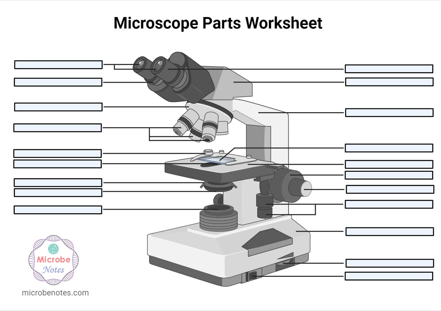
1. Illuminator (Light Source)2. Diaphragm (Iris)3. Condenser4. Condenser Focus Knob5. Rack Stop6. Stage7. Stage Control Knobs8. Nose Piece9. Objective Lens10. Tube (Head)11. Eyepiece (Ocular Lens)12. Diopter Adjustment13. Adjustment Knobs (Fine Adjustment Knob and Coarse Adjustment Knob)14. Arm15. Base16. Light Switch17. Brightness Adjustment
Thanks alot of your help and knowI can draw it well in my exams and write the functions.Thankyou very much for your help
Having been constructed in the 16th Century, microscopes have revolutionized science with their ability to magnify small objects such as microbial cells, producing images with definitive structures that are identifiable and characterizable.
Microscopes are made up of lenses for magnification, each with its own magnification powers. Depending on the type of lens, it will magnify the specimen according to its focal strength.
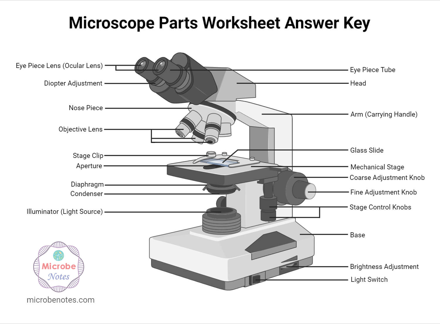
1. Ocular Lens (Eye Piece)2. Diopter Adjustment3. Head4. Nose Piece5. Objective Lens6. Arm (Carrying Handle)7. Mechanical Stage8. Stage Clip9. Aperture10. Diaphragm11. Condenser12. Coarse Adjustment13. Fine Adjustment14. Illuminator (Light Source)15. Stage Controls16. Base17. Brightness Adjustment18. Light Switch
1. which objective lens focuses closest to object 2. what controls the light entering the binocular lenses 3. how can you enhance the resolving power of a microscope 4. what is useful and false magnification PLEASE CAN YOU HELP ME IN ASWERING THOSE QUESTIONS
Find the best Lamps & Magnifiers for your project. We offer the 10X Magnifying Jewelers Loupe Magnifier Eye Tool New for $11.34 with free shipping ...
You can achieve a deep DoF by setting the aperture to f/16 or lower. And, if you want to make your image even clearer, try placing the camera as far away from the image as possible.
The optical parts of the microscope are used to view, magnify, and produce an image from a specimen placed on a slide. These parts include:
In shallow DoF, only a small part of your image is visible. Viewers are inherently attracted to the area in focus and will ignore everything else, thus making the subject of the image stand out. This technique is especially suitable for taking commercial photographs since the subject of the ad gets all the attention.
A beam of light is passed through the condenser to the specimen. The light transmitted from the specimen enters the objective lens. While passing through the objectives, the transmitted rays are spread so that they appear to come from the bigger objects.
Sometimes you get such awesome scenery that you want everything to be in focus in your image. Say, for example, you're taking pictures of a landscape, an architectural scene, or even a simple family photo, you want everything to be seen in order to give your image more context, and that's what you get with deep DoF.
I did NOT like this website sourse. Wanna know why I didn’t like it??? I don’t like it BECAUSE my school wants me to use this website sourse. My new science teacher wants us to answer those 10 questions. I think its pretty dumb. No Offensen to anyne out there, because I am a nice person not a mean one.
Thank you so much for the note that you have given to me i was so grateful to know that you are so bright people that extend your help to a student
The light is then focused on the eyepiece lens. This lens further magnifies the pre-magnified image coming from the objectives.
Thorlabs' plano-convex lenses have a positive focal length and can be used to focus collimated light, to collimate a point source, or reduce the divergence ...
Thank you for the support u have done may the Holy Spirit from the Almighty shine upon you to have more knowledge 2 continue making more notes from various topics in microbiology????✍️
Ans. A microscope is an optical instrument with one or more lens systems that are used to get a clear, magnified image of minute objects or structures that can’t be viewed by the naked eye.
Thanks for helping me to know the parts and functions of a light microscope. THANKS AGAIN AND I HOPE THAT I WILL DRAW IT IN MY EXAM
it very good website i use in 4 grade right after i plai amog us and they vote me out using orang strat witch mad me sad 🙁
Ans. The eyepiece, also known as the ocular is the part used to look through the microscope. Its found at the top of the microscope. Its standard magnification is 10x with an optional eyepiece having magnifications from 5X – 30X. Objective Lens are the major lenses used for specimen visualization. They have a magnification power of 40x-100x. There are about 1- 4 objective lenses placed on one microscope, in that some are rare facing and others face forward.
In deep depth of field (also known as large depth of field), both the background and foreground are sharp and clear. The viewer can also see all the details in the frame. This can make it quite challenging to create a well-balanced composition, but with proper graphics design software like CorelDraw, you can bring the image to life.
Image of HTENG VISHI GigE Ethernet 0.4MP 1/2.9" Color Industrial Camera Machine · Image of HTENG VISHI GigE Ethernet 1.3MP 1/3" Color Industrial Camera Machine.
Plastic Optics · Plastic Optics · Footer Start. Connect With Us. Facebook · Twitter ...
That being said, you should remember that using a large aperture puts your image at risk of overexposure. So, to avoid this, try using a fast shutter speed and low ISO.
BZBGEAR offers PTZ camera mounting brackets tailored for secure attachment to either walls or ceilings. These mounts have undergone rigorous testing to ensure ...
In any image, there is a zone that appears sharp and in focus. That region is referred to as the depth of field (DoF). DoF allows you to determine the areas you want to focus on in your image. You can either draw a viewer's attention to or away from certain areas within the image.
There are different types of microscopes like light microscope, dark-field microscope, phase contrast microscope, electron microscope, fluorescent microscope, etc.
WG70530-E3 - Ø1/2in ZnSe Broadband Precision Window, AR Coated: 7 - 12 µm.


 Ms.Cici
 8618319014500