
The Complete Guide to Hex Keys - alum wrench
The most important parameter of a microscope objective is the numerical aperture (NA). NA measures the microscope objective’s ability to gather light and determines the resolution of a microscopy system.
What is objective lens in microscope
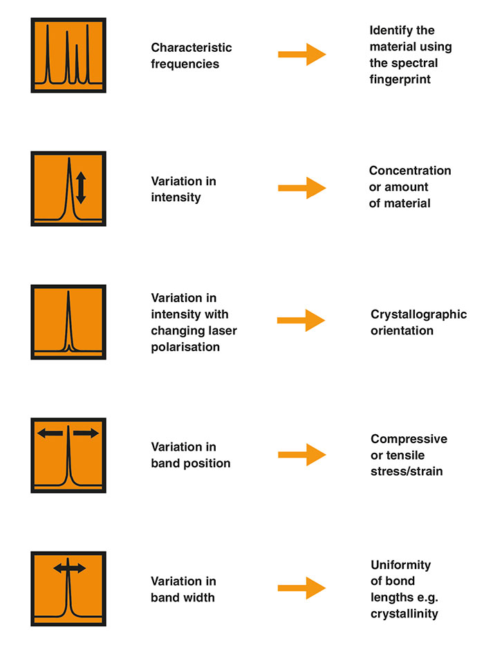
A Raman spectrum therefore consists of a number of bands, each associated with a vibrational mode. The spectrum is unique to the material and enables you to identify it. Some researchers aim to fully understand each Raman band and how it relates to vibrational modes. However, most analysts simply identify samples using a spectral library.
Continue your exploration of Raman and photoluminescence (PL) spectroscopy. We answer your questions on Raman microscopy, fast Raman imaging, data analysis, fluorescence and complementary analytical techniques.
The optical aberration corrections determine the optical performance of an objective lens. According to the degrees of the aberration corrections, objective lenses are typically classified into five basic types: Achromat, Plan Achromat, Plan Fluorite (Plan Semi-Apochromat), Plan Apochromat, and Super Apochromat. Choosing an objective with a proper aberration correction level will help you build a microscopy system at a reasonable cost.
Stagemicroscopefunction
In the polystyrene spectrum, we see the high frequency carbon-hydrogen (C-H) vibrations at about 3000 cm-1. The low frequency carbon-carbon (C-C) vibrations are at around 800 cm-1. The C-H vibrations have a higher frequency than the C-C vibrations because hydrogen is lighter than carbon.
Room 609, 6/F, Global Gateway Tower, No.63 Wing Hong Street, Cheung Sha Wan, Kowloon, Hong Kong +852-54993705 info@shanghai-optics.com
Ideally, you would use a Raman instrument with high spectral resolution across the whole Raman range. This gives you better chemical specificity. You can then identify, differentiate and investigate a wider range of materials.
Whatarethe3objectivelenses on amicroscope
The most common immersion media are air, water, oil, and silicone. Choosing the appropriate objective designed for your immersion medium will result in higher resolution images.
A dry objective is designed to work with the air medium between the specimen and the objective lens, while an immersion objective requires a liquid medium to occupy the space between the object and the front element of the objective for enabling a high NA and high resolution. Figure 4 shows the oil immersion objective, which can collect more light (i.e., have a higher NA) compared to a dry objective.
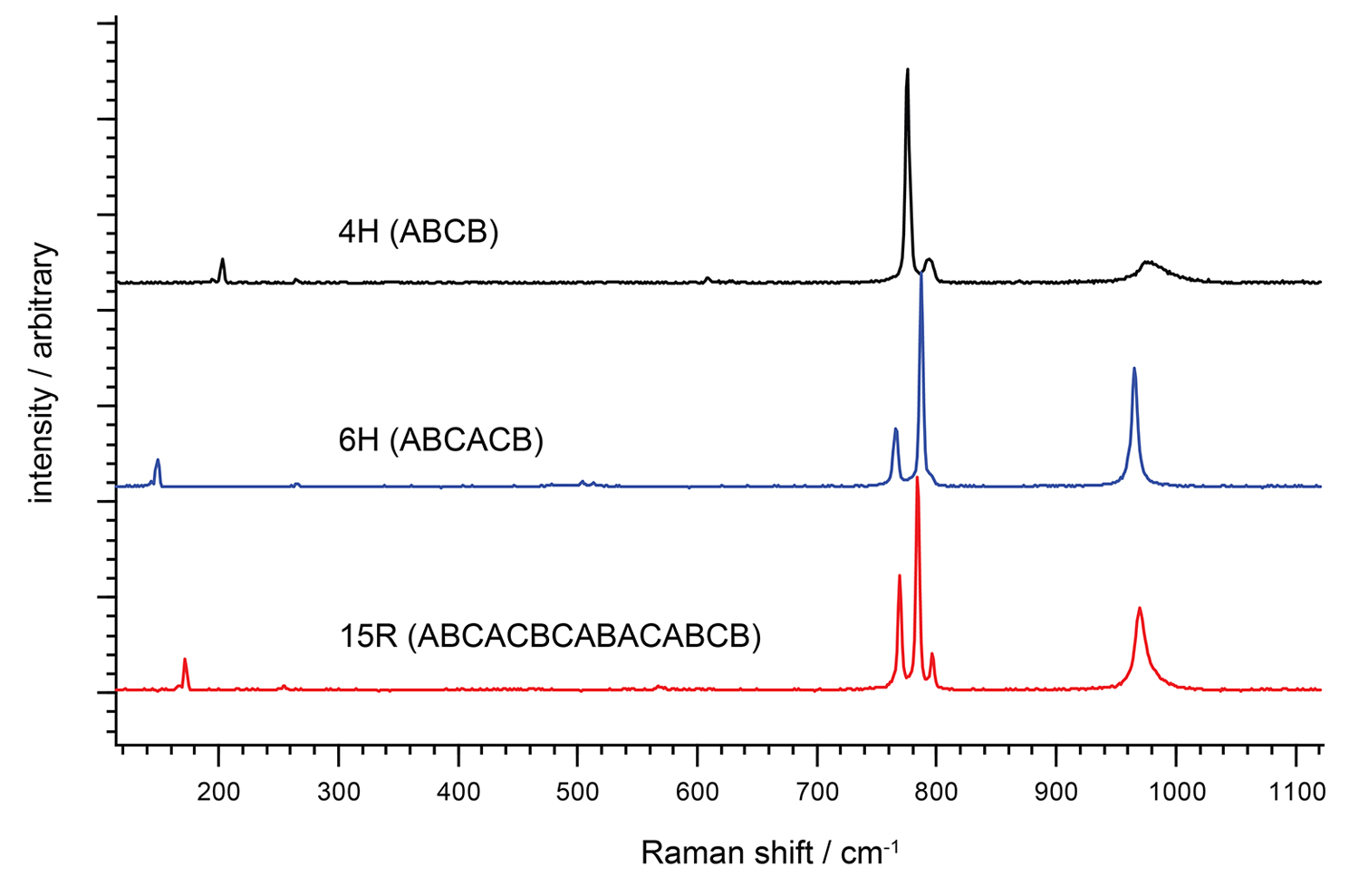
You can study differences in material structure by comparing their Raman spectra. You could quantify the degree of crystallinity and distinguish similar crystal forms (polymorphism) of the same chemical. To do this, you would need a Raman spectrometer with high spectral resolution, such as an inVia™ confocal Raman microscope.
In contrast, the Raman spectrum of polystyrene is more complex. The molecule is less symmetric and has hydrogen atoms in addition to carbon atoms. There are also different bond types connecting the atoms.
Usually the working distance (WD) refers the distance from the front lens element of the objective to the observed object when the object is in sharp focus. Objective lenses with long working distance are needed for many scientific research applications such as atom trapping and analyzing fluid samples that require putting an object in a chamber. The resolution of a microscopy system can be significantly affected if the observed object is not placed on the designed object plane, especially for an objective with high NA.
Functionofcondenserin microscope
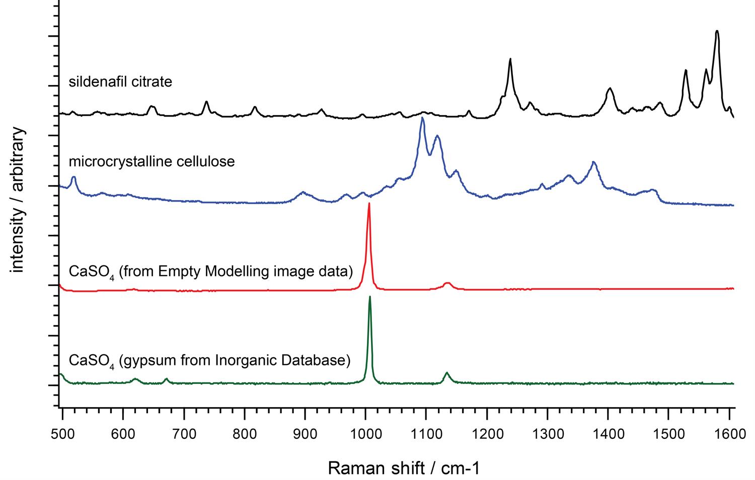
We can identify unknown materials from their unique Raman spectral fingerprints, typically using software searches of spectral libraries. We use the Raman bands in the fingerprint region (from 300 cm-1 to 1900 cm-1) to identify molecules.
Infinity-corrected objectives are ideal for research-grade biomedical industrial applications especially when additional components (such as filters, dichroic mirrors, polarizers) are needed in the microscopy system. Adding optical plate components in the infinity space (shown in the Fig.2 labelled as “Parallel Optical Path) between the infinity-corrected objective and tube lens will not introduce spherical aberration, or change the objective’s working distance.
Characteristic vibrational frequencies of chemical bondsThe frequencies of vibration depend on the masses of the atoms involved and the strength of the bonds between them. Heavy atoms and weak bonds have low Raman shifts. Light atoms and strong bonds have high Raman shifts.
Many objective lenses are corrected for infinite conjugate distance, while others are designed for finite conjugate distance applications. Compared to infinite conjugate objectives which need a secondary lens (also called tube lens), a finite conjugate objective can generate an image of a specimen by itself. A finite conjugate objective, as shown in Figure 1, is a good, economical choice for a simple microscopy system.
Objective lensfunction
We graphically display the results of our Raman spectroscopy measurements as Raman spectra. The y-axis represents the intensity of the scattered light, while the x-axis represents the energy (frequency) of light. We are interested in the shift in frequency of the Raman-scattered light, so we plot the x-axis frequencies relative to that of the laser. We label the x-axis as the Raman shift (shown by the units cm-1).
Objective lensmagnification
Low-frequency Raman bandsYou can also study molecular vibrational and rotational modes with low-frequency Raman shifts, below 100 cm-1. These originate from very heavy atoms or very large-scale vibrations, such as the whole crystal lattice vibrating. Renishaw's Raman instruments enable you to study these modes. You can explore a wide range of materials and crystals, easily distinguishing between different crystalline forms (polymorphs) and layered structures.
Objective lenses are used to magnify an image. In addition to numerical aperture, magnification is also an important parameter. The objective magnification typically ranges from 4X to 100X. As the image sensor size or eye observed area is fixed, the field of view of a microscopy system changes with the magnification of the objective lens. Typically a lower magnification objective lens will have a larger field of view and lower resolution, and a higher magnification objective lens will have a smaller field of view and higher resolution. The diameter of the FOV can be calculated by using the following formula: FOV= FN/Mag The field number (FN) in microscopy is defined as the diameter of the area in the image plane that can be observed through the eyepiece or image sensor.
Raman spectra of diamond and polystyrene. The Raman spectrum of polystrene is more complex than that of diamond due to differing bond types.
High powerobjective microscopefunction
Raman spectra of two polyethylene samples showing differences in intensities and band widths. These spectral differences are due to varying degrees of crystallinity.
Similarly, we see the vibrations of two carbon atoms linked by strong double bonds (C=C) at around 1600 cm-1. This is at a higher frequency than two carbon atoms linked by a weaker single bond (C-C, 800 cm-1).
NA is commonly expressed as NA = n × sinθa where θa is the maximum 1/2 acceptance angle of the objective, and n is the index of refraction of the immersion medium. The limit of resolution of a microscope objective refers to its ability to distinguish two closely spaced Airy disks. Resolution (r) = λ/(2NA) Where r is resolution (the smallest resolvable distance between two objects), and λ is the imaging wavelength. The higher the NA, the better the objective resolution.
You can view the vibrations of a complex molecule as partly consisting of many simple diatomic vibrations. However, you should also consider the vibrations of larger groups of atoms to get a full understanding of the Raman spectrum. For example, the Raman spectrum of polystyrene has a band at 1000 cm-1. This is due to the expanding and contracting ‘breathing mode' of the aromatic carbon ring in polystyrene.
Typesof objectivelenses
Raman shifts are sensitive to neighbouring bondsYou can see more subtle effects if you inspect Raman spectra closely. For example, the C-H vibrations of polystyrene appear in two bands, at approximately 2900 cm-1 and 3050 cm-1. The carbons in the former band are part of aliphatic carbon chains, whereas the carbons in the latter band form part of aromatic carbon rings.
Objective lenses are used in microscopy systems for a range of scientific research, industrial, and general lab applications. A microscope objective is typically composed of multiple lens elements and located closest to the object. There are so many types of microscope objectives available, choosing the right objective can help you produce good quality images at a reasonable cost. When choosing a microscope objective, we will need to consider a number of factors including conjugate distance, numerical aperture (NA), magnification, working distance, immersion medium, cover glass thickness, and optical aberration corrections. In this article, we will discuss how to choose the right microscope objective.
You can interpret Raman spectra to identify chemicals and get structural information. Raman scattering results from the interaction of light with molecular vibrations. These vibrations are very sensitive to changes in chemistry and structure, so you can spot subtle differences in molecular environment. Generally, all materials produce Raman spectra, except for pure metals.
One way of understanding a Raman spectrum is to consider the molecular functional groups as distinct units. This makes it easy to interpret the Raman spectrum of crystals with a regular array of identical atoms, all in the same configuration. For example, diamond contains carbon atoms in a regular tetrahedral network. In these cases, you often see just one dominant Raman band because there is just one molecular environment of the crystal.
Alpha Industrial Park, Tu Thon Village, Ly Thuong Kiet Commune, Yen My District, Hung Yen Province Vietnam 17721 +84 221-730-8668 rfqvn@shanghai-optics.com
SO offers a wide range of objective designs, which provide various degrees of optical aberration corrections for supporting different needs, such as achromatic objectives (the cheaper objectives) for laboratory microscope applications and long working distance apochromats (expensive objectives) for biological and scientific research applications. We can help you choose or design a properly corrected objective lens for meeting your application requirements.


 Ms.Cici
 8618319014500