
TASK Quick Support Rods - support rod
l Elimination of Air Gap: Like other optical bonding methods, LOCA bonding eliminates the air gap between the layers, minimizing internal reflections and increasing the amount of light that reaches the viewer's eyes. This leads to improved readability, especially in bright ambient conditions.
A reflected darkfield objective works for darkfield microscopy. This technique produces a dark background with a strong contrast to aid in the visibility of translucent specimens. This object is designed to observe samples not dropped inside a covered slide. Reflected darkfield objectives typically have signs like BD, Neo, or BF/DF to help you identify them.
Microscope lenses are pieces of glass that work in a microscope to aid magnification. Based on the lens type and power, you can magnify a specimen by up to 200x or more. How these tools work is straightforward, and this article will cover everything you need to know about them.
l Easy Repairability: LOCA bonding allows for relatively easier repair or replacement of components compared to some other bond.
l Touch Sensitivity: LOCA bonding can enhance touch sensitivity and accuracy by reducing the parallax effect, which is the misalignment between the touch input and the actual display content. With a reduced air gap and tighter alignment, LOCA bonding ensures more precise touch response and a better user experience.
l Optical Clarity: LOCA adhesives are designed to have excellent transparency, ensuring minimal impact on the optical properties of the display. They have a high refractive index, closely matching that of glass, which helps reduce reflections and increase contrast, resulting in improved visual quality.
LOCA glue remover
Long-working distance objectives are made so you can see specimens even when they are farther away than usual. This is usually needed when a sample is stuck in a thick slide or is under a thick glass plate.
The simplest types of microscopes are magnifying glasses with a single convex lens (meaning both sides are curved outward). This kind of lens usually makes items look 5–10 times bigger by changing how the light gets into the human eye. Compound microscopes are used in schools, homes, and professional labs. They have at least two lenses that work together to magnify an image.

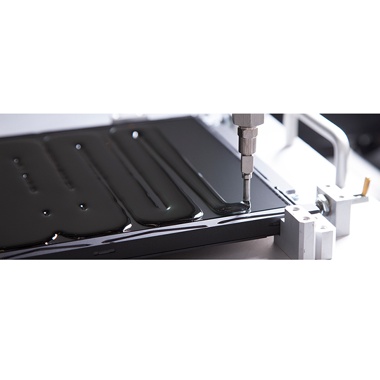
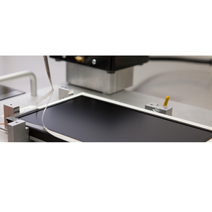
Liquid optically clear adhesive (LOCA) bonding is a specific type of optical bonding technique used to join display components together. It involves the application of a special optically clear adhesive between the display panel and the cover glass or touch sensor layer. The adhesive is in liquid form during the bonding process and cures or solidifies afterward.
You can identify a high magnification lens by the blue band around the housing of the lens. Typically, compound microscopes come with a 40x lens. However, there are cases when this is not true. For example, you might buy a microscope with a high magnification lens of 60x or more.
l Bond Strength: LOCA adhesives create a strong bond between the display panel and the cover glass or touch sensor layer. This enhances the overall durability and mechanical strength of the display assembly, making it more resistant to impact, vibrations, and environmental stresses.
Most basic microscopes do not come with an oil immersion lens, and this is because most leisure microscopy experiments do not require them. These lenses can reach up to 200x or more magnification with a 10x eyepiece lens and a 200x objective lens. You can find this lens by a white or cream-colored band around the lens.
The use of differential interference contrast (DIC) lenses in brightfield microscopy helps to visualize transparent samples better. By providing contrast without the need for staining, DIC objectives reduce the amount of staining performed. In most cases, a DIC lens will not be present on a compound microscope for school or home use.
LOCA UV Glue
There is one lens above the object, called the objective lens. Also, there’s another one close to your eye (eyepiece). In some cases, each type of lens consists of various lenses. Compound microscopes can typically magnify by 10x, 20x, 40x, or 100x. However, you can find professional ones that can reach up to 200x magnification or more. There are also modern microscopes like the electron microscope for those who want higher magnification.
Low magnification objective lens typically ranges from 2x to 20x. Using a 10x or 20x eyepiece will magnify objects by 100x or 200x. This lens lets you view tiny specimens such as skin, hair, and fly legs. Furthermore, it has a yellow band that encircles the housing of the lens.
Phase contrast microscopy makes translucent specimens easier to see by making the difference between the background and the foreground stronger. In a phase contrast objective, a black ring around the lens is used to control and translate changes in the phase of light rays into changes in their amplitude. In addition, the way the light rays are bent and focused gives the image seen through the eyepiece a lot of contrast.
Due to the difference between the glass slide and the refractive indices of air, a specific oil is required to help fill the space. Without this oil, the objective lens won’t function correctly. Hence, you won’t get the appropriate magnification and resolution, leaving you with too much distortion.
Microscope objective lenses work by changing how light goes through them. Essentially, when light shines on an object underneath a microscope, this light travels through the lens and bends toward your eyes, which makes the object bigger than it is. Remember that magnification power varies based on the type of lens and microscope, with magnification reaching 1000x and above. You can also find specialized objective lenses for advanced experiments.
An optical microscope comes with lenses that change how rays of light travel through them. When light bounces off an object under a microscope and goes through the lens, it deflects toward the eye. This makes the item seem bigger than it is.
l Moisture Resistance: The adhesive used in LOCA bonding can provide a moisture barrier, helping to prevent the ingress of water or moisture into the display assembly. This is particularly beneficial in outdoor or high-humidity environments where moisture can damage the internal components and cause fogging or image distortion.
LOCA glue for Tempered glass
Hot tags:optical bonding, optical bonding touch screen, optical bonding display, optical bonding vs air bonding, optical contact bonding technology, lcd display with optical bonding, touch screen display with oca bonding, optical bonding process, dry optical bonding, oca vacuum bonding process, china, customized, suppliers, factory, manufacturers
VIETNAM:Alpha Industrial Park, Tu ThonVillage, Yen My District, HungYen Province 17721+84 221-730-8668sales-vn@avantierinc.com
Microscope lenses come in different types that vary based on the magnification’s power. Here are the types of microscope objective lenses.
You can purchase certain specialized microscope objectives when you want to perform advanced microscopy experiments. Here are some of the most common lenses to buy.
Optically Clear Adhesivefilm
This lens, in conjunction with the eyepiece lens, will provide the smallest magnification possible. For example, a microscope with a 10x eyepiece lens and a 4x objective lens will have a magnification factor of 40x. The magnification you get from this lens is similar to what you would from a stereo microscope, allowing you to study specimens like leaves and feathers. Also, the lens has a red band that encircles the housing of the lens. Scanning object lenses have low power and are typically used to scan a specimen before using higher magnifications.
This type of lens is usually used for smaller specimens, such as cells and bacteria, which cannot be seen with just the human eye. This includes molds, tardigrades, germs, and others.
Utilizing this microscope objective lens is pretty simple. Firstly, you need to adjust the scanning lens to properly focus and center the specimen. Afterward, you need to turn the objective turret clockwise to face the low magnification lens. Lastly, re-center your specimen after you’ve fine-tuned the focus with the coarse focus knob.


 Ms.Cici
 8618319014500