
Silicon rod, polycrystalline, unpolished, 1.6cm (0.6in) dia, ... - silica rod
PVD can cause a person to develop vitreous traction. This is a condition that occurs when part of the vitreous remains stuck to the macula during PVD. This part of the vitreous then pulls on the macula, causing symptoms of vitreous traction.
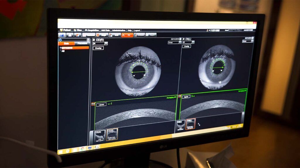
During the scan, a person may focus on a target within the scanner. They may also see a red line appear during the scan.
Choose products to compare anywhere you see 'Add to Compare' or 'Compare' options displayed. Compare All Close
OCT can also help medical professionals diagnose optic nerve disorders. This is because an ophthalmologist can use OCT to spot changes to the fibers of the optic nerve.
Gaussianbeamprofile
Laser beams are often propagated through an optical system consisting of lenses, mirrors, dielectric interfaces, or other optical elements. Fortunately, under most conditions, a Gaussian beam remains a Gaussian beam after encountering these elements, ensuring that the propagation equations used above remain valid. An optical element modifies the input beam by changing the position and size of its beam waist. Knowledge of the input beam parameters and the properties of the optical element can be used to determine the new values using a variety of propagation methods. A thin focusing lens is probably the most important optical element that impacts a laser beam. Figure 2 illustrates beam propagation through a thin lens resulting in a repositioning and resizing of the beam waist (as well as zR and θ). When a collimated beam (i.e., 2.zR >> f) encounters a lens, the resulting beam waist is simply the product of the focal length and original divergence (see full angular width divergence equation) with the waist positioned at the focal point of the lens.
Central serous retinopathy is a condition that occurs when fluid builds up under the retina. Medical professionals may also refer to this condition as central serous chorioretinopathy.
Without treatment, diabetic retinopathy can lead to blindness. OCT can help a medical professional diagnose diabetic retinopathy.
Gaussianbeamcalculator
While many laser systems operate with near-Gaussian beams, other laser systems possess non-Gaussian beams that propagate differently and exhibit significantly different spatial distributions (see Figure 4 for examples). In some cases, a laser resonator emits a beam with a higher-order TEMmn mode. Depending on the resonator geometry, these modes can be cylindrical in nature and are called Laguerre-Gaussian beams or rectangular and are called Hermite-Gaussian beams. In other cases, a laser beam is modified by an optical system to such an extent that its profile and propagation can no longer be approximated using the Gaussian beam analysis. Flat-top beams are one such example where a beam exhibits a nearly constant irradiance over its beam width (see Figure 4). Given the steep edges of the beam profile, the diameters of these beams are often characterized by their full-width at half-maximum (FWHM) values as opposed to the HW1/e2 radius values used for Gaussian beams. Such flat-top beams are important for laser-based material processing where a constant irradiance provides more uniform material modification. The propagation of these beams can be quite complicated and is often encountered when a laser beam overfills a focusing objective in order to generate a very small spot size in high-resolution microscopy.
Gaussianbeamsoftware
OCT can help an ophthalmologist see the different layers of the retina. This helps them map and measure the thickness of these layers.
A commonly-available helium-neon laser emits a near-ideal Gaussian beam with a value of M2 < 1.1. For many solid-state lasers, M. is in the range of 1.1-1.3. Collimated laser diodes that emit fundamental TEM00 modes possess M. of 1.1 to 1.7, whereas high-energy multimode lasers can generate M2 factors as large as 10 to 100. Spatial filtering can improve beam quality using Fourier optics. Finally, additional beam quality metrics include the Beam Propagation Factor (K) where K = 1/M2 as well as the Beam Parameter Product (BPP), which is defined as BPP = M2λ/π = 2WM.M2
A person may not experience any symptoms if they have early stage glaucoma. Over time, a person may slowly experience vision loss.
Measuring the thickness of the different layers of a person’s retina can help an ophthalmologist diagnose a number of medical conditions, such as age-related macular degeneration (AMD) and glaucoma.
By allowing an ophthalmologist to do all the above, OCT can help them diagnose certain eye disorders and guide specific treatments.
Gaussianbeamdivergence
If a person has received dilating eye drops during OCT, their eye may be sensitive to light for several hours after the procedure.
The macula is a small area in the center of the retina. It plays a role in helping people clearly see the details of objects.
Gaussianbeamradius
A medical professional can use OCT to help diagnose AMD. It can be suitable for helping diagnose both early and advanced-stage AMD.
Prolonged high blood sugar levels can damage the retina. This occurs when diabetes damages the blood vessels that supply the retina with blood.
Yes, opt-in. By checking this box, you agree to receive our newsletters, announcements, surveys and marketing offers in accordance with our privacy policy
Gaussianbeampdf
This illustrates that the beam shape remains Gaussian at any point along the axis and changes only in its width and amplitude. The beam radius W is defined as the radius at which the irradiance decreases to 1/e2 or 0.135 of the peak on-axis (r = 0) value. This is sometimes referred to as the half-width 1/e2 (HW1/e2) value. As shown in Figure 1, W gradually increases as the distance from the minimum beam radius (known as the beam waist, W0 gets larger. Since E is the power per unit area, as indicated by above equation, the irradiance decreases as one moves away from the beam waist. Integration of the irradiance over the entire radial plane (at any axial position) results in the total optical power. In other words, φ remains constant along the axis. From a practical standpoint, integrating the irradiance within a circle of radius 1.5.W results in 99% of the total power. This is relevant when measuring the optical power of a Gaussian beam.
In some cases, an ophthalmologist may administer dilating eye drops before the procedure. This is to help widen the pupil to make imaging easier.
The equation describing the evolution of the beam radius along the axis where z is the distance from the beam waist is given by:
Vitreous is the substance that fills the middle of the eye. Posterior vitreous detachment (PVD) occurs when the vitreous pulls away from the retina.
zR is known as the Rayleigh range and represents the distance from the waist where the radius increases by a factor of . If above equation is substituted into irradiance distribution equation, then zR is the distance at which the irradiance has decreased by a factor of √2 from its peak value at the waist. Twice the Rayleigh range is called the confocal parameter (or depth-of-focus) and is a rough estimate of the collimation of a beam. As shown in the above equation, the beam size increases slowly with axial distance from the waist. As z >> zR, the beam size then increases linearly with z with a slope of W0/zR. This slope represents the full angular width divergence (θ) of the beam given by:
Diabetic retinopathy is a complication of diabetes that causes damage to the blood vessels in the retina. Learn about its causes, symptoms, and…
During a CT scan, a medical professional aims the X-rays at a part of the person’s body and quickly rotates them around the body. The CT scanner processes the signals that these X-rays produce. The CT scanner then creates cross-sectional images.
Gaussianbeamintensity formula
An ophthalmologist may use OCT to help confirm a diagnosis if a person is displaying symptoms of certain eye conditions. Below are the symptoms that may lead an ophthalmologist to use OCT.
During OCT, an ophthalmologist uses a beam of light to scan an area of a person’s eye. The OCT device then measures the light that the structures within the eye have reflected back.
The irradiance distribution of a laser beam is determined by the transverse modes that exit the laser cavity. Typically, the lowest-order transverse mode (TEM00) is selected for emission since it propagates with the least beam divergence and can be focused to the tightest spot. The irradiance distribution of this TEM00 mode is described by a Gaussian function and therefore much of this section details a Gaussian beam's profile as well as its evolution with distance. Gaussian beam propagation is well understood and even laser beams that do not possess a TEM00 mode are often described using a modified Gaussian mode analysis using its M-Squared value. Finally, a brief description of non-Gaussian beams is also given.
Glaucoma is a group of eye diseases that damage the optic nerve. Glaucoma can lead to a person experiencing vision loss and blindness.
In its early stages, diabetic retinopathy usually does not cause symptoms. Some people may notice changes in their vision, such as problems seeing things that are far away.
Optical coherence tomography (OCT) is a form of noninvasive imaging test that creates high-resolution, cross-sectional images of the inside of a person’s eye.
The evolution of a Gaussian beam as it propagates along its axial direction (z) is shown in Figure 1. The irradiance has a radial (r) distribution that is circularly symmetric in any plane orthogonal to the axis and the beam's power (φ) is concentrated close to the axis. The functional form of this irradiance distribution (E) is given by:
The OCT device then uses these measurements to create images of a person’s retina that are cross-sectional and three-dimensional.
Read on to learn more about how OCT works. This article also explains the conditions that OCT can help with and what happens during the procedure.
OCT is not the same as a computed tomography (CT) scan. OCT uses light rays to create images, and CT scans use X-ray beams.
There is no research to prove that exercises can help with drooping eyelids. Learn more about this here and find out about other treatment options.
One practical consequence of this definition is that an ideal Gaussian beam (i.e., M2 = 1) can be focused to a minimum spot diameter, whereas beams of higher M. values focus to larger spot diameters in proportion to the M2 value. As a result, M2 provides meaningful information about lasers, particularly if their application involves small focused spot sizes. While WM and θM are sufficient for determining M2, these values often cannot be measured directly. By focusing the beam with a lens of known focal length (like Figure 2), the characteristics of the artificially created beam waist and divergence can be measured. To provide an accurate calculation of M2, the International Organization for Standards (ISO) requires at least 5 measurements in the focused beam waist region and at least 5 measurements in the far fields (two Rayleigh ranges away from the waist area), as shown in Figure 3. These multiple measurements ensure that the minimum beam width is found while a "curve fit" improves the accuracy by minimizing measurement error at any single point.
OCT can help an ophthalmologist diagnose a number of medical conditions including problems with the macula, AMD, glaucoma, and different types of retinopathy.
The TEM00 mode of even a well-designed laser system is not a perfect Gaussian beam. The M2 ("M-Squared") analysis was developed to characterize the quality of a laser beam. That is, how close it is to an ideal Gaussian beam. M2 is defined as the product of the spot size (2WM) and divergence (θM) of the real beam divided by the spot-size-divergence product of an ideal Gaussian beam:
A clear reciprocal relationship exists between the spot size (2W0) and the divergence. In addition, for a Gaussian beam at a given wavelength, the product of the spot size and the divergence is a constant at 4λ/π. This is important in defining how much a beam deviates from a perfect Gaussian beam.
Gaussianbeamformula
For additional insights into photonics topics like this, download our free MKS Instruments Handbook: Principles & Applications in Photonics Technologies
Optical coherence tomography (OCT) is a type of noninvasive imaging test. Ophthalmologists use OCT to create high-resolution, cross-sectional images of the inside of a person’s eye.
Researchers say the drug aflibercept performed better in a clinical trial than bevacizumab in helping wean people off treatments for wet age-related…
They must then remain still and follow the instructions of the person carrying out the procedure. These instructions will direct the person to look in certain directions throughout the scan.


 Ms.Cici
 8618319014500