
Security Cameras - Wired & Wireless Home Surveillance ... - cameas
Each microscope objective is itself a complex assembly of lenses, and besides contributing to the magnification, it is the objective lens which determines the resolution power of the microscope. An objective lens can also provide optical aberration corrections. A reflective objective, for instance, includes two mirrors within the assembly. These mirrors can focus laser light as well as provide chromatic corrections.
Opticians need to understand light polarization to provide the best advice on eyewear selection. The advantages of polarized lenses can be substantial for patients with specific needs, such as those involved in particular sports or professions. Different lens materials affect the polarization of light in varying ways. Knowledge of this is essential when recommending lens materials or coatings. For instance, some lenses may have a polarizing filter or coating that can reduce glare and increase visual comfort.[35]
While polarized lenses are generally beneficial in reducing glare and enhancing visual comfort, they can interfere with the visibility of certain displays, such as computer and liquid crystal display (LCD) screens, that use polarization to display images. This is due to the orientation of the polarizing filters in the lenses, which may block or distort the light emitted from these screens. Users may experience a darkening or color shift when viewing computer monitors, smartphones, tablets, or other devices with LCD screens while wearing polarized eyewear.[28] Additionally, polarized lenses can interfere with the visibility of certain instruments or displays in vehicles and aircraft, such as GPS systems or heads-up displays, potentially affecting the performance of these devices.
Circular polarization filters are incorporated into many slit lamps and ophthalmoscopes. Corneal reflections can hinder the clinician's ability to view ocular structures, especially when examining the anterior segment. When circularly polarized light interacts with the cornea, the reflected light changes its polarization state. A circular polarizing filter placed in the observation pathway, oriented perpendicular to the first filter on the light source, effectively filters out the altered polarization state of the corneal reflections. This process results in a clearer and more detailed view of other ocular structures.[23]
PS-OCT combines the principles of optical coherence tomography (OCT) with polarization-sensitive detection. PS-OCT provides depth-resolved, high-resolution images of the retina, choroid, and optic nerve head, along with information on their polarization properties. This enables the assessment of structural changes and the birefringence of the tissues, which can indicate various pathological conditions.[13] Compared with OCT, PS-OCT offers improved contrast between different retinal layers and structures and can evaluate birefringent tissues, such as the RNFL, Henle fiber layer, and RPE. Compared with SLP, PS-OCT allows for a comprehensive evaluation of the retina, choroid, and optic nerve head, making it useful for diagnosing and monitoring a range of ocular conditions.[22]
Unpolarized light, possessing random polarization, is passed through a linear polarizer to create linearly polarized light, then passed through a quarter-wave plate to create circularly polarized light. Contributed by Garrett Manion
Two major lens components—the objective lens and the ocular lens, or eyepiece—work together to project the image of the specimen onto a sensor. This may be the human eye or a digital sensor, depending on the microscope setup.
The development of specialized instrumentation and devices has facilitated the utilization of polarized light in clinical practice, allowing for assessing and managing various ocular conditions. Further advancements in polarized light-based technologies have the potential to contribute to the evolution of patient care, offering more accurate and noninvasive diagnostic modalities, as well as innovative treatment approaches.
Polarized light is utilized in various diagnostic and therapeutic techniques in ophthalmology, such as anterior segment imaging, retinal imaging, photodynamic therapy, low-level light therapy, and laser surgery. The unique properties of polarized light offer numerous benefits for enhancing patient care, from improving visual performance to facilitating the diagnosis and management of various ocular conditions.
While the simplest of microscopes is simply a magnifying glass with a single lens, compound microscopes used today are highly complex devices with a carefully designed series of lenses, filters, polarizers, beamsplitters, sensors, and perhaps even illumination sources. The exact combination of optical components used will depend on the application of the microscope; the wavelength of light with which it is intended to be used, and the resolution and magnification required in the final image.
Objective lenses can be classified based on the objective construction, field of use, microscopy method, performance (optical aberration corrections), and magnification. Many microscope objective manufacturers offer a wide range of objective designs, which provide various degrees of optical aberration corrections for supporting different needs. Mirrors or reflective elements are used in objective lenses for the applications that requires chromatic aberration over board spectral ranges. Most traditional microscopy systems use refractive objectives such as achromatic objectives (the cheaper objectives) for laboratory microscope applications and plan apochromats (expensive objectives) for biological and science research microscope applications.
Titmus Fly stereotest, Randot stereotest, and Lang stereotest are among the commonly used stereo tests in clinical practice.[26][27] These tests use polarized glasses to present a series of images or patterns with different disparities, requiring the integration of visual information from both eyes to correctly perceive the depth or three-dimensional structure in the images or patterns. In the Titmus Fly Stereotest, for example, patients wearing polarized glasses are asked to identify a three-dimensional fly. Similarly, in the Randot stereotest, patients must identify three-dimensional shapes among two-dimensional images.
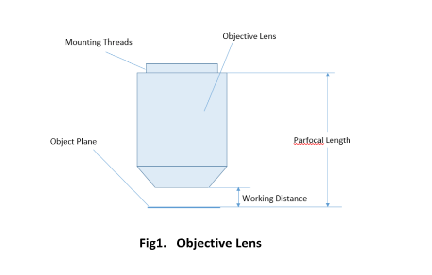
For keeping the objective at the proper position, there are mounting threads on almost all objectives. Commonly used mounting threads include RMS, M25 x 0.75, M26X 0.706, M32 x 0.75.
Unpolarized lightexamples
Alpha Industrial Park, Tu Thon Village, Ly Thuong Kiet Commune, Yen My District, Hung Yen Province Vietnam 17721 +84 221-730-8668 rfqvn@shanghai-optics.com
Elliptically polarized light is a type in which the electric field vector traces out an ellipse as it propagates, with varying amplitude and phase. This type of polarization can be considered a combination of two linearly polarized waves of unequal amplitude and perpendicular polarization directions, with a phase difference between them that is not necessarily 90 degrees.[8] Elliptically polarized light can be generated by passing linearly polarized light through a material that induces a phase shift between the two perpendicular polarization directions, such as a birefringent crystal.[9]
A simple magnifier (magnifying glass), works when the object to be examined is situated within focal length of the magnifier lens, enabling larger virtual image is produced. This type of magnifier is very limited in both resolution and magnification. A compound microscope, on the other hand, uses a relay lens system instead of the single lens, and since each lens component can contribute magnifying power, the result is greatly increased capability.
All light possesses polarization. Light, commonly termed "unpolarized," lacks organized polarization, instead exhibiting randomized polarization. Randomly polarized light is a type of light in which the electric field vector oscillates in random directions perpendicular to the direction of the light's propagation. In other words, the light waves are not aligned in any particular direction and are constantly changing orientation.[3] As a result, the light cannot be blocked or transmitted by a polarizer, which is a material that only allows light waves of a specific polarization orientation to pass through.
A microscope objective is an important component of a microscopy or imaging system for a range of science research, biological, industrial, and general lab applications.. An objective lens determines the basic performance of an optical microscope or imaging systems and is designed for various performance needs and applications. It is located closest to the object and is an important component in imaging an object onto the human eye or an image sensor.
Examples ofpolarizedandunpolarized light
Room 609, 6/F, Global Gateway Tower, No.63 Wing Hong Street, Cheung Sha Wan, Kowloon, Hong Kong +852-54993705 info@shanghai-optics.com
The glaucoma diagnostics (GDx) test utilizes SLP to measure RNFL thickness, allowing for the detection of glaucomatous damage and monitoring of disease progression. The GDx test has two main iterations: the GDx Nerve Fiber Analyzer and the GDx VCC (variable corneal compensation). The GDx Nerve Fiber Analyzer was the first-generation device that used a fixed corneal compensation algorithm to account for corneal birefringence. However, this fixed compensation could not accurately account for the variability in corneal birefringence between individuals, leading to potential measurement errors. To address this limitation, the GDx VCC was developed, incorporating a variable corneal compensation algorithm that individually measures and compensates for the corneal birefringence of each patient. This improvement allows for more accurate RNFL measurements, making the GDx VCC a valuable tool in glaucoma assessment.[21]
SLP is a noninvasive imaging technique that utilizes polarized light to measure the thickness of the retinal nerve fiber layer (RNFL). The RNFL consists of axons of retinal ganglion cells responsible for transmitting visual information from the retina to the brain. The change in polarization as light moves through the layer is directly proportional to the thickness of the RNFL. SLP is beneficial as it is a rapid and quantitative assessment of the RNFL in high resolution. However, ocular media opacities, such as cataracts or corneal irregularities, can affect SLP, which may influence the accuracy of RNFL thickness measurements. In addition, the birefringence of the RNFL may vary among individuals, which can impact the accuracy of SLP measurements.[20]
Stereo testing, also known as stereopsis or depth perception testing, is a critical element in the comprehensive eye examinations conducted in optometry and ophthalmology.[24] Stereopsis is the visual perception of depth and three-dimensional structure. This perception is primarily due to the slight differences in the images projected onto the retinas of the two eyes, a concept known as binocular disparity.[25]
LLLT uses polarized light at specific wavelengths to modulate cellular processes, such as mitochondrial respiration, and promote tissue healing. This noninvasive therapy has shown promising results in managing ocular conditions, such as dry eye syndrome, by reducing inflammation and stimulating the production of tear film components. Additionally, LLLT has been explored as a potential treatment for retinal diseases, such as diabetic retinopathy and AMD, by promoting cellular repair mechanisms and reducing oxidative stress.[30]
The parfocal length is the distance between the objective mounting plane and the specimen / object. This is another specification that can often vary by manufacturer.
Trained technicians and nurses must be knowledgeable about operating these machines and obtaining quality results to provide the most accurate diagnostic information for the patient's condition. The interprofessional team's comprehensive understanding of light polarization can significantly improve patient outcomes and satisfaction. Effective coordination among ophthalmologists, optometrists, opticians, and optical technologists or nurses ensures patients receive the most appropriate and beneficial ocular care.
Unpolarized lightdiagram
As our understanding of the principles underlying light polarization and its applications in ophthalmology and optometry continues to grow, healthcare professionals must remain informed of the latest developments and consider integrating these technologies into their clinical practice when appropriate. Interprofessional collaboration and education are essential in leveraging the benefits of polarized light to enhance the patient experience and contribute to the ongoing advancement of the fields of ophthalmology and optometry.
Since indirect backlight illumination is generally more effective than direct illumination, most microscopes do not include an internal light source. Instead, they rely on daylight or on background illumination such as a lightbulb. In brightfield illumination, also known as Koehler illumination, two convex lenses saturate the specimen with external light admitted from behind. These two lenses, the collector lens and condenser lens, work together to provide a bright, even, and constant light throughout the system: on the image plane as well as on the object plane. This system of illumination is used in many compound microscopes, including student microscopes and those found in many research labs.
What isunpolarized lightin physics
This book is distributed under the terms of the Creative Commons Attribution-NonCommercial-NoDerivatives 4.0 International (CC BY-NC-ND 4.0) ( http://creativecommons.org/licenses/by-nc-nd/4.0/ ), which permits others to distribute the work, provided that the article is not altered or used commercially. You are not required to obtain permission to distribute this article, provided that you credit the author and journal.
Polarization oflightnotes PDF
In stereo testing, the role of polarization is significant. Polarized glasses are employed to deliver different images to each eye, simulating the binocular disparity that is inherent in natural vision. This process effectively tests the patient's ability to perceive depth and integrate binocular visual information. Polarized glasses used in these tests have differently oriented filters for each eye, allowing different images to be presented to each eye simultaneously.
Ophthalmic laser systems increasingly utilize polarized light to ensure precise tissue ablation or photocoagulation during various surgical procedures. The controlled delivery of polarized light in these surgeries improves the accuracy and precision of tissue targeting. Some of the key ophthalmic procedures that involve the use of polarized light in laser surgery include:
Important specifications are marked on the barrel of the objective, so students or researchers can easily identify the properties of an objective and determine the optical performance and working conditions for proper use. Figure 1 shows a diagram of an objective lens. A detailed discussion of the objection specifications is provided below.
At Shanghai Optics, we design and manufacture custom objectives and imaging systems to support our customers’ needs in many industries, including medical, biomedical, machine version, scientific research, and metrology, etc. Taking the client’s budget and precision requirements into consideration, our experienced engineering team ensure that each design can be manufactured at a reasonable cost and the optical performance is being met based on fabrication, assembly, and alignment tolerance analysis.
Most objectives are designed to image specimens with air as the medium between the objective and the cover glass. However, for achieving higher working numerical apertures, some objectives are designed to image the specimen through another medium such as special oil with a refractive index of 1.51.
Polarized vs unpolarized lightreddit
Magnification is one important parameter. Magnification is usually denoted by an X next to a numeric value. Objectives are available in a range of magnifications from 2X to 200X.
In PDT, polarized light activates photosensitizing agents that selectively target pathological tissues, such as neovascular membranes in age-related macular degeneration (AMD) or tumor cells in ocular malignancies. The photosensitizing agents absorb the polarized light, which triggers a series of chemical reactions that produce reactive oxygen species (ROS). The ROS then cause localized cellular damage and death, ultimately destroying the targeted tissue. Polarized light in PDT ensures that the light is precisely delivered to the target tissue, minimizing damage to surrounding healthy structures.[29]
Difference betweenpolarizedandunpolarized lightsunglasses
The .gov means it's official. Federal government websites often end in .gov or .mil. Before sharing sensitive information, make sure you're on a federal government site.
Field of View is the area of the object that can be imaged by a microscopy system. The size of the field of view is determined by the objective magnification or focal length of the tube lens for an infinite-corrected objective. In a camera system, the field of view of the objective is related to the sensor size.
Polarization of light is a fundamental optical phenomenon with significant implications in ophthalmology and optometry, enhancing diagnostic and therapeutic techniques for various ocular conditions.
Since the objective is closest to the specimen being examined, it will relay a real image to the ocular lens. While doing so, it contributes a base magnification of anywhere from 4x (for a scanning objective lens, typically used to provide an overview of a sample) to 100x (for oil immersion objectives).
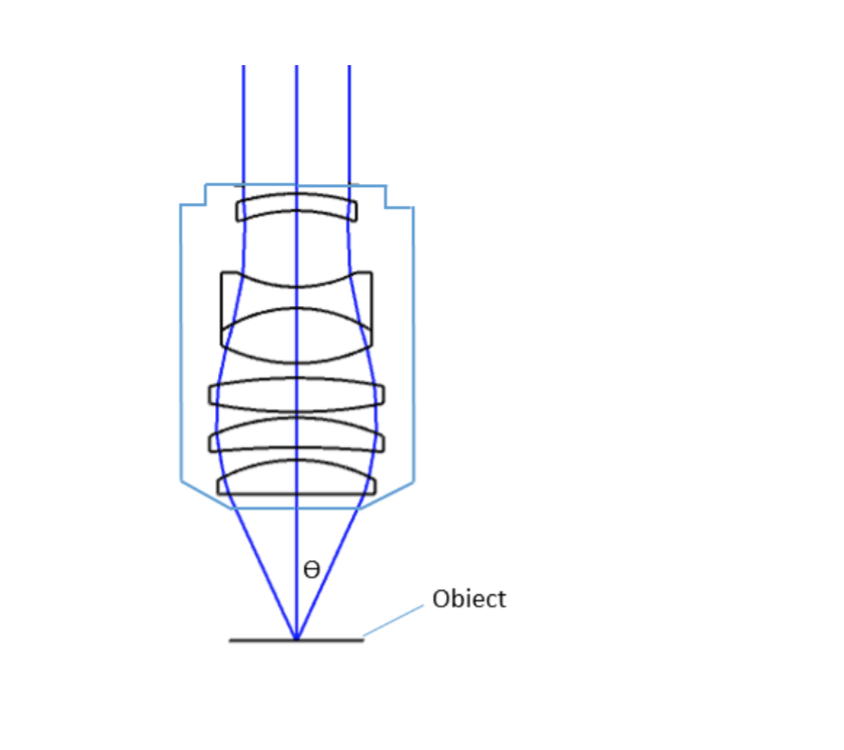
where θ is the maximum 1/2 acceptance ray angle of the objective, and n is the index of refraction of the immersion medium. Figure 2 shows the ray angle θ of an infinity-corrected objective.
Light polarization plays a vital role for the interprofessional healthcare team to ensure the best patient outcomes and satisfaction with their vision. The understanding and practical application of polarized light requires collaborative efforts from ophthalmologists, optometrists, opticians, and optical technologists or nurses. Opticians and optical technologists play a key role in assisting patients with the selection of polarized eyewear. These lenses can reduce glare and improve visual comfort, particularly in bright conditions or during outdoor activities.
Difference betweenunpolarizedand planepolarized light
The optical aberration correction determines the optical performance of an objective lens and plays a central role in the image quality and measurement accuracy of imaging or microscopy systems. According to the degrees of the aberration corrections, objective lenses are generally classified into five basic types: Achromat, Plan Achromat, Plan Fluorite (Plan Semi-Apochromat), Plan Apochromat, and Super Apochromat.
Polarization of light has been an area of interest in ophthalmology and optometry due to its potential diagnostic and therapeutic applications. The unique properties of polarized light enable various applications, such as eyewear with contrast sensitivity and glare reduction, advanced imaging techniques, and enhanced treatment options.
The interprofessional team should be prepared to explain these benefits to patients, assisting them in making informed decisions about their eyewear. Further, using polarized light in advanced imaging techniques such as optical coherence tomography (OCT) is essential. OCT is a non-invasive imaging test that uses light waves to take cross-section pictures of the retina.
Microscope Objectives or Objective lenses are in many ways the heart of the microscope, and are typically mounted on a rotating nosepiece or turret to enable easy selection. Many microscopes will be equipped with a scanning objective (4x), a low power objective (10x), a high power objective (40x), and perhaps even an oil immersion objective lens.
A microscope is a special optical device designed to magnify the image of an object. Depending on the type of microscope, it may project the image either onto a human eye or onto a recording or video device. As an example, consider the photographs of cells that can be found in a science textbook. These photographs have all been taken by a specialized microscope, and may be called micrographs.
Polarization of light refers to the process by which the oscillations of the electric field vector within an electromagnetic (EM) wave become restricted to a single plane.[1] The plane of polarization is determined by the direction of propagation and the orientation of the electric-field oscillations. Different types of polarization, such as linear, circular, and elliptical, exhibit distinct characteristics and behaviors.[2]
The site is secure. The https:// ensures that you are connecting to the official website and that any information you provide is encrypted and transmitted securely.
Many objectives are designed to be used with a cover glass. Using an incorrect coverslip thickness can greatly reduce the optical performance of a microscopy system.
Polarized eyewear reduces glare, enhances visual acuity and contrast sensitivity, and improves overall visual comfort. Applications of polarized light in eyewear extend beyond sunglasses to include various types of eyewear, such as swimming and ski goggles. Polarization is also applied to non-eyewear surfaces such as windshields and windows.
The ocular lens, or eyepiece, is also an optical assembly rather than a single lens, but it is typically more simple than the objective. Often it is composed of two lenses: a field lens and an eye lens. The design of the ocular lens determines the field of view of the microscope, as well as contributing to the total magnification of the system.
Linearly polarized light occurs when the electric field vector oscillates in a single plane perpendicular to the direction of the light's propagation. The direction of polarization is typically denoted as either vertical or horizontal but can be at any angle relative to the viewer. Light can be linearly polarized with a polarizer that selectively transmits light waves in a desired polarization direction while blocking others.[4] Various methods can be employed to generate polarized light, including reflection, refraction, scattering, and absorption.[5]
Birefringence refers to the phenomenon where a material causes the polarization state of light to change as it passes through. This occurs because the material has different refractive indices along different axes, causing the components of the light wave polarized parallel to each axis to travel at different speeds, resulting in a phase shift. Several ocular structures exhibit birefringence due to their cellular arrangement.
Circularly polarized light occurs when the electric field vector rotates around the direction of the light's propagation in a circle, with a constant amplitude and frequency; it can be thought of as a combination of two linearly polarized waves of equal amplitude and perpendicular polarization directions, with a phase difference of 90 degrees between them.[6] Circularly polarized light can be generated by passing linearly polarized light through a quarter-wave plate or a circular polarizer.[7]
The ocular lens, located at the top of a standard microscope and close to the sensor (receiving eye) receives the real image from the ocular lens, magnifies the image received and relays a virtual image to the sensor. While most eyepieces magnify 10x, there are some which provide no magnification and others which magnify as much as 30x. The magnification power of the microscope can be calculated by multiplying the magnification power of the eyepiece, or ocular lens, by the magnification power of the objective lens. For example, an objective lens with a magnification of 10x used in combination with a standard eyepiece (magnification 10x) would project an image of the specimen magnified 100x.
Polarized light is particularly valuable in enhancing the contrast of anterior segment structures, making it easier to visualize and diagnose various ocular conditions. For instance, polarized light can improve the visualization of the trabecular meshwork in gonioscopy.[18] Similarly, AS-OCT and Scheimpflug imaging can use polarized light to provide high-resolution images of the cornea, iris, and anterior chamber, enabling clinicians to detect and monitor conditions like corneal dystrophies, pterygium, and iridocorneal endothelial syndromes.[19]
Objectives are complex multi-element lenses. For any given application, careful consideration of the optical parameters and specifications is necessary. In many cases, custom-designed objective assemblies provide the best-fit solution for meeting all the requirements of a specialized application. Custom parameters may include antireflection coatings, chromatic focus shift, working distance, image quality (MTF and spot size), lens mount, glass window thickness, and field of view, among others.
The use of polarized light in corneal examination allows for the visualization of corneal stress patterns, which can indicate structural changes in the cornea.[15] These patterns can help clinicians diagnose and monitor conditions such as keratoconus, post-LASIK ectasia, and other corneal thinning disorders.[16] Additionally, evaluating corneal stress patterns can help assess the success of corneal cross-linking procedures, which strengthen and stabilize the cornea in patients with progressive corneal ectasia.[17]


 Ms.Cici
 8618319014500