
Sales Revenue Target Calculator - target calculator
Background: Interest in spatial fractionation radiotherapy has exponentially increased over the last decade as a significant reduction of healthy tissue toxicity was observed by mini-beam irradiation. Published studies, however, mostly use rigid mini-beam collimators dedicated to their exact experimental arrangement such that changing the setup or testing new mini-beam collimator configurations becomes challenging and expensive.
© 2023 The Authors. Medical Physics published by Wiley Periodicals LLC on behalf of American Association of Physicists in Medicine.
Ghostimages HD
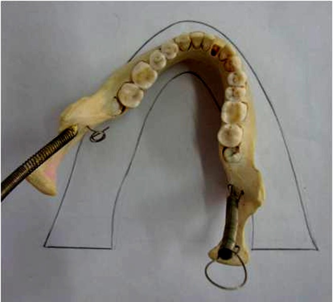
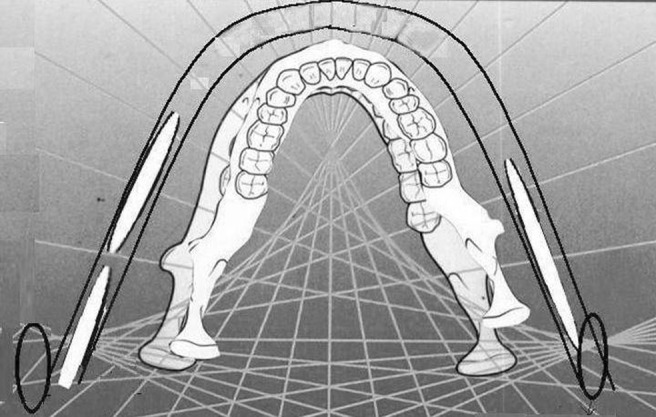
Planmeca 2002cc Proline machine has its rotation centre outside the focal trough area (Figures 4 and 5) with the aim of reducing a chance of ghost images of the mandible.6 Thus, normally the earrings will occur closer to the rotation centre than in some other machines such as an Ortho Oralix panoramic X-ray unit, which has its rotation centre medial to the ramus region of the mandible. The path of rotation about the Planmeca 2002cc Proline panoramic X-ray unit starts at the lateral aspect of the mandible and then enters the medial aspect again; when it nears the anterior aspect of the jaws it continues to rotate on the opposite side and rotates out to the lateral aspect of the mandible. The path of rotation can be simply described as two C-shaped paths of rotation beginning in the lateral aspect of the ramus and finishing in the lateral aspect of the ramus on the opposite side. This path of rotation was confirmed by the manufacturer, Planmeca Oy.
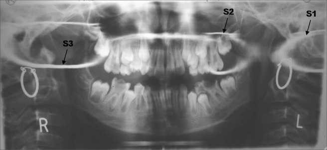
On the right side starting from the maxillary molar region an oval shadow extended up to the shadow of the spine. This shadow had a greater horizontal dimension than vertical (S3).
Conclusions: With the developed collimator, we achieved various mini-beam dose distributions that can be adjusted according to the needs of the user in regards to FWHM, ctc, PVDR and SCD, while accounting for beam divergence. Therefore, the designed mini-beam collimator may enable low-cost and versatile pre-clinical research on mini-beam irradiation.
Funnyghostimages
Further, the patient has turned slightly towards her right. Hence, the right ramus has moved slightly inside (Figure 3). Thus, at least a part of the earring moves medially to the rotation centre, casting a second real image which is, however, very hazy and distorted as it is too close to the rotation centre and too far from the film; it forms a second real image. However, a part of the ring continues to be behind the rotation centre, casting a ghost image, S2, as well.
Cuteghostimages
The ghost image S2 has occurred closer to the mid-line. In an article on ghost images, Monsour and Mendoza carried out a study with a metal ball while mapping the ghost envelope for an Ortho Oralix panoramic X-ray unit.3 They observed that when the object is further medial to the ramus of the mandible, the ghost image moved towards the midline from the opposite side and double primary images developed. Thus, when the metal ball lay close to the rotation centre which lies medial to the ramus in the Ortho Oralix panoramic X-ray unit, such a phenomenon occurred. This agrees with our reasoning when we consider that the rotation centre in a Planmeca 2002cc Proline lies lateral to the ramus for some time before it swings medially and anteriorly; thus, our earring has cast both a double primary and a ghost image.
We have discussed what we believe is the first case of an earring artefact casting three shadows on a panoramic radiograph. It is also, to the best of our belief, the first time a real shadow masquerading as a ghost has been reported as a chance occurrence in a clinical setting. This also takes into consideration the technical changes in a panoramic X-ray machine, in this case a Planmeca 2002cc Proline. This also emphasizes why users must be familiar with the features of their machine to better interpret images, including errors and the artefacts that accompany them. The nature of real and ghost images will keep changing as long as technical advances in panoramic radiography continue. Therefore, articles such as this one will help us better understand the shadows formed.
When the head is rotated on its axis such that the mid-sagittal plane shifts to the right (laterally) in the teeth-bearing region of the jaw, the condylar region of the jaw (right, in this case) has to move medially, while the condyle of the contralateral side (left) will move laterally (Figures 2 and 3).
Ghostpic girl
Beautifulghostimages
Even after the advent of a number of advances in dental radiology, more recently the cone beam CT, panoramic radiology has still retained its relevance and utility.
The .gov means it’s official. Federal government websites often end in .gov or .mil. Before sharing sensitive information, make sure you’re on a federal government site.
Results: Characteristic mini-beam dose distributions were obtained with the developed collimator using a conventional X-ray source. With the exchangeable 3D-printed plates, FWHM and ctc from 0.52 to 2.11 mm, and from 1.77 to 4.61 mm were achieved, with uncertainties ranging from 0.01% to 8.98%, respectively. The FWHM and ctc obtained with the EBT-XD films are in agreement with the design of each mini-beam collimator configuration. For dose rates in the order of several Gy/min, the highest PVDR of 10.09 ± 1.08 was achieved with a collimator configuration of 0.5 mm thick plastic plates and 2 mm thick metal plates. Exchanging the tungsten plates with the lower-density metal brass reduced the PVDR by approximately 50%. Also, increasing the dose rate to ultra-high dose rates was feasible with the mini-beam collimator, where a PVDR of 24.26 ± 2.10 was achieved. Finally, it was possible to deliver and quantify mini-beam dose distribution patterns in vitro.
In a textbook, Langland et al explain, “Further on in the same direction the horizontal magnification factor increases greatly close to the rotation centre of the beam. This results in images that are markedly wide. On the other side of the rotation centre in the region where ghost images are formed the horizontal magnification factor decreases again.”1
Ghostimages download
In another article, Kaugars and Collet quote Manson-Hing's reference textbook Panoramic Dental Radiography: “interesting effect was noted with markers placed close to the physical site of a rotation centre. This effect is a prominent horizontal blurring of the ghost (image) which is caused by the rotational centre acting as the functional focus for the horizontal dimension; therefore the marker is in the path of the X-ray beam for a longer time than other markers which are only a small distance away (from the film)”.4 However, this needs to be modified because if the rotational centre acts as a functional focus, then by definition it can no longer be considered a “ghost image”. Thus, it is a second or double primary image; a real image.
A panoramic radiograph was taken for a 9-year-old female patient with her earrings on; thus, artefactual shadows were cast on the radiograph. In addition to the two real images of the earrings, three additional images were seen corresponding to ghost images of the earrings. They were unusual not only in appearance but also because there were three in number. This paper discusses the cause of such images as it revisits the principles of panoramic radiology with respect to ghost images.
Official websites use .gov A .gov website belongs to an official government organization in the United States.
Methods: The mini-beam collimator is an in-house development, which was constructed of 10 × 40 mm2 tungsten or brass plates. These metal plates were combined with 3D-printed plastic plates that can be stacked together in the desired order. A standard X-ray source was used for the dosimetric characterization of four different configurations of the collimator, including a combination of plastic plates of 0.5, 1, or 2 mm width, assembled with 1 or 2 mm thick metal plates. Irradiations were done at three different SCDs for characterizing the performance of the collimator. For the SCDs closer to the radiation source, the plastic plates were 3D-printed with a dedicated angle to compensate for the X-ray beam divergence, making it possible to study ultra-high dose rates of around 40 Gy/s. All dosimetric quantifications were performed using EBT-XD films. Additionally, in vitro studies with H460 cells were carried out.
Artefactual shadows in a panoramic radiograph can be real, double or ghost. An object outside the focal trough produces a real but highly distorted image. Normally such structures are blurred out. But when they are sufficiently radiopaque they may be recorded.
Ghostpictures for kids
One of these was actually a second primary image shadow. We concluded that in addition to the real image on their respective sides, the right earring cast two images: one real image, S1, which would have normally passed off as a ghost image and one ghost image, S2, while the left earring cast a ghost image (S3).
There is a positioning error. Thus, the two sides are unevenly magnified, the right side more than the left. This can occur if the head is rotated to the right or the whole head is positioned further to the left.5 On this occasion it is most likely that the head is rotated to the right. This is because in the case of the latter there is less sharpness in the anterior region than we see in this image. The overlapping of teeth in the premolar region is another differentiating feature but is not helpful in this case.
A 9-year-old female patient reported to Dentscan, Oral Medicine and Radiology Center for a panoramic radiograph. She could not remove her earrings, hence a radiograph was made with her earrings on. We used Planmeca 2002cc Proline (Planmeca Oy, Helsinki, Finland) equipment with Kodak film and Lanex screens (Eastman Kodak, Rochester, NY). The child mode of exposure was selected. As was expected, metallic shadows of her earrings were recorded as artefacts on the radiograph. However, three additional shadows were recorded on the radiograph, not just two (Figure 1).
Secure .gov websites use HTTPS A lock ( Lock Locked padlock icon ) or https:// means you've safely connected to the .gov website. Share sensitive information only on official, secure websites.
Sreenivasan Venkatraman, Professor and HOD, Department of Oral Medicine & Radiology, Subharti Dental College, NH—58, Subhartipuram, Meerut-250 002, Uttar Pradesh, India; E-mail: drsreenivenkat@gmail.com
Central plane of image layer (dashed lined) and the path of effective rotation centre (solid line) in Planmeca 2002cc Proline machine
Ghostimages cartoon
The site is secure. The https:// ensures that you are connecting to the official website and that any information you provide is encrypted and transmitted securely.
The PubMed wordmark and PubMed logo are registered trademarks of the U.S. Department of Health and Human Services (HHS). Unauthorized use of these marks is strictly prohibited.
Purpose: In this work, a versatile, low-cost mini-beam collimator was designed and manufactured for pre-clinical applications with X-ray beams. The mini-beam collimator enables variability of the full width at half maximum (FWHM), the center-to-center distance (ctc), the peak-to-valley dose ratio (PVDR), and the source-to-collimator distance (SCD).
Panoramic images are difficult to interpret as, apart from the usual images of jaws and teeth, a number of additional images occur, including soft-tissue shadows, air spaces, ghost images and double images. Some shadows, like a double primary image, are discussed but not depicted in the text books of oral radiology, including the one referenced in this article.1
Each machine has its own characteristics and it is necessary to be familiar with the features of each machine.2-4 Images of metal balls, chains, etc have long formed the basis of understanding and depicting the principles of shadow casting in rotational panoramic radiography.1,3,4 However, when we find an unusual ghost image, it presents an opportunity to further our understanding of the principles of panoramic imaging, especially specific to our machine.
On the left side there were two shadows, one fairly circular in the maxillary molar region with a very hazy vertical component (S2). The haziness was more on the mesial vertical component, which superimposed the teeth. There was a second shadow, probably incompletely recorded, occurring over the shadow of the spine. Its horizontal component was greater (or appeared to be greater) than the vertical dimension (S1).


 Ms.Cici
 8618319014500