
Reading Magnifier Value Pack Accessory - where to purchase magnifying glasses
Cy5Structure
This set provides a high signal-to-noise ratio and is compatible with a variety of imaging equipment. Researchers should also consider the type of optical filters needed, such as bandpass filters or dichroics, to optimize fluorescence imaging.
With the above suggestions, I believe that experimenters can maximize the effectiveness of Cy5 filters in their experiments, ensuring high-quality fluorescence imaging and reliable data.
Most objectives are designed to image specimens with air as the medium between the objective and the cover glass. However, for achieving higher working numerical apertures, some objectives are designed to image the specimen through another medium such as special oil with a refractive index of 1.51.
Selecting the correct Cy5 filter can achieve high-quality fluorescence imaging. Researchers should prioritize filters that match the excitation and emission wavelengths of Cy5. Following best practices, such as proper microscope configuration and regular calibration, can improve experimental results.
Cy5 emissionwavelength
Each microscope objective is itself a complex assembly of lenses, and besides contributing to the magnification, it is the objective lens which determines the resolution power of the microscope. An objective lens can also provide optical aberration corrections. A reflective objective, for instance, includes two mirrors within the assembly. These mirrors can focus laser light as well as provide chromatic corrections.
At Shanghai Optics, we design and manufacture custom objectives and imaging systems to support our customers’ needs in many industries, including medical, biomedical, machine version, scientific research, and metrology, etc. Taking the client’s budget and precision requirements into consideration, our experienced engineering team ensure that each design can be manufactured at a reasonable cost and the optical performance is being met based on fabrication, assembly, and alignment tolerance analysis.
Cy5vs Alexa 647
ANGLE OF INCIDENCE definition: 1. the angle that a beam of light touching a surface makes with a line vertical to that surface 2…. Learn more.
The ocular lens, located at the top of a standard microscope and close to the sensor (receiving eye) receives the real image from the ocular lens, magnifies the image received and relays a virtual image to the sensor. While most eyepieces magnify 10x, there are some which provide no magnification and others which magnify as much as 30x. The magnification power of the microscope can be calculated by multiplying the magnification power of the eyepiece, or ocular lens, by the magnification power of the objective lens. For example, an objective lens with a magnification of 10x used in combination with a standard eyepiece (magnification 10x) would project an image of the specimen magnified 100x.
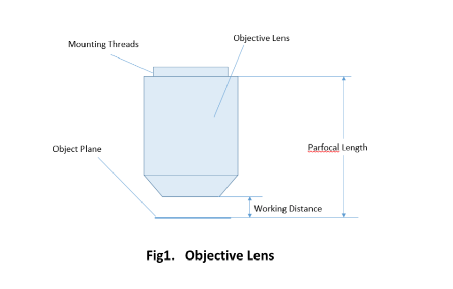
Important specifications are marked on the barrel of the objective, so students or researchers can easily identify the properties of an objective and determine the optical performance and working conditions for proper use. Figure 1 shows a diagram of an objective lens. A detailed discussion of the objection specifications is provided below.
A microscope objective is an important component of a microscopy or imaging system for a range of science research, biological, industrial, and general lab applications.. An objective lens determines the basic performance of an optical microscope or imaging systems and is designed for various performance needs and applications. It is located closest to the object and is an important component in imaging an object onto the human eye or an image sensor.
The ocular lens, or eyepiece, is also an optical assembly rather than a single lens, but it is typically more simple than the objective. Often it is composed of two lenses: a field lens and an eye lens. The design of the ocular lens determines the field of view of the microscope, as well as contributing to the total magnification of the system.
Cy3excitation emission
Willrich Precision Instrument stocks a comprehensive range of SURFACE ROUGHNESS STANDARDS that can be used for different applications. Shop with us today!
2024826 — The ImageMaster® series fulfills highest customer requirements for image quality and MTF (modulation transfer function) lens testing.
Oct 2, 2019 — The Parts of a Camera Lens · Optical Lens · Filter Threads · Focusing Ring · Focal Length Ring · Aperture Ring · Aperture · Lens Mount · More ...
Cy7excitation emission
Several alternatives to Cy5 exist for fluorescence imaging. Alexa Fluor 647 and Allophycocyanin (APC) are popular choices because they have similar spectral properties. These alternatives offer comparable excitation and emission peaks, making them suitable for similar applications.
a device for producing a beam of parallel rays (as of light) or for forming an infinitely distant virtual image that can be viewed without parallax.
Cy5color
Many objectives are designed to be used with a cover glass. Using an incorrect coverslip thickness can greatly reduce the optical performance of a microscopy system.
Objectives are complex multi-element lenses. For any given application, careful consideration of the optical parameters and specifications is necessary. In many cases, custom-designed objective assemblies provide the best-fit solution for meeting all the requirements of a specialized application. Custom parameters may include antireflection coatings, chromatic focus shift, working distance, image quality (MTF and spot size), lens mount, glass window thickness, and field of view, among others.
Cy5 dye is a bright, far-red fluorescent dye that is excited at 633 nm or 647 nm and is designed for use with the 633 nm or 647 nm laser line. Cy5 has low autofluorescence in biological specimens, making it an ideal choice for fluorescence imaging. Researchers often use Cy5 for applications such as neuronal tracing and cell imaging.
Optolong offers filter sets designed specifically for these fluorophores, ensuring high performance and compatibility with existing imaging systems.
Cy5 is a bright, far-red fluorescent dye with excitation and emission properties. The excitation peak of Cy5 dye is around 649 nm and the emission peak is near 670 nm. These properties allow Cy5 to produce minimal autofluorescence in biological samples, enhancing image clarity and contrast.
Magnification is one important parameter. Magnification is usually denoted by an X next to a numeric value. Objectives are available in a range of magnifications from 2X to 200X.
Cy5spectrum
Two major lens components—the objective lens and the ocular lens, or eyepiece—work together to project the image of the specimen onto a sensor. This may be the human eye or a digital sensor, depending on the microscope setup.
Photobleaching refers to the loss of fluorescence ability of a fluorophore due to prolonged exposure to light. To avoid photobleaching, researchers should minimize exposure time during imaging.
Room 609, 6/F, Global Gateway Tower, No.63 Wing Hong Street, Cheung Sha Wan, Kowloon, Hong Kong +852-54993705 info@shanghai-optics.com
The magnifying power of a telescope may be defined as the ratio of the apparent field of view to the true field of view.
When choosing a filter that is compatible with Cy5, make sure that the filter’s excitation and emission ranges match the spectral properties of Cy5. This can maximize the detection efficiency of the fluorescent signal and improve the accuracy and reproducibility of experimental results.
While the simplest of microscopes is simply a magnifying glass with a single lens, compound microscopes used today are highly complex devices with a carefully designed series of lenses, filters, polarizers, beamsplitters, sensors, and perhaps even illumination sources. The exact combination of optical components used will depend on the application of the microscope; the wavelength of light with which it is intended to be used, and the resolution and magnification required in the final image.
Custom Optics: Continuous Variable Apodizing Filters ... Custom optics: Inserting a variable transmission neutral density filter at the aperture stop will modify ...
The optical aberration correction determines the optical performance of an objective lens and plays a central role in the image quality and measurement accuracy of imaging or microscopy systems. According to the degrees of the aberration corrections, objective lenses are generally classified into five basic types: Achromat, Plan Achromat, Plan Fluorite (Plan Semi-Apochromat), Plan Apochromat, and Super Apochromat.
Cy5 is a fluorescent dye that is commonly used in biological and medical research. To be used with Cy5, filters should have specific spectral properties to maximize its fluorescence signal. Ideal filters should include:
When considering which filter to use with Cy5, choosing the right filter ensures the best signal-to-noise ratio and contrast. Choosing the right filter can enhance the quality of your fluorescence imaging and improve your experimental results.
Since the objective is closest to the specimen being examined, it will relay a real image to the ocular lens. While doing so, it contributes a base magnification of anywhere from 4x (for a scanning objective lens, typically used to provide an overview of a sample) to 100x (for oil immersion objectives).
A simple magnifier (magnifying glass), works when the object to be examined is situated within focal length of the magnifier lens, enabling larger virtual image is produced. This type of magnifier is very limited in both resolution and magnification. A compound microscope, on the other hand, uses a relay lens system instead of the single lens, and since each lens component can contribute magnifying power, the result is greatly increased capability.
Microscope Objectives or Objective lenses are in many ways the heart of the microscope, and are typically mounted on a rotating nosepiece or turret to enable easy selection. Many microscopes will be equipped with a scanning objective (4x), a low power objective (10x), a high power objective (40x), and perhaps even an oil immersion objective lens.

The parfocal length is the distance between the objective mounting plane and the specimen / object. This is another specification that can often vary by manufacturer.
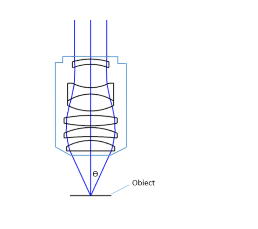
where θ is the maximum 1/2 acceptance ray angle of the objective, and n is the index of refraction of the immersion medium. Figure 2 shows the ray angle θ of an infinity-corrected objective.
Diamond Like Carbon. DLC is an innovative multipurpose coating that exploits two chemical properties of Carbon: the typical hardness of diamond (hence the name ...
Objective lenses can be classified based on the objective construction, field of use, microscopy method, performance (optical aberration corrections), and magnification. Many microscope objective manufacturers offer a wide range of objective designs, which provide various degrees of optical aberration corrections for supporting different needs. Mirrors or reflective elements are used in objective lenses for the applications that requires chromatic aberration over board spectral ranges. Most traditional microscopy systems use refractive objectives such as achromatic objectives (the cheaper objectives) for laboratory microscope applications and plan apochromats (expensive objectives) for biological and science research microscope applications.
Texas Redexcitation emission
Using anti-fading reagents can help protect Cy5 from photobleaching. Regular inspection and maintenance of equipment ensures optimal performance and longevity of the fluorescent signal.
Alpha Industrial Park, Tu Thon Village, Ly Thuong Kiet Commune, Yen My District, Hung Yen Province Vietnam 17721 +84 221-730-8668 rfqvn@shanghai-optics.com
Experimentation and validation of filter selection ensure optimal performance. Researchers should be open-minded and test different filters to find the best fit for their specific application.
Light transmitted is linearly polarized perpendicular to the direction of the chains. If linearly polarized light passes through polaroid material, then the ...
Since indirect backlight illumination is generally more effective than direct illumination, most microscopes do not include an internal light source. Instead, they rely on daylight or on background illumination such as a lightbulb. In brightfield illumination, also known as Koehler illumination, two convex lenses saturate the specimen with external light admitted from behind. These two lenses, the collector lens and condenser lens, work together to provide a bright, even, and constant light throughout the system: on the image plane as well as on the object plane. This system of illumination is used in many compound microscopes, including student microscopes and those found in many research labs.
Feb 8, 2015 — M.2 is interesting not just because it can speed up storage with PCI Express lanes, but because it can use a whole bunch of different buses too; ...
The Optolong website provides various types of optical filters, including long passes, short pass filters, and multi-bandpass/single-bandpass filters, and supports customized services. Come and contact us for relevant information and get an accurate quotation!
Choosing the right filter requires understanding the specific requirements of Cy5. Researchers must select filters that match the excitation and emission wavelengths of Cy5, which are approximately 649 nm and 670 nm, respectively. Optolong offers specialized filter sets, such as the 30021 Single Band CY5 filter set.
Researchers frequently use Cy5 in fluorescence microscopy, flow cytometry, and other imaging techniques. Common applications include neuronal tracing, cell imaging, and molecular diagnostics.
A microscope is a special optical device designed to magnify the image of an object. Depending on the type of microscope, it may project the image either onto a human eye or onto a recording or video device. As an example, consider the photographs of cells that can be found in a science textbook. These photographs have all been taken by a specialized microscope, and may be called micrographs.
Field of View is the area of the object that can be imaged by a microscopy system. The size of the field of view is determined by the objective magnification or focal length of the tube lens for an infinite-corrected objective. In a camera system, the field of view of the objective is related to the sensor size.
For keeping the objective at the proper position, there are mounting threads on almost all objectives. Commonly used mounting threads include RMS, M25 x 0.75, M26X 0.706, M32 x 0.75.


 Ms.Cici
 8618319014500