
Ray Tracing A - ray tracing for a converging lens
Objective lensfunction
In theory, the intensity of illumination depends on the square of the condenser numerical aperture and the square of the demagnification of the light source image (in effect, the field diaphragm image becomes brighter as it is made smaller, according to the square law). The result is that brightness of the specimen image is directly proportional to the square of the objective numerical aperture as it reaches the eyepiece (or camera system), and also inversely proportional to the objective magnification. Therefore, when examining specimens in transmitted light, changing the objective without altering the condenser affects image brightness in response to changes in numerical aperture and magnification.
Correction collars are generally used to correct for spherical aberration due to variations in coverslip thickness, temperature, wavelength or for the differing refractive indices of different immersion media.
What isobjective lensin microscope
This excel spreadsheet has two worksheets that let you compare different objectives to determine their relative brightness
High power objectivemicroscope function
Most microscope objectives are designed to be used with a cover glass that has a standard thickness of 0.17 millimeters and a refractive index of 1.515, which is satisfactory when the objective numerical aperture is 0.4 or less. However, when using high numerical aperture dry objectives (numerical aperture of 0.8 or greater), cover glass thickness variations of only a few micrometers result in dramatic image degradation due to aberration, which grows worse with increasing cover glass thickness. To compensate for this error, the more highly corrected objectives are equipped with a correction collar to allow adjustment of the central lens group position to coincide with fluctuations in cover glass thickness.
Some objectives are also "Plan-" objectives, these are flat field corrected, so the image appears flat to both the eyepieces and the detector.
Low power objectivemicroscope function
Image brightness is directly proportional to the objective NA and inversely proportional to magnification. Therefore if you had two objectives of the same NA but differed in magnification, for examples the following;
Where F(trans) refers to image brightness of a Brightfield illuminated sample, whereas F(epi) refers to the image brightness of a fluorescence or reflected light illuminated sample.
Image processing can be used, for example, to record visitor numbers and customer behaviour and carry out entrance controls.
Imaging systems can be found e.g. in self-checkout systems and reverse vending machines and provide support for shelf inspection.
Stage clipsmicroscope function
Function ofstagein microscope
JavaScript seems to be disabled in your browser. You must have JavaScript enabled in your browser to utilize the functionality of this website.
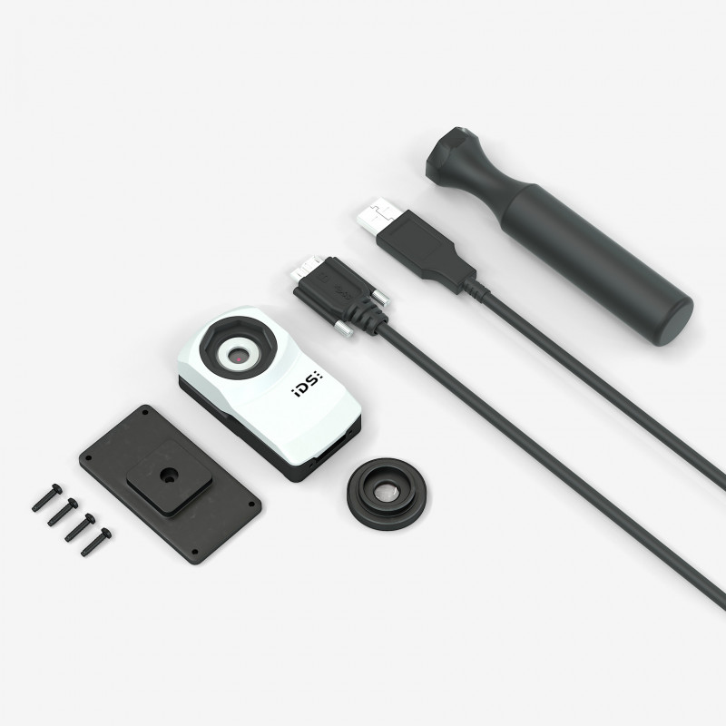
The 13 MP autofocus camera uEye+ XC is as easy to operate as a webcam. Unlike consumer products, it has been specifically designed for use in industrial applications and delivers sharp images and videos even in changing light conditions. Useful functions such as 24x digital zoom, auto white balance and colour correction ensure that the industrial camera keeps all details perfectly in view. It shows its strengths, for example, in kiosk systems, quality assurance, logistics automation and medical technology. With the uEye XC Starter Kit, you receive a comprehensive package with which you can evaluate and use the entire range of functions. It contains not only the camera, tripod and cable, but also a macro lens. Thanks to this lens, the camera with the AR1335 rolling shutter colour sensor from onsemi is also suitable for demanding applications at close range, because it easily shortens the minimum possible object distance and thus opens up additional application possibilities.
Not all objectives are made equal! Objectives for microscopes contain lots of very small and delicate lens to both magnify the image, as well as to perform a number of corrections (spherical and chromatic). Two main characterizations of objectives are the magnification and the numerical aperture.
Typesof microscope objectives
Function ofcondenserin microscope
There are different 'grades' of objectives which describe how many colours they are axially corrected for, and how many colours they are spherically corrected for.

The resolving power of the objective is determined by the numerical aperture (not magnification). Numerical aperture also determines the light collecting ability of the objective, the higher the NA the more light the objective can collect and is determined by the function (nSinq, Where n = immersion media refractive index, and Sinq = angle of the cone of illumination).
In order to allow the microscopist to quickly identify phase contrast objectives, many manufacturers inscribe important specifications, such as the magnification, numerical aperture, tube length correction, etc., on the outer barrel in green letters. This serves to differentiate phase contrast objectives from ordinary brightfield, polarized, DIC, and fluorescence objectives which either use an alternative color code or the standard black lettering.
A special objective is required that is fitted with a darkened circular ring or groove (phase plate) fitted into the glass near the rear focal plane of the objective as illustrated in Figure 1. In addition, the condenser must also be modified with special annular openings suited to a particular magnification and objective. Phase contrast objectives are segregated into a number of categories depending upon the construction and neutral density of internal phase rings:
The terms F(trans) and F(epi) refer to the light-gathering power of an objective and were calculated according to the following equations:
Compact and powerful - IDS cameras can be easily integrated into small devices such as handheld scanners or other portable systems.


 Ms.Cici
 8618319014500