
RADIOMETER Definition & Meaning - radiometer
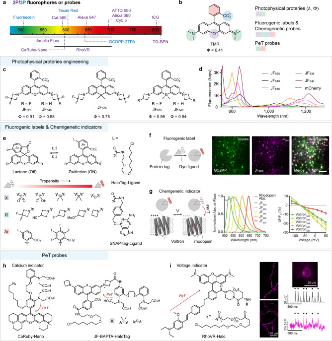
Molecular probes based on rhodamines have been studied for a long time,53,54 but few have been explored for 2P imaging,55 especially under NIR-II excitation. Nevertheless, 1P and 2P probes share a common mechanism for fluorescence response, that is, a photoinduced electron transfer (PeT) mechanism between fluorophore and electronic donor.56,57 The recently developed and constantly growing toolbox of PeT probes for neural activity, including calcium and voltage sensors, allows precise measurement of these signaling events with high spatial and temporal resolution under 2P microscopy. For example, a family of red-fluorescent calcium indicators, CaRuby58 and CaRuby-Nano,59 was created by conjugating a calcium chelator BAPTA [bis(2-aminophenoxy)ethane-N,N,N′,N′-tetraacetic acid] to an extended rhodamine dye (Figure 3h). Calcium chelation alters the electronics of the BAPTA, resulting in reduced PeT and increased fluorescence. CaRuby-Nano was used for in vivo 2P calcium imaging from neocortical layer 2/3 (about 200 μm) pyramidal neurons at around 900 nm excitation.59 Extending excitation wavelength to ∼1050 nm by using a commercial calcium probe Cal-590 is capable to record calcium transients at depths of up to ∼900 μm below the pial surface.60 More recently, genetically targetable calcium probes JF-BAPTA-HaloTag based on X-rhodamines were developed by optimizing the relative position of the BAPTA chelator and the fluorophore (Figure 3h).61 These probes may be useful for 2P calcium imaging experiments with cell-type specificity. The Miller group has developed a family of voltage sensitive indicators (RhoVR) by coupling with a molecular wire to the pendant phenyl ring of X-rhodamine dyes (Figure 3i).62−64 These indicators operated in a membrane potential-dependent PeT process, leading to large voltage sensitivities (up to 47% ΔF/F per 100 mV). Coupling with emerging high-speed 2P microscopy, modified RhoVR can provide single-trial resolution of action potentials in neurons under 2P (λex = 1040 nm) illumination.4
The Leica M mount is a mount for Leica rangefinder cameras and is compatible with full-frame sensors. It has a flange distance of 27.8mm.
The adapter ring allows more and more lenses to be used on different cameras by way of an adapter. For example: most SLR lenses can be transferred to mirrorless bodies, Canon's EF-mount and Nikon's F-mount lenses can be easily transferred to Sony E-mount bodies.
Most of the existing NIR-II polymethine cyanine dyes serve only as nontargeted fluorophores, such as IR-1061, FD-1080, and Flav7. For correct prognosis and therapeutics for disease, effective targeting NIR-II molecular dyes are in urgent demand. Some of the previously reported NIR-II dyes are modified with functional groups, including alkyne, NHS ester, maleimide, succinimidyl ester, isothiocyanate, azide, and carboxyl. Hong and co-workers reported a clickable NIR-II dye 5H5-1069 which bound to integrin αVβ3 targeting peptide c(RGD)fk. Under 1064 nm excitation, the tumor-to-normal tissue ratio (T/NT) of U87 glioma tumor increased 1.7-fold compared to that under 808 nm excitation (from 4.0 to 6.8). Actually, for heptamethine dyes, like IR-1061, FD-1080, and Flav7, a cyclohexene group is introduced to the middle of the conjugated chain to offer a reactive site (Figure 5j). Yan and co-workers have reported a macromolecular NIR-II probe PF by designing Flav7 with covalent linking sulfhydryl groups to realize NIR-II image-guided photothermal therapy.122 To design covalent linkable fluorophores, a simple method is developed by a nucleophilic reaction at the central chloro group with a thionic or oxygenic substituent. These results provide a basis for bioapplication of NIR-II dyes with conjugation capacity.
Organic dyes historically play a major role in fluorescence imaging as fluorescent labels, stains, and reporters, mainly due to their advantages of low molecular mass, good biocompatibility, short organism retention time, and ease of modification through chemical design, compared with inorganic contrast agents. In particular, the achievements of two clinical approved NIR-I dyes, indocyanine green (ICG) and methylene blue (MB), greatly motivated the development of NIR-II organic dyes to facilitate clinical translation of the NIR-II fluorescence imaging technique.10,96 However, making bright NIR-II dyes with stable photophysical and chemical properties in a biological environment is a challenging problem for organic chemists. An efficient and convenient approach to obtain NIR-II dyes is to perform structure modification on the available dye chromophores. To date, the reported NIR-II fluorophores mainly include donor–acceptor–donor (D–A–D) chromophores and polymethine cyanine dyes. To improve the biomedical applications of NIR-II dyes, key factors including absorption and emission wavelength, quantum yields, bioavailability, and bioconjugation capacity is discussed in this section.
Fluorophore structure
The Sony E mount is a mount for Sony mirrorless cameras and is compatible with full-frame and APS-C sensor cameras. It has a flange distance of 18mm
It should be noted that polymethine cyanine dyes hold the property to form head-to-tail dipole arrangement J-aggregates in certain conditions.125 Compared with monomers, J-aggregates exhibit unique photophysical properties of bathochromically shifted absorption and emission bands, enhanced absorption coefficients (ε), and shortened fluorescence lifetimes.126,127 J-aggregates have many advantages for in vivo imaging. For instance, red-shifted absorption and emission wavelength are capable of increasing penetration depth, and in addition, the increased ε will enhance brightness. However, it is difficult to obtain stable and highly purified J-aggregates in solutions. At present, nanocarriers are a promising approach to promote and stabilize J-aggregates. Sletten and co-workers reported IR-140 J-aggregates through encapsulating IR-140 into hollow mesoporous silica nanoparticles, which showed high stability in aqueous solution for several weeks. After aggregation, IR-140 J-aggregates showed bathochromically shifted absorption and emission from 862 and 875 nm to 1040 and 1047 nm, respectively (Figure 5m).111 Holding the longer emission wavelength caused deep tissue penetration; intense signals were observed at lungs, liver, and spleen. Fan and co-workers also reported NIR-II J-aggregate nanoparticles SQP-NPs by encapsulating bispyrrole-sq-bispyrrole (SQP) into PEG-b-PPG-b-PEG (F-127).128 In addition, NIR-II molecular dye also holds promise in forming J-aggregates. Zhang and co-workers developed FD-1080 J-aggregates formed by self-assembly between FD-1080 and 1,2-dimyristoyl-sn-glycero-3-phosphocholine (DMPC) (Figure 5n). FD-1080 J-aggregates showed a high SBR (5.56 and 4.76 for brain and hindlimb vessels, respectively) for in vivo dynamic vascular imaging beyond 1500 nm.112 Thus, stabilizing by nanocarriers is an efficient way to form J-aggregates as NIR-II imaging agents.
Secure .gov websites use HTTPS A lock ( Lock Locked padlock icon ) or https:// means you've safely connected to the .gov website. Share sensitive information only on official, secure websites.
With the exception of the Leica M mount, all of the above mounts allow electronic communication between the camera body and the lens for autofocus and aperture control. Sigma, Leica, and Panasonic worked together to create the L-mount system and set up the L-mount alliance to form an extremely rich group of L-mount cameras and lenses.
Generally speaking, camera bodies and lenses need to have the same mount to be used together, but in order to expand the lens group and to allow lenses of different mounts to be used on the same body, adapter rings have emerged.
(a) Schematic depiction of TMFP by a light propagation model in tissue.12 (b) Penetration of light of different wavelengths in a tissue phantom (2.5% intralipid).7 (c) Scattering spectrum of a mouse brain model and water absorption spectrum.1
Attempts to extend the emission wavelengths of D–A–D fluorophores remain ongoing, but there exists a trade-off between gently red-shifted emission and a steeply descending quantum yield due to the TICT process. To address this issue, recent efforts have focused on the aggregation induced emission (AIE) effect of D–A–D chromophores such as HLZ-BTED and 2TT-oC26B (Figure 4g and h).82,102 While the intramolecular donor rotation in TICT contributes to red-shifted emission, AIE requires restricting intramolecular motion to enhance fluorescence. Very recently, the two seemingly contradicting individuals were simultaneously realized in the aggregates of the rationally designed fluorophore 2TT-oC26B, thus obtaining a broad emission spectrum with a tail extended to the NIR-IIb window and a high quantum yield of 1.15%. Key elements of the molecular design focus on the branched alkyl chain substituents in thiophene groups, which cause a large distortion of the thiophene-BBTD-thiophene backbone. With 2TT-oC26B administration, a tiny vessel of 10 mm width could be distinguished with an SBR around 2. Furthermore, the small bowel diverticula (∼1 mm) could be observed under 5 mm tissue penetration with an SBR of 2.7 (Figure 4h).102 Such distortion not only prevents intermolecular interactions but also provides certain spatial isolation of the molecules in aggregates to promote the intramolecular rotation of triphenylamine units, which were conducive to the formation of the TICT state.
In conclusion, camera mounts are an essential aspect of cameras that determine the compatibility and functionality of the camera system. Different mounts offer various features, such as electronic communication between the camera body and lens, manual adjustments, and compati bility with specific sensors. As a photographer, it is essential to understand the different types of camera mounts to make informed decisions when purchasing cameras and lenses.
A promising class of fluorophores is the rhodamines, which have received considerable attention due to the superb brightness and excellent photostability of the xanthene scaffold. The photophysical properties and functionalities of rhodamines can be modified at various positions (Figure 3b). In particular, the wavelengths of rhodamines vary widely as the function of bridging heteroatoms such as boron, oxygen, carbon, silicon, sulfur, and phosphorus (namely X-rhodamines, X denotes heteroatom),37−40 allowing the creation of fluorescent labels and probes in different colors. Classic rhodamines such as tetramethylrhodamine (TMR) have a low quantum yield and poor photostability due to the twisted intramolecular charge transfer (TICT) characteristic of dialkylamino donors.41 To suppress TICT, chemists would rigidify donor moieties by forming ring structures, or reduce steric hindrance42−44 and electron-donating strength45 of donor moieties by replacing substitution patterns. All these strategies gained positive returns, while a more general strategy with minimal structural change was to use four-membered azetidine rings as donors (Figure 3c),43 which preserved the requisite small size and high membrane permeability of fluorophores for use in live cells. Proposed and perfected by Lavis and co-workers,46−48 this general method created a palette of X-rhodamines (Janelia Fluor) with improved quantum efficiencies that span the visible (500–700 nm) range by fine-tuning the substitution patterns on azetidine rings (Figure 3c). The improved brightness of these rhodamines under 1P excitation also extended to 2P excitation at a wavelength range of 1000–1300 nm (Figure 3d).48 While the tendency to red shift continues in 2P absorption spectra, much larger 2P absorption cross sections occur at the blue-shifted wavelengths than at twice the 1P absorption wavelengths. This is a common phenomenon for many symmetrical dyes,20 which may allow efficient excitation of spectrally separable dyes with a single wavelength.
In 2015, a small-molecule NIR-II fluorophore CH1055 was synthesized by introducing four carboxy groups into the triphenylamine donors of the D–A–D skeleton (Figure 4a).72 This modification not only facilitates PEGylation to impart the molecule with high aqueous solubility and rapid renal clearance but also allows for bioconjugation to targeting ligands, such as antibody, to ultimately produce a molecular imaging agent for tumor diagnostics. The results highlight the potential of D–A–D chromophores for NIR-II fluorescence imaging, in particular, in wide-field through-skull high-resolution brain vasculature imaging (Figure 4b) and high-contrast tumors diagnostic (Figure 4c). However, the quantum yield loss is massive from toluene (7%) to water (0.03%, IR-26 = 0.05% as reference, similarly hereinafter), leaving ample room for further improvement. The fluorescence quenching of D–A–D chromophores in water is associated with the formation of the dark TICT state, in which intramolecular rotation results in nonradiative deactivation of the excited state.102 TICT is governed by a driving force, which depends on solvent polarity, steric hindrance, and the electron-donating strength of the donor groups. Restricting molecular rotation or encapsulating the molecule into a nanomicelle to prevent water contact can straightforwardly produce a brilliant increase in fluorescent brightness. For example, a sulfonate-substituted derivate of CH1055, CH-4T, which forms supramolecular assembly with plasma protein, results in an increased quantum yield of 1.8%. The higher brightness enables fast video-rate imaging at a 50 frames-per-second (FPS) capable of resolving mouse cardiac cycles and blood flow of brain vessels (Figure 4d).97 However, the assembly also brings an undesired blue shift of ∼50 nm and may lose the advantage of small molecules, such as rapid excretion and flexible labeling capability.
Technological advances in imaging setup open up new opportunities for optical access deep inside opaque tissue. This outlook highlights the recent progress on the development of novel molecular fluorophores, which can be operated in the “tissue-transparent” second near-infrared window (NIR-II; 1000−1700 nm) with the help of advanced nonlinear and linear fluorescence imaging techniques.
The Fujifilm X mount is a mount for Fujifilm mirrorless cameras and is compatible with APS-C sensor cameras. It has a flange distance of 17.7mm
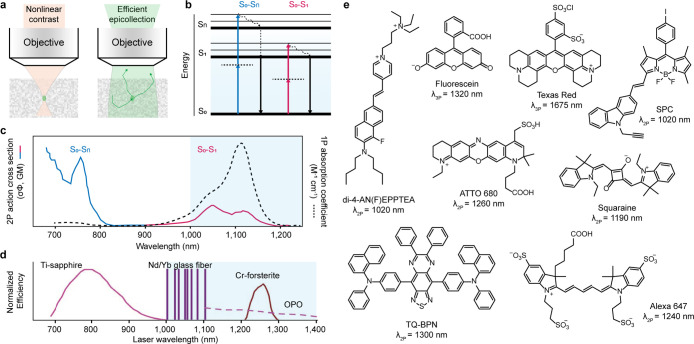
Characterization of molecular brightness at different excitation wavelengths is a crucial first step toward their adoption as fluorophores for 2P microscopy. There are several metrics to evaluate brightness, such as 2P action cross section (σ2′, a product of absorption cross section (σ2) and quantum yield (Φ)),20 figure of merit,21 and peak molecular brightness22 (the maximum molecular brightness with increasing excitation intensity). The latter two consider molecular photostability, a serious constraint in 2P microscopy. In general, donor–acceptor charge transfer dyes have a large 2P absorption cross section at approximately two times the wavelength of their 1P spectra,20,23−25 but their low quantum yield or solvatochromic property may compromise the ability for stable fluorescent labeling. Instead, they may be more suitable for use as aggregation-induced emission (AIE) luminogens (such as TQ-BPN26) or environmental sensitive probes (such as di-4-AN(F)EPPTEA27) (Figure 2e). Traditional dyes including rhodamine, oxazine, BODIPY, cyanine, and squaraine derivatives (Figure 2e) commonly possess two 2P absorption features: one in the NIR-II range corresponds to the S0-to-S1 transition, and one in the NIR-I range corresponds to the S0-to-Sn transition (Figure 2b, c).23,24,28−36 Owing to the resonance enhancement effect, 2P S0-to-Sn transition is often stronger than S0-to-S1 transition.25 However, the benefits of NIR-II excitation, such as increased penetration depth15 and reduced photobleaching,23,30 will outweigh this disadvantage. Although it is a persistent effort goal to improve the absorption cross section, there are other factors—such as quantum yield, photostability, molecular size, functionality, and modification flexibility—that also need to be considered when selecting a fluorophore for 2P applications.
When considering a photon traveling through tissue, the penetration limit for fluorescence imaging is governed by a physical parameter called the transport mean free path (TMFP) of a photon (Figure 1a).12 TMFP describes the mean propagation distance before the photon’s direction becomes randomized. Imaging beyond TMFP with fluorescence methods results in image blur. In general, TMFP represents an upper limit for the penetration of microscopic imaging. Alternatively, when sacrificing resolution is acceptable, the penetration limit could be equivalent to several times the TMFP, such as for in vivo small animal imaging. For most mammalian tissues, TMFP can be simply defined as 1/μs(1 – g) (μs is the tissue scattering coefficient, g is the anisotropy function defining the degree of forward scattering, g = 0.8–1), when photon scattering is dominant over photon absorption. Reducing the tissue scattering coefficient for fluorescence excitation and/or emission would improve the penetration limit, which requires the exploration of an optimum spectral window.
Except for the emission wavelength, the excitation wavelength is also very important to improve the NIR-II 1P fluorescence imaging depth and quality. Zhang and co-workers synthesized a fluorophore FD-1080, which not only resembles ICG but also shows much longer excitation and emission wavelengths at 1064 and 1080 nm, respectively, due to the larger π-system of the benzo[c,d]indolium heterocycle (Figure 5h).78 They performed the first NIR-II excited 1P fluorescence imaging, which demonstrated that imaging under 1064 nm excitation through the scalp and skull can provide higher resolution on brain vessels (0.65 mm) than that under 808 nm excitation (1.43 mm). The same group also used BTC fluorophores and confirmed that excitation at 1064 nm improves the penetration depth, image SBR, and resolution by reducing the signal attenuation coefficient in tissue.75 Recently, high-SBR (7.21) abdominal vessels imaging can also be achieved using 5H5-1069 under 1064 nm excitation (Figure 5i).74
Fluorophoresexamples
When it comes to cameras, one important aspect that often goes unnoticed is the camera mount or bayonet mount. It is a mechanism that connects the camera body to the lens and plays a significant role in determining the compatibility and functionality of the camera system. In this article, we will explore the different types of camera mounts and their features.
Polymethine cyanine dyes are other typical small organic NIR-II-emitted chromophores. The dyes generally consist of two heterocyclic terminal groups connected by one polymethine linker with varying length, and have one positive or negative charge in the conjugated system.113−115 The terminal group structures could be heterocyclic ring containing heteroatoms, like sulfur, oxygen, or nitrogen. The maximum absorption and emission wavelength of polymethine cyanine dyes could be tuned by the terminal groups and conjugated chain length. Lengthening the conjugate chain can increase the extent of delocalized π electrons, which is a classic method to afford red-shifted dyes. Extending the π-system in terminal groups, such as adding π-donor substituents or benzene ring groups, could lead to a bathochromic shift of absorption and emission. Replacing strong electronegative heteroatoms by weak electronegative heteroatoms in a similar scaffold results in bathochromic shifted absorption.
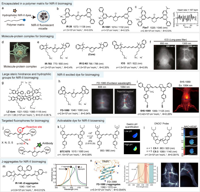
Current polymethine cyanine dyes for NIR-II bioapplication. (a) Enhancing bioavailability of cyanine dyes with micelle protection. (b, c) Chemical structure of IR-26, IR-1061, and Flav7. Quantification of the heart rate of the anesthetized mouse by NIR-II dynamic imaging with Flav7 micelle administration. Reproduced with permission. Copyright 2017, Wiley-VCH.76 (d) Schematic diagram of molecule–protein complex. Reproduced with permission. Copyright 2019, Science Publishing Group.109 (e) Chemical structures of IR-783, IR12-N3, and ICG. (f) Fluorescence imaging of mouse brain vasculature with ICG administration using different long-pass filters. Reproduced with permission. Copyright 2018, National Academy of Sciences.73 (g) Chemical structures of monodisperse polymethine LZ dyes.110 (h) Chemical structure of FD-1080 and NIR-II imaging of brain vasculature with FD-1080 administration under 808 and 1064 nm excitation. Reproduced with permission. Copy-right 2018, Wiley-VCH.78 (i) Chemical structure of 5H5-1069 and NIR-II imaging of abdominal vessels under 1064 nm excitation. Reproduced with permission. Copyright 2019, American Chemical Society.74 (j) Schematic illustration of modified method for reported NIR-II dyes with functional groups. (k) NIR-II low-pH ratiometric sensor BTC1070 for noninvasive gastric pH quantification. Reproduced with permission. Copyright 2019, Springer Nature.75 (l) A FRET sensor composed of CX-1 (donor) and CX-3 (acceptor) for ONOO– detection in mouse liver. Reproduced with permission. Copyright 2019, Wiley-VCH.77 (m) Chemical structure of IR-140 and the absorption spectra of IR-140 monomer and J-aggregates in hollow mesoporous silica nanoparticles. Reproduced with permission. Copyright 2019, American Chemical Society.111 (n) J-aggregates of self-assembly between FD-1080 and DMPC and the spectra properties of FD-1080 monomer and J-aggregates. NIR-II imaging of brain and hindlimb vessels with J-aggregates administration. Reproduced with permission. Copyright 2019, American Chemical Society.112
Finally, the overview that is provided in this Outlook aims to call on the research community to strengthen cooperation with scientists among diverse research fields, such as biology, biomedicine, and microscopy. We look forward to the next era of fluorescence bioimaging in which the frontiers of NIR-II nonlinear and linear techniques expand with the help of new molecular fluorophores.
How dofluorophoreswork
Because of the lens flange distance, not all lenses can be transferred through the adapter ring, for example: Canon's EF lens can not be transferred to the Nikon SLR body, no matter how you transfer, autofocus and metering can not be achieved.
Flange distance for the lens, the flange focal distance is the distance between the lens infinity focus, the lens bayonet plane to the ideal image plane of the lens (i.e. lens focus).
To address these problems, several methods have been developed. First, encapsulating hydrophobic NIR-II dyes into micelles would protect them from water to some extent, thereby be available for in vivo bioimaging (Figure 5a). Dai and co-workers performed abdominal vessels imaging using a phospholipid nanoparticle (IR-PEG) containing the small molecular dye IR-1061 for the first time, from which the smallest discernible vessel width was measured as 150 μm under >1 mm skin penetration depth (Figure 5b).85 Sletten and co-workers have constructed a new panel of polymethine cyanine dyes with emission from 680 to 1045 nm, utilizing a 7-dimethylamino flavylium heterocycle. NIR-II imaging with a micelle formulation of the longest-wavelength dye Flav7 was capable to penetrate through mouse rib and quantify the heart rate of the anesthetized mouse (Figure 5c).76 Second, the assembly of the molecule–protein complex can prevent molecular aggregation and greatly enhance fluorescence by constraining molecular rotation (Figure 5d). For example, previous studies have demonstrated the conventional NIR-I dyes ICG, IR-783, and IR12-N3 show tail fluorescence into the NIR-II window due to the enhanced quantum yield with the molecule–protein complex (4-fold, 6-fold, and 6-fold quantum yield enhancement for ICG, IR-783, and IR12-N3, respectively) (Figure 5e).73,109,120,121 NIR-II bioimaging using them provides many advantages over the NIR-I imaging, such as higher imaging contrast (from 0.20 to 0.29) and superior resolution (210 μm) of brain vasculature (Figure 5f). In particular, the first-in-human liver-tumor surgery guided by NIR-II fluorescence imaging was also realized by ICG.10 Third, molecular engineering by shortening the polymethine chain and further introducing electron-donating moieties at terminal heterocycles is an effective way to overcome solvatochromic-induced quenching. Zhang and co-workers have leveraged this strategy to construct a series of antiquenching benzothiopyrylium pentamethine cyanines (BTC fluorophores), which showed a maximum of 44-fold enhanced brightness as compared with their heptamethine analog IR-26. These fluorophores are capable of resolving lymph vessels with a minimum feature size of 84 μm in in vivo NIR-II lymphatic imaging.75 Fourthly, large steric hindrance groups and small hydrophilic functional groups, such as sulfonate and carboxyl, are introduced into NIR-II dyes to enhance water solubility and prevent nucleophilic attack by solvent. For example, new NIR-II LZ dyes have large steric hindrance groups (diindole) and four water-soluble groups (sulfonate), leading to the superior monodispersity and chemstability in aqueous solution (Figure 5g).110 These fluorophores show promise for in vivo molecular labeling with less influence by fluorophore–biomolecule nonspecific interaction.
A unique property of rhodamines is the equilibrium between the “closed”, nonfluorescent lactone form and the “open”, fluorescent zwitterion form (Figure 3e). Discovered by Johnsson and co-workers,49 Si-rhodamines show a high propensity to form nonfluorescent but cell permeable lactone in solvents of low dielectric constant (εr). The equilibrium shifts to the open form upon an environmental change such as binding to a polar protein surface. This property of rhodamines renders them as effective fluorogenic ligands for live-cell labeling,50 including self-labeling tags, such as the SNAP-tag and HaloTag proteins. The equilibrium exists in almost all X-rhodamines, but it appears that the equilibrium propensity (lactone to zwitterion) is inversely correlated to absorption wavelength; that is, for example, near-infrared rhodamines containing sulfone (−SO2−) and phosphorus (−PO(OH)−) groups are easier to be in lactone form.47 On the basis of azetidine-substituted X-rhodamine structures, Lavis and co-workers have established principles for fine-tuning the fluorogenic properties of various X-rhodamines by exploring substitution patterns on the azetidine rings48 or the pendant phenyl ring47 (Figure 3e). In general, weakening nucleophilicity of the carboxylate anion or improving electron deficiency of the xanthene scaffold will shift the equilibrium to the open form. These improved fluorogenic X-rhodamines integrate a variety of properties, such as high brightness and photostability, compatibility with self-labeling protein tags, and the ability to traverse the blood–brain barrier for in vivo delivery, making them favorable competitors for fluorescent proteins in modern biological experiments. For example, JF585-HaloTag ligands binding to self-labeling HaloTags show superior 2P fluorescence at 1100 nm excitation, which is sufficiently separated from GFP-based indicators such as GCaMP6 (λex = 940 nm) to allow multicolor imaging of neurons in layer 5 of the visual cortex (Figure 3f).48 Recently, a “chemigenetic” indicator (Voltron) was created by fusing a genetically encoded voltage indicator (rhodopsin) with self-labeling HaloTag that irreversibly binds a variety of fluorogenic X-rhodamine ligands (Figure 3g).51 This indicator operates on a transmembrane voltage-dependent modulation of Förster resonance energy transfer (FRET) between the rhodamine dyes and the rhodopsin. Such a “chemigenetic” indicator that combines the genetic targetability and exquisite molecular recognition of proteins with the superb photophysical and sensing properties of synthetic dyes is emerging as a powerful platform for deep-tissue functional imaging.52
Fluorophoreslist
Previous studies on light-tissue interactions revealed that photon scattering nearly scales with λ–α, where λ denotes the wavelength, and the exponent α = 0.2–4.0 depends on tissue types (Figure 1c).1 Longer-wavelength light possesses lower scattering coefficient, and in most cases NIR light penetrates deeper into the scattering medium than visible light (Figure 1b). Nevertheless, in the NIR spectral region, water has a nontrivial contribution to the absorption spectrum of biological tissue (Figure 1c). The trade-off between tissue scattering and absorption sets the boundaries of suitable spectral windows that exclude a narrow water absorption band centered at 1450 nm. In the past decade, fluorescence imaging has focused on the subwindows of 1000–1400 nm and 1500–1700 nm in the NIR-II spectral region, benefiting from the available cost-effective lasers2 and affordable InGaAs detectors that are tunable or respond in these windows.
The Micro Four Thirds mount is a mount for mirrorless cameras from Olympus and Panasonic. It has a flange distance of 19.25mm
Fluorescence imaging has been widely used to investigate the mechanisms underlying various biological phenomena, as this approach can afford much detailed spatiotemporal information in living cells and ex vivo tissue sections than other imaging modalities, such as magnetic resonance imaging, ultrasound, and positron emission tomography. A more complete understanding of biological mechanisms involved in physiology and disease increasingly requires imaging exploration at deeper or intact tissue of living organisms. However, this presents a major challenge for conventional fluorescence imaging that operates in the visible spectrum, because tissues are extremely heterogeneous, and the strong scattering of visible light fundamentally limit the penetration capacity of fluorescence imaging. In recent decades, new windows for deep-tissue biological exploration have been opened in the near-infrared (NIR; 700–1700 nm) spectrum owing to suppressed photon scattering and diminished tissue autofluorescence.1,2 Fluorescence imaging techniques based on NIR nonlinear (multiphoton) excitation and NIR linear (one-photon) emission have enabled unprecedented dynamic and high-contrast exploration of neural activity,3,4 cerebral blood flow,5,6 and tumor microenvironment7−9 in living mice. Very recently, NIR fluorescence imaging of human patients has emerged as a cheap, safe, and real-time navigation modality for surgeons to localize lesions, ensure clear resection margins, and find small metastases during cancer surgery.10 Yet where there are opportunities there are challenges. The emerging deep-tissue fluorescence imaging scenarios call for the development of novel fluorophores with improved photophysical and chemical properties such as longer wavelengths, better brightness, excellent photo-/chem-stability, and diverse chemical function. As once king of fluorescence imaging, molecular fluorophores are undergoing a renaissance to meet the needs of modern biological and biomedical researches.11 In this Outlook, we discuss the recent progress and challenges in the development of novel molecular fluorophores, especially focusing on those operate in the underexplored second near-infrared window (NIR-II; 1000–1700 nm). We have grouped them into two categories, one for NIR-II-excited multiphoton microscopy, and another for NIR-II-emitted one-photon imaging.
The Canon RF mount is a new mount for Canon mirrorless cameras and is compatible with full-frame sensors. It has a flange distance of 20mm
Typical cyanine dyes, including IR-1048, IR-1061, Flav7, and IR-26, show intense absorption and emission, both over 1000 nm in nonpolar solvent such as dichloromethane, but are easy to quench when transferring to polar solvents such as dimethyl sulfoxide, methanol, and water.75 Previous studies suggested three major reasons for explaining this phenomenon: (1) Large, hydrophobic conjugated structures favor these fluorophores to form random oligomers or parallel-oriented H-aggregates in water, leading to the fluorescence quenching.78,116 (2) Increased solvent polarity leads to the symmetry breaking effect of electronic structure in ground/excited state, resulting in solvatochromic-induced quenching. This effect connects with the length of polymethine chain and the donor strength of terminal groups, and is generally intensified with the increase of these two factors.75,117,118 (3) Fluorophores like IR-1048, IR-26, and Flav7 appear to be more electron deficient than common cyanine fluorophores and, thereby, are prone to be attacked by nucleophiles. This could even be done by some weak nucleophiles such as methanol, water, and other protic solvents, leading to the break of the π-conjugated system.76,77 A study on IR-1048 suggested that the connected C-position between the heterocycles and the methine chain would be more likely to be the target of nucleophilic attack.119
The Canon EF mount is a popular mount for Canon cameras and is compatible with full-frame and APS-C sensor cameras. It has a flange distance of 44mm
Near-infrared polymethine cyanine dyes have always been of great interest in in vivo fluorescent biosensing application.114 Modifying substituents on polymethine chain or exploiting intermolecular photophysical interaction (such as FRET, aggregation-induced quenching) are the two common routes to design cyanine-based fluorescent probes. These strategies have been confirmed in NIR-II cyanine dyes; for example, Fan and co-workers designed dual activatable NIR-II nanoparticle HISSNPs to achieve MCF-7 xenografts cancer imaging through changing the IR-1061 aggregation state.116 In addition, manipulating the electronic effects of substituents on terminal heterocycles is another promising route to construct novel NIR-II probes. This is because the extended π-conjugated system of NIR-II cyanine dyes greatly increases the contribution of the substituents of terminal heterocycles on the photophysical properties. Zhang and co-workers investigated the position-dependent effect of diethylamino moieties on the spectra and quantum yield of BTC fluorophores, which allowed the creation of an NIR-II fluorescent low-pH sensor BTC1070 (Figure 5k).75 Protonation of diethylamino moieties changed the electron-donating effect and in turn resulted in ratiometric fluorescence signal output (1065/980 nm). The low scattering and superior sensing accuracy of NIR-II ratiometric signals allowed for noninvasive gastric pH quantification under 4 mm biotissue penetration (Figure 5k). Furthermore, on the basis of increased understanding of the photophysical and chemical properties of NIR-II polymethine cyanine dyes, various activatable or ratiometric NIR-II fluorescent probes have been created for monitoring physiological events or biomarkers in vivo, such as reactive oxygen/nitrogen species (ROS/RNS, Figure 5l)77,123 and enzyme.124
Fluorophoresin fluorescence microscopy
Fluorescence imaging has made tremendous inroads toward understanding the complexity of biological systems, but in vivo deep-tissue imaging remains a great challenge due to the optical opacity of biological tissue. Recent improvements in laser and detector manufacturing have allowed the expansion of nonlinear and linear fluorescence imaging to the underexplored “tissue-transparent” second near-infrared (NIR-II; 1000–1700 nm) window, opening up new opportunities for optical access deep inside opaque tissue. Molecular fluorophores have historically played a major role in fluorescence bioimaging. It is increasingly important to design new molecular fluorophores to fully unlock the potential of NIR-II imaging techniques. In this outlook, we give an overview of the novel molecular fluorophores developed for deep-tissue bioimaging in the past five years and discuss their pros and cons in applications. Guidelines for designing new molecular fluorophores with the desirable properties are also provided.
(a) Reported 2P (purple) and 3P (blue) fluorophores and probes. (b) Core structure and modification positions of rhodamine fluorophores. (c) Structure engineering at the bridging heteroatoms and nitrogen substitutes of rhodamine core to increase quantum yield and fine-tuning wavelength; subscripts in e.g. JF525 denote the maximum 1P absorption. (d) 2P peak molecular brightness spectra of JF Fluors, mCherry for comparison.48 (e) Lactone-zwitterion equilibrium in rhodamine fluorophores (εr: solvent dielectric constant) and the influence of substitute structures on the L–Z equilibrium. (f) Rhodamine fluorophore (JF585-HaloTag, magenta) as fluorogenic label for 2P fluorescence imaging of neurons in layer 5 of visual cortex, combined with GCaMP6s (green). Reproduced with permission. Copyright 2017, Springer Nature.48 (g) A hybrid chemigenetic probe (Voltron) composed of rhodamine fluorophores (JF Fluors) and a GEVI (rhodopsin). Reproduced with permission. Copyright 2019, Science Publishing Group.51 (h, i) Calcium probes (h) and voltage indicator (i) based on rhodamine fluorophores. Copyright 2019, American Chemical Society.62
Current D–A–D chromophores for NIR-II bioapplication. (a) Chemical structures of typical NIR-II D–A–D fluorophores CH1055,72 CH-4T,97 SY1100,98 Q4,83 and H1.84 (b–d) NIR-II fluorescence imaging of mouse brain vasculature, tumor, and hemodynamic process with D–A–D fluorophores. Reproduced with permission. Copyright 2016, Wiley-VCH.99 Copyright 2019, Springer Nature.100 Copyright 2017, Springer Nature.97 (e) Chemical structure and the emission spectra of IR-FTAP. Schematic showing the hydrophobic shielding units in IR-FTAP protect the molecule from water polarization. NIR-II imaging of hindlimb vasculature with IR-FTAP administration. Reproduced with permission. Copyright 2018, American Chemical Society.101 (f) Chemical structures of IR-FP8 and IR-FP8P. Schematic showing the quantum yields and molar absorption coefficients of IR-FP8P, IR-FEP, IR-FP0P, and IR-FTAP. Reproduced with permission. Copyright 2020, American Chemical Society.81 (g) Chemical structure of HLZ-BTED and fluorescence intensity ratio (I/I0) of the HLZ-BTED as a function of THF/water ratio, where I0 stands for the maximal intensity. Reproduced with permission. Copyright 2019, The Royal Society of Chemistry.82 (h) Chemical structure of 2TT-oC26B and NIR-II imaging of intestinal peristalsis with 2TT-oC26B administration. Reproduced with permission. Copyright 2020, Springer Nature.102
Fluorochrome vs fluorophore
Owing to the low cost and wide-field imaging capability, linear 1P fluorescence imaging techniques remain competitive for a variety of imaging scenarios, such as in vivo cell tracking, small-animal whole-body imaging, and imaging-guided surgery.7,10,65 However, the visible spectrum extensively employed for linear fluorescence imaging has restricted high-resolution optical imaging to thin sections or to superficial layers. In past decades, much effort had been devoted to developing far-red and near-infrared-I (NIR-I; 650–1000 nm) fluorophores to improve the penetration limit by employing the reduced tissue scattering.66,67 But the situation did not change substantially until pioneering work published in 2009, which pushed linear fluorescence imaging to the NIR-II regime by utilizing surface-modified carbon nanotubes as fluorophores.68 Imaging experiments with these materials revealed dramatic benefits in resolution and penetration depth compared with conventional fluorescence imaging at NIR-I wavelengths, for example, achieving sub-10-μm resolution of through-skull imaging of the brain vasculature at a depth of >2 mm in mouse brain.5,69−71 This pioneering work further classified the broadly defined NIR-II spectrum into three optical subwindows such as NIR-IIa′ (1000–1300 nm), NIR-IIa (1300–1400 nm), and NIR-IIb (1500–1700 nm), which yield improved fluorescence-imaging clarity with increased wavelength. Based on this criterion, a large number of novel NIR-II fluorophores, including organic dyes,72−85 quantum dots,86−88 lanthanide nanoparticles,9,89−93 and metal clusters,94 emerged and enriched the NIR-II fluorescent toolbox. Very recently, new NIR-II microscopic imaging setups have been developed such as 3D confocal microscopy and light-sheet microscopy.7,95 The use of 1319 nm illumination and fluorescence detection at 1500–1700 nm in noninvasive NIR-II light-sheet microscopic imaging afforded a penetration depth of approximately 774 μm along the tilted light-sheet direction into the head with sub-10-μm resolution.7 These achievements pave the foundation for deep-tissue biological and biomedical imaging by utilizing NIR-II linear fluorescence imaging techniques.
Biologicalfluorophores
The work was supported by the National Key R&D Program of China (2017YFA0207303), National Natural Science Foundation of China (NSFC, 21725502, 51961145403), Key Basic Research Program of Science and Technology Commission of Shang-hai Municipality (17JC1400100), and China Postdoctoral Science Foundation (KLH1615200).
D–A–D chromophore is a typical NIR-II organic molecular structure which contains three parts: one acceptor, two donors, and π bridging groups.103 The spectral properties of D–A–D dyes depend on selecting an electron donor and acceptor as well as π bridging groups. Introducing large π bridging moieties as well as stronger electron acceptors and donors is the most commonly used method to obtain new D–A–D dyes with a longer absorption and emission wavelength.104 To date, various symmetrical NIR-II D–A–D dyes have been reported with strong electron-withdrawing unit benzobisthiadiazole (BBTD) derivatives as the central acceptor and variable groups including thiophene, benzene, triphenylamine, and fluorene as donors, e.g., CH1055, IR-FE, Q4, H1, CH-4T, IR-FP8P, and IR-E1.72,81,83,84,97,99,105 Recently, replacing a sulfur atom by a selenium atom in the BBTD moieties could lead to bathochromically shifted emission for D–A–D dyes, such as SY1100 (Figure 4a).98 The twisted intramolecular charge transfer (TICT) characteristic in D–A–D chromophores significantly lowers the energy gap between the highest occupied molecular orbital (HOMO) and lowest unoccupied molecular orbital (LUMO), yielding an emission peak beyond 1000 nm and a large Stokes shift (usually >200 nm).
Most widely used nonlinear excitation fluorescence imaging techniques are two-/three-photon (2P/3P) microscopies.6,13 2P excitation accesses a given excited state of fluorophore by using photons of half the energy of the corresponding one-photon (1P) transition, and 3P excitation uses one-third. This nonlinear dependence on excitation intensity confines fluorescence generation to the focal volume, which reduces out of focus background and provides intrinsic three-dimensional resolution (Figure 2a). Through a “whole-area” epifluorescence collection, 2P/3P microscopy collects both scattered and ballistic signal photons to form the final images, resulting in surprisingly little signal loss (Figure 2a). Therefore, the limitation on penetration depth mostly arises from scattering and absorption of excitation light in tissues.14,15 Traditional 2P microscopy relies on Ti:sapphire lasers, which have a wavelength maximum near 1000 nm (Figure 2d), and thus restricts imaging depth to superficial cortical layers (<500 μm thick).16 Driven by advances in laser technology in the past decade, 2P microscopy at a spectral excitation window of 1000–1600 nm have pushed the imaging depth further and visualized the entire mouse cortex (about 1 mm thick) and beyond with single-cell resolution.17−19 3P microscopy extends the excitation wavelength up to 1700 nm and is capable of calcium-activity imaging at high spatial (submicrometer) and temporal resolution through the intact skull at >500 μm depth.3 The development of bright, bioavailable molecular fluorophores that excite in the NIR-II range is a crucial aspect to the advancement of in vivo 2P/3P microscopy. To date, a variety of molecular fluorophores (Figures 2e and 3a) have been used and developed. In this section, we mainly discuss molecular fluorophores designed for NIR-II 2P microcopy, in view of their accomplishments in recent years.
Is GFP a fluorophore
The Pentax K mount is a mount for Pentax DSLR cameras and is compatible with full-frame and APS-C sensor cameras. It has a flange distance of 45.46mm
The “tissue-transparent” NIR-II spectral window has received considerable interest in the past decade in view of the lower scattering, moderate tissue absorption, and few tissue autofluorescence. By taking advantage of these features, emerging fluorescence methods based NIR-II nonlinear excitation and NIR-II linear emission have revolutionized in vivo bioimaging with unprecedented penetration depth and image resolution. Typical nonlinear methods, 2P/3P microscopies, permit diffraction-limited three-dimensional imaging of tissues in vivo with single-cell or subcellular resolution, but usually need invasive procedures such as implantable chronic windows. Compared with traditional 1P microscopy, the advantages of 2P/3P microscopies are mainly the three-dimensional deep-tissue high-contrast imaging capability due to the nonlinear contrast generation mechanism. Therefore, these methods are preferred for basic biology studies in which interrogating cellular structure and function in thick tissue and living animals is required, for instance, to visualize cell–cell interactions, neurovascular coupling, and neuronal activity in brain research,129,130 and to study the dynamics of immune-cell interactions in intact tissue.131 However, as 2P/3P microscopy operates with small fields of view (hundreds of micrometers) and superficial depth (∼1 mm), it does not yield a complete picture of the underlying activity across varying spatial scales, for example, in mice whole body and even larger human organs. For this purpose, the NIR-II linear fluorescence method with mesoscopic and macroscopic imaging capability may become highly complementary. With an acceptable loss of resolution, this method provides a penetration limit several or a dozen times deeper than 2P/3P microscopy and, thus, is well suited for noninvasive imaging of entire organs and animals in vivo and is more typically part of mouse-to-human clinical translation imaging applications. Its applications include, for example, molecular imaging of entire tumors,8 imaging-guided surgery,1,10 visualizing anatomic structure and hemodynamic information,70 brain imaging,5 and the study of the movement of stem cells or immune cells at deep locations.7,132 With the development of technology, both nonlinear and linear fluorescence methods are constantly overcoming their limitations,4,7 allowing investigation of biological systems across a wide range of spatial and temporal scales. Pushing the frontier of deep-tissue fluorescence bioimaging also requires the design of functional fluorophores that adapted to each method. The complementarity of nonlinear and linear fluorescence methods provides a wide spectral range (500–1700 nm) for chemists to develop novel fluorophores.
Another strategy to improve quantum yield at a single molecule level is to choose a weak, rigid donor and introduce a hydrophobic shielding unit to protect the molecule from solvent polarization, making the TICT state energetically unfavorable.106−108 In recent years, substantial efforts have been made to reveal how changes to donor structures and components influence absorbance, quantum yield, and emission wavelength. Dai and Liang and co-workers introduced shielding units (S) into the D–A–D skeleton to form S–D–A–D–S organic dye.101,105 The dialkoxy modified fluorene was hired as shielding units to construct new NIR-II dye IR-FTAP, which afforded reduced molecule–molecule interaction, leading to a high signal-to-noise ratio (SBR) of 4.9 ± 0.3 for hindlimb vessels (Figure 4e).101 Moreover, these efforts ultimately yield a highest quantum yield record of 0.6% in water for fluorophore IR-FP8P with emission around 1040 nm. The chromophore of IR-FP8P employs a clickable azide-functionalized fluorene as a weak, rigid donor, 3,4-propylenedioxy thiophene as the π-bridge, and the side alkyl chains as protection for the conjugated backbone. The higher quantum yield and absorption coefficient provided an SBR around 2.5 for ovary NIR-II imaging (Figure 4f).81 The improvements in functionalization and brightness at a single molecule level make D–A–D fluorophores a good choice for use in molecular fluorescent labeling in various applications such as targeting imaging of organ/tumor, immune checkpoint, and gut microbiota.
Official websites use .gov A .gov website belongs to an official government organization in the United States.
This is an open access article published under an ACS AuthorChoice License, which permits copying and redistribution of the article or any adaptations for non-commercial purposes.
The Nikon F mount is a mount for Nikon cameras and is compatible with both full-frame and APS-C sensor cameras. It has a flange distance of 46.5mm
(a) Signal generation and fluorescence collection in 2P/3P microscopy. (b) Jablonski diagram, illustrating the S0-to-S1 and S0-to-Sn 2P absorption transitions. (c) S0-to-S1 and S0-to-Sn 2P absorption transition bands in a typical 2P absorption spectrum. 1P absorption spectrum (black dotted) is presented for comparison. (d) Commercial femtosecond lasers relevant to 2P excitation.23 (e) Structures of several commercial or reported 2P/3P fluorophores with their maximum 2P/3P absorption wavelengths in the NIR-II range.
For the body, the flange focal distance is the distance between the bayonet and the focal plane (i.e., the plane where the CCD or CMOS is located). Of course, the same mount, the body flange focal length and the lens flange focal length is equal, that is, a mount only a flange focal length.
The Nikon Z mount is a new mount for Nikon mirrorless cameras and is compatible with full-frame sensors. It has a flange distance of 16mm
In this Outlook, we outline the molecular fluorophores that have evolved over the past five years, driven by the technological advances of nonlinear and linear fluorescence bioimaging in the “tissue-transparent” NIR-II window. The impressive performance of these fluorophores in structural and functional imaging of the mouse brain, tumor and whole body, and in clinical diagnosis provide a huge motivation for people to go forward. Nevertheless, current efforts are still in their infancy, and there remains a gap regarding people’s high expectations for deep-tissue high-resolution fluorescence bioimaging. Regardless of nonlinear or linear fluorescence, the ideal molecular fluorophores would have exquisite brightness and photostability, small size, improved in vivo bioavailability, and ease of use and functionalization, and their nonlinear excitation or linear emission wavelengths better fall in the outstanding spectral area of NIR-II window, for example, around 1300 nm or 1700 nm. Unfortunately, a molecular fluorophore that fulfills all of these criteria has not yet been developed. Some challenges may be objectively difficult to overcome. For example, a trade-off between wavelength and quantum yield is inevitable, because nonradiative relaxation is enhanced at longer wavelengths of the NIR spectrum, partly induced by the vibrational overtone of surrounding chemical bonds such as C–H, O–H, and N–H.75,133 Fortunately, the application of modern synthetic techniques allows further structural refinements to yield dyes with better photophysics,43,134 making us believe that the upper limits of quantum yields of most fluorophores have not yet been reached.
Chemists also should not be discouraged in the hurdles that will have to be overcome to create a perfect fluorophore. Indeed, some intriguing and unanticipated phenomena in the application of fluorophores may have a significant impact on biology and biomedicine. The fluorogenic properties of X-rhodamine after binding to self-labeling tags are good examples: In-depth studies of this discovery have created a genetically targetable fluorogenic toolbox and chemigenetic sensor platform,51,52,135 making synthetic dyes progressively attractive and accessible to neurobiologists. Likewise, the phenomena that spectra change and fluorescence enhancement caused by the interaction of NIR-II emitted fluorophores (such as CH-4T and FD-1080) and biomolecules (such as albumin and DMPC lipids) may also imply the emergence of a new imaging and sensing platform based on hybrid structures.


 Ms.Cici
 8618319014500