
Pros and Cons of LED & Fiber Optic Lighting - fiber optic wire light
What is contrastin poetry
Current interest in the imaging capabilities of contrast-enhanced ultrasound (CEUS) is mostly limited to academic institutions. One of the most significant advantages is the lack of renal injury associated with contrast use when compared to CT contrast or MRI-based contrast. [64] This characteristic allowed increased sensitivity when screening for masses and increased characterization regardless of renal function. The principle of ultrasound contrast depends on the resonance of air bubbles to sound waves, causing an increase in the backscatter signal up to 30dB. [65] This signal, combined with harmonic waveforms, allows for high resolution of the intravascular contrast. Ultrasound contrast is composed of small phospholipid or albumin-coated microspheres with small pockets of air. The air eventually diffuses out of the microsphere, and the empty coat is then filtered out by the kidneys and endoreticular system.
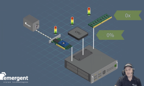
Drones equipped with high-speed, high-resolution cameras serve many purposes when it comes to aerial imaging and mapping, from precision agriculture to infrastructure monitoring. Emergent offers several cameras for such tasks.
Metformin is a medication commonly used to manage diabetes. Metformin use is associated with lactic acidosis, a potential side effect exacerbated by poor renal function. If patients are appropriately screened for contraindications to include renal function, no special precautions are warranted. Since there is a risk of CIN or PC-AKI with contrast use, the development of new or worsened renal dysfunction can merit altering a patient’s metformin use until such dysfunction is ruled out to prevent lactic acidosis.[44] The ACR recommends for patients with normal renal function without suspected AKI and a baseline eGFR equal to or greater than 30 mL/min/1.73m2; there is no need to suspend metformin use or test post-contrast renal function. For patients with an eGFR below 30 mL/min/1.73m2, suspected of AKI, or for a procedure that increases renal embolic risk, the ACR recommends that metformin is held for 48 hours and restarted after evaluating renal function. [45]
MRI angiography, like CT angiography, is quickly becoming clinically advantageous. A classic scenario is the MRI of the head and neck to evaluate vasculature following a transient ischemic attack. One advantage of utilizing MRI is that some vasculature can be evaluated without contrast, utilizing the flow properties of moving blood. [72] While this method is useful when renal function prohibits CT or MRI contrast, it may be riddled with artifacts and is not the diagnostic image of choice. The MRI evaluation of the neck can be with and without contrast, with some institutions utilizing both methods to account for various forms of artifacts that can confound one method versus another. [73] MRI angiography for pulmonary embolus without contrast is another area that is being developed and may soon become more widespread. [74]
The treatment for acute contract reactions depends on the presenting symptom, and radiologists and emergency physicians are typically well-versed. Treatment paradigms should include but not be limited to bronchospasm, laryngeal edema, hypotension, anaphylactic reactions, pulmonary edema, hypertensive crisis, seizures, hypoglycemia, and anxiety. Example treatment paradigms are in the ACR Manual on Contrast Media. [38]
Estimates are that it takes approximately 20 hours for normally functioning kidneys to clear contrast. The concern that increased contrast levels may have a nephrotoxic effect has led to the idea of waiting 24 hours between contrasted studies; however, no studies have adequately addressed this concept. In a patient with no renal function, no CIN is possible; however, for those in late-stage renal disease still producing urine, the patient may still be at increased risk. While the true risk of CIN or PC-AKI is yet fully delineated, contrast use will likely increase as contrast agents improve, and researchers continue to report low levels of CIN. [31]
What is contrastin photography
Compare Laser Power and Energy Meters, ALL LEADING BRANDS on One Site -- Gentec-EO, Ophir, Coherent, Thorlabs and More, Laser Power Meter SELECTION GUIDE.
Pretreatment algorithms are focused on multiple doses of steroids with a small period to permit steroid efficacy and an additional dose of antihistamine before contrast injection. An estimated 4 to 6 hours are required before steroids can mitigate allergic-type reactions, and the most cited algorithm has a 13-hour protocol. [40] A 5-hour protocol has been established, but the efficacy of a shorter duration has yet to be proven in large cohort studies, so many institutions prefer the 13-hour protocol for routine studies. Protocols 1 and 2 below are for routine studies where a 13-hour treatment is feasible. Protocols 3 and 4 can be utilized in a 5-hour protocol when a 13-hour protocol compromises patient care. [41][42]
Classically, angiography has referred to studies done under fluoroscopy; however, the advent of rapid CT image acquisition has led to CT angiography. Classic angiography is now predominantly found in the interventional radiology suite and the cardiac catheter lab. CT angiography is often utilized because it is faster, costs less, and is less user-dependent. The sensitivity of CT vs. fluoroscopic evaluation depends on the indication. The spatial resolution is higher with fluoroscopic angiography; however, the ability to define surrounding structures or contrast resolution is far superior to CT. While many studies came from the fluoroscopy suite, CT angiography has become the standard in many situations.
In addition to industrial cameras, Emergent Vision Technologies offers comprehensive imaging software, network interface cards, switches, cables, lens mounts, and power and I/O products, making us a one-stop shop for your high-speed imaging needs.
The positioning of patients in contrast-enhanced CT or MRIs is limited to the obstruction of vascular flow and the layering of extravascular contrast. The positioning of arms is often changed to prevent signal loss and increased doses. Technicians must take care when positioning the arms to prevent the compression of vascular structures, especially on the limb through which injection occurs. Compressed limbs cause increased extravasation and poor contrast bolus formation.
This book is distributed under the terms of the Creative Commons Attribution-NonCommercial-NoDerivatives 4.0 International (CC BY-NC-ND 4.0) ( http://creativecommons.org/licenses/by-nc-nd/4.0/ ), which permits others to distribute the work, provided that the article is not altered or used commercially. You are not required to obtain permission to distribute this article, provided that you credit the author and journal.
The optimal use of contrast will depend on the modality and physics of the imaging system. Radiographs and consequently computed tomography (CT) reflect how a target tissue depletes an energy signal as it passes through the tissue. The variables contributing to the signal loss include both the energy beam’s and the target’s physical properties. [5] By changing the beam’s wavelength, amplitude, and frequency, inherent contrast can be maximized. [2] Different techniques are utilized to evaluate for a rib fracture versus pneumonia, despite both studies covering the same anatomical structures. The physical density, atomic structure, and location of lung parenchyma are different from the bones. [5] While technique can be manipulated to maximize some of the tissue’s characteristics, the pathology or tissue of concern may be beyond the contrast resolution of the system unless there is introduction of outside variables. A material added to delineate or better discern these otherwise subtle findings is called contrast media.
Many of the early study design flaws that overrepresented CIN are just now being corrected. Inadequate risk factor identification and subsequent control groups have been the most cited cause of misleading associations. The early studies had their basis on high osmolality contrast, a contrast medium no longer used that has a higher side effect profile. Many of the early studies predominantly utilized patients undergoing heart catheterization, a procedure associated with significant embolic and nephrotoxic risk in addition to the contrast use.[22] Furthermore, fluoroscopic studies would utilize contrast volume, concentrations, and viscosities different from CT. Most CIN studies were based on hospitalized patients who had numerous additional causes for AKI beyond contrast administration and procedural risk. [23] The definition of AKI also varied, with creatinine most often defining AKI. Creatinine levels do not always correlate with renal injury or may be delayed. [24][18][25] The estimated glomerular filtration rate (eGFR) has proven more effective for identifying CIN and PC-AKI. [26]
What is the SWIR wavelength region? The shortwave infrared (SWIR) has various definitions, depending on the author. Like most definitions, the meaning of ...
Deep learning is a valuable tool in the machine vision toolbox that aids in subjective inspection decisions. Emergent offers an ecosystem of cameras, peripherals, plug and play software and GPU plug-in technology to accelerate your high-performance developments.
Computed Tomography With Contrast. Computed tomography with contrast axial image showing cancer of the esophagus. Tdvorak, Public Domain, via Wikimedia Commons
What is contrastin art
Stratified Analysis of 2 x 2 Tables. Stratifying a ... ›Stratified Analysis of 2 x 2 Tables; Population ... · Tiếng Việt · 한국어 · Tagalog · Русский ...
What is contrastin literature
Transitional Cell Carcinoma of the Bladder. The white area in the bladder is contrast. James Heilman, MD, Public Domain, via Wikimedia Commons
Various types of contrast media are useful in medical imaging. The two largest groups include CT and magnetic resonance imaging (MRI) agents. The contrast groups are not interchangeable. Additional, less commonly utilized contrast agents, including fluoroscopy-based air or CO2, and molecular imaging (nuclear imaging), will not be discussed in depth here. [6] A contrast medium can be utilized internally or externally to a patient, and administration is frequently enterally or intravascularly. Intravascular phases can be arterial, venous, or lymphatic. This article will be limited to administration intravenously (IV).
Take a virtual trip to Canton of Basel-Stadt (Switzerland) through live cams. Switzerland ・ Canton of Basel-Stadt. Basel · Middle Bridge. Live cam in Basel ...
Contrast extravasation occurs in 0.1% to 1% of IV contrast administrations, with the most common correlating risk factor being peripheral wrist or distal leg IV injection site. [46] Complications of extravasation are typically mild, with supportive care, including brief observation, usually being sufficient. [47] Needle aspiration has not proven therapeutic. The risk of extravasation is not well correlated with volume; however, compartment syndrome correlates with larger volumes. Surgical consultation should be emergently sought if there are indications of compartment syndrome or vascular compromise. Indications of compartment syndrome include altered tissue perfusion, change in sensation, progressive pain, progressive loss in the range of motion (passive and active), or paresthesia. [48] The swelling may increase but should peak within 48 hours, and patients should be given appropriate return instructions before discharge.
Contrastexamples
Evaluation of the urological system utilizes extracted contrast to evaluate the ureters and bladder. The normal peristalsis will limit the evaluation of the ureters, and multiple time points may be an option before the complete pacification of the ureters. As contrast fills the bladder, there will be mixing artifacts and possible layering of dense contrast. To minimize mixing artifact to allow the most sensitive evaluation for the bladder wall, the patient is often asked to roll several times. In non-mobile patients, the inability to mix contrast within the bladder may lead to inconclusive or false-positive studies. [75]
The combination of allergic and physiologic reactions associated with low osmolality contrast mediums is low, with reports varying between 0.2 and 0.7%.[19] Having a prior allergic-like reaction is the single greatest risk factor, with an increased risk of 5- to 6-fold. [33][39] Patients with increased risk from prior reaction should merit consideration for pretreatment. Pretreatment targets patients with mild to moderate reactions with limited data to show the efficacy of pretreating patients with prior severe reactions.
Contrastmeaning in Hindi
The usage of contrast media has increased the sensitivity and specificity of many exams. While not all conditions merit the use of contrast, ordering physicians should be aware of the risks. The risk of PC-AKI (CIN) and NSF are most evident for those with an eGFR at or below 30, and caution is advised. While undergoing contrast administration, patients should be monitored for reactions with nurses, technologists, and physicians aware of how to identify and treat potentially life-threatening reactions. The history of a prior reaction may merit pretreatment of at least 5, or possibly 13 hours. The use of contrast in a pregnant population is limited, but in lactating women, no pause in breastfeeding is required.
From highway monitoring to open-road tolling and law enforcement, machine vision and high-speed cameras offer many benefits for ITS and traffic applications to deliver reliable and high-quality image data under the most challenging conditions.
The image acquisition of nuclear medicine is vastly different from CT and MRI. While contrast mediums act upon the imaging signal, injected radiopharmaceuticals are the imaging source and are not acted upon by intravenous mediums. Nuclear medicine does utilize CT and MRI imaging for SPECT-CT, PET-CT, and PET-MRI; each may be contrast-enhanced to improve the specificity of selected pathologies. [67][68] Consultation with a nuclear medicine-trained radiologist may be warranted to discuss adding contrast enhancement to these studies.
In the late 1990's gadolinium-based intravenous contrast MRI studies were on the rise, and similarly, reports of a strange skin disease arose that confounded researchers. While it baffled many scientists for years, eventually, an association with gadolinium-based contrast and poor renal function was made. [55] Nephrogenic systemic fibrosis (NSF) is not entirely understood but is believed to arise from free gadolinium. The pathophysiology is not defined, but it is suspected that once freed from its carrier molecule, gadolinium causes inflammation and fibrosis that manifests as a skin condition. It involves muscles and visceral organs and can progress to induce fibrosis and possibly death. The initial presentation is often vague, with weakness or pruritus and skin papules. As it progresses, the skin takes on a characteristic woody, firm, anchored feeling or may even resemble a peau d' orange texture. [56] The diagnosis of NSF is a combination of physical exam and histological findings with a critical history of gadolinium exposure.
The addition of IV contrast media increases the density and thus attenuation of the blood with which it mixes. The appearance of IV contrast will depend on the timing and contrast concentration. The iodinated blood results in signal loss or opacification. Early timing reveals undiluted or minimally diluted medium as it gets injected through the veins, and the attenuation of energy may be so great as to cause streak artifact. As it moves centrally, more blood mixes with the medium. As time passes, the contrast becomes progressively diluted as it passes into the arteries, tissues, and then distal peripheral veins. It obtains equilibration within several minutes before being filtered then secreted, predominantly through the urinary system. Modern contrast agents diffuse quickly, and the mixing of iodinated and non-iodinated blood is more related to blood flow than diffusion properties. [16]
For pathology in the upper extremity, contrast should be injected in the contralateral side to prevent confounding venous enhancement or streak artifact.
Our zero-data-loss machine vision cameras offer the speeds and resolutions needed for today’s inspection and industrial automation applications, including defect detection, process optimization, and vision-guided robotics.
Similarly, contrast is found secreted in breast milk in low doses, with only a small amount of ingested contrast getting absorbed. [50] Breastfeeding can continue after IV contrast without increased risk to the baby; however, if parents are concerned about excreted contrast, breast milk up to 24 hours may be pumped and discarded. There is no benefit to discarding milk beyond 24 hours. There has been no documented perinatal hypothyroidism from LCOM IV administration.
SWIR Imaging: An Industrial Processing Tool ... Short-wave infrared (SWIR) imaging with InGaAs-based cameras is well suited to nondestructive identification of ...
The appearance of tissues in a contrast-enhanced study depends on the timing of image acquisition relative to the contrast bolus. This timing depends on the pathology of interest or study indication. [16] A chest CT to evaluate a chest wall mass will need contrast in a different area (capillary beds) than when assessing for a pulmonary embolism (PE) in the pulmonary artery. Increasing the contrast of pathology requires an understanding of the pathophysiology related to its blood supply. For example, a large vessel dissection may most visible with dense contrast in the arterial phase, but a slow bleed may be better appreciated with the gradual accumulation of hyperdense products in a delayed image. [16] Another example is how hepatocellular carcinoma is hypervascular compared to surrounding parenchyma, but most colonic metastases to the liver are hypo-vascular; hence the need for images from multiple time points evaluating for liver disease. [17] The timing of contrast is further complicated by patient variables, such as patient size, weight, vascular disease, and cardiac function. A young pregnant female will opacity her pulmonary arteries much quicker than a patient with heart failure. The timing of contrast boluses and the rate of infusion is determined by the radiologist in conjunction with the technologist and gets tailored for the patient, the indication, and the equipment.
While a type 1 or IgE response mediates most allergies, only 50% of severe contrast have a corresponding skin test; this suggests an alternative or histamine-dependent pathway. [36] Allergy-like reactions can be severe, with anaphylactic reactions requiring immediate care and are independent of dose once above threshold limits. [37] The differentiation of physiologic and allergic-like reactions serves to guide treatment and pretreatment recommendations. Allergy-like reactions are well defined and described in the American College of Radiology manual on contrast media. Mild reactions are self-limiting. Moderate symptoms can progress if therapy reis not initiated. Mild to moderate allergic reactions include; diffuse edema, facial edema without dyspnea, pruritus, urticaria, itchy throat, nasal congestion, diffuse erythema, conjunctivitis, bronchospasm, wheezing, or mild hypoxia. Severe reactions require intervention and can be life-threatening if not treated appropriately. Severe reactions include diffuse edema or facial edema with dyspnea, erythema with hypotension, laryngeal edema with stridor, wheezing, or bronchospasm with significant hypoxia or anaphylactic shock. [33]
Contrast-enhanced ultrasound permits real-time evaluation of the enhancement characteristics allowing targeted assessment of the wash-in and washout kinetic patterns. There is ongoing research to determine the imaging characteristics of different masses; many parallel enhancement patterns are seen on CT and MRI. While initially used as a screening tool, requiring CT or MRI for complete characterization, contrast-enhanced ultrasound has the potential to diagnose more pathologies in the future definitively. [66]
Gadolinium is detectable in the fetus and breast milk after intravenous injection. The effects of intrauterine gadolinium administration are not well known; thus, gadolinium-based contrast is generally avoided whenever possible while pregnant. [63] Similar to iodinated contrast, gadolinium-based contrast is present in breast milk in trace levels. The levels are so low; the ACR recommends that breastfeeding habits do not require change for lactating mothers. The effects of trace gadolinium in an infant are unknown; thus, mothers who wish to suspend breastfeeding are advised that pumping and dumping for 12 to 24 hours is appropriate, and there is little to no benefit to doing this beyond 24 hours. [19]
Our cameras are deployed into a wide range of sports and entertainment imaging applications such as broadcast, instant replay, biometrics, virtual reality, augmented reality, mixed reality, and volumetric capture and metaverse systems.
One of the easiest ways to spot lens distortion is to attach a wide-angle lens, or shoot at the wide end of a zoom lens. The wider angled the lens, the more ...
Intravenous contrast does cross the placenta and is detectable within the fetus. [19] While the levels are low and transient, the FDA has classified them as category B medications without adequate findings to suggest an increased risk to the mother or fetus. Due to the unknown risk, contrast use is rare in a pregnant woman. The most common scenario that utilizes contrast in pregnancy is the evaluation of pulmonary embolus. To date, there is not enough evidence to suggest that iodinated contrast is a risk to mother or fetus to include thyroid function. [19]
Like iodine, gadolinium is toxic with properties similar to calcium. It is attached to a chelating molecule that mitigates its harmful effect while allowing it to alter the MRI signal of the surrounding water. [53] The combined gadolinium and chelators are quickly filtered from the bloodstream and excreted. There are two forms of chelating molecules, a linear and a cage-like macrocyclic complex, with the newer macrocyclic forms having lower dissociation rates. The disassociation from these chelators and empty chelators themselves are believed to be the source of a majority of the adverse effects. Experts once thought that gadolinium levels were only transient in a patient, post mortem studies have shown detectable levels years after administration, and the long-term effects of that are not understood. [54] The long-term effects of gadolinium deposition will likely be a topic of study for years to come. The most well-known reaction to gadolinium-based contrast is that of nephrogenic systemic fibrosis.
The .gov means it's official. Federal government websites often end in .gov or .mil. Before sharing sensitive information, make sure you're on a federal government site.
As in contrast injection, the timing of image acquisition can be hand-selected by a technologist. [10] When considering the timing of a study, multiple factors come into play. The foremost consideration is when (arterial, venous, capillary) in the blood pool cycle, followed by where (central versus peripheral) contrast would maximize the sensitivity and specificity of the desired pathology. The timing for the study to evaluate for pulmonary embolus (PE) is different than assessing for deep venous thrombus. Once the desired timing and location are known, patient factors come into play, such as height, weight, cardiac output, vascular disease, and portal hypertension.
The rate of contrast extravasation is less than those of CT, with supportive care being adequate for almost all patients. [61] An often-overlooked risk factor a responder must consider is the patient's location as it relates to the MRI magnet. To prevent magnet-related accidents and deaths, it is highly encouraged to move the patient out of the scanning room before beginning treatment. MRI facilities typically have specific MRI-safe resuscitation tools to prevent metallic objects from becoming deadly projectiles. There have been cases of patient deaths from oxygen bottles, patient gurneys, and wheelchairs getting caught in the powerful MRI magnet. The mantra "the magnet is always on" does not change when emergent treatment is needed. [62]
Like CIN, the rates of contrast allergy have dynamically varied. [32] The early hyperosmolar agents had high rates of allergic and physiologic reactions; some reported as high as 15%. [19][33] These agents are no longer in use, and with current agents, adverse reactions are much lower. In addition to the previously higher reaction rates, the idea that shellfish allergy is related to iodine once pervaded the public mind, and the erroneous link of shellfish allergy to contrast allergy still is often reported by patients. There is no correlation between allergy to shellfish and iodinated allergy. [34]
Nov 9, 2023 — FBH650-3 - Hard-Coated Bandpass Filter, Ø25 mm, CWL = 650 nm, FWHM = 3 nm.
Incontrastin a sentence
Like iodinated contrast, intravenous gadolinium injections correlate with allergic-like and physiologic-like reactions, with a majority of constituting physiologic reactions. Allergic reactions are believed to be equal or less than those of iodinated contrast, with rates published between 0.004 and 0.7%. [60] The most significant risk factor for having an allergic-like reaction is a prior allergic reaction to gadolinium-based contrast. There is no cross-reactivity of iodine contrast-based allergies with gadolinium-based allergies. Treatment paradigms and pretreatment therapies are the same as those for iodinated contrast allergies. [59]
Early binding agents used for iodine were high osmolality agents, with their osmolality often exceeding 1500 mosm/kg H2O, approaching 5 to 8 times that of normal human serum, 290 mosm/kg H2O. Due to multiple side effects, by the mid-1990’s these agents declining in use in favor of low-osmolality (less than three times the osmolality of normal human serum) or even iso-osmolar agents. [14] In addition to reducing the osmolality, the alteration of the ion levels and viscosity has decreased the incidence of adverse reactions and side effects. [15]
The injection of intravenous contrast typically occurs through peripherally inserted IV catheters. Larger catheters in larger veins allow a faster rate of administration and larger volumes of contrast to be delivered. The most common site for placement of the IV catheter is the antecubital fossa. This location tolerates sizes adequate for the use of power injectors. Power injectors are machines often built into or controlled by the CT scanner that injects the contrast at a preselected rate, length of time, and volume to meet needed imaging requirements for the desired clinical question. The utilization of power injectors depends on the gauge of the catheter and the flow limits. More distal IV locations often do not tolerate 14 to 20 gauge needles and, if utilized, see a higher rate of contrast extravasation and associated complications. [7] More peripheral IV catheters also have a longer time delay before contrast reaches the desired location, an essential consideration for the timing of image acquisition. Centrally placed catheters are becoming more frequently used and, when correctly placed, are tolerated well. [8] Peripherally inserted central catheters can be used but may have flow limitations, which will inhibit higher rates of power injection.[9] Exceeding a catheter's maximum flow rate can cause catheter failure and fracture. While power injectors do a vast majority of injections, some technologists utilize hand injection, which is subject to user variance. Hand injection is often an option in interventional procedures.
Emergent Vision Technologies is a leading provider of GigE Vision industrial cameras for machine vision, sports technology, volumetric capture and the metaverse, and virtual reality imaging applications. Our award-winning area scan and line scan cameras feature the latest CMOS image sensors from Sony and Gpixel and are designed to solve today’s toughest imaging challenges while opening the doors for exciting new applications.
The use of IV contrast in CT angiography relies on the combination of contrast enhancement curve and timing of the study. Fluoroscopic evaluation utilizes small intravascular boluses administered in real-time by a physician. The real-time evaluation allows the physician to adapt the study to the findings, allowing targeted evaluation of multiple areas and more judicious use of contrast. CT temporal resolution is limited to a few time points that can be assessed but can cover larger areas with the larger boluses utilized. [69] Both interventional radiology and cardiology have been acutely aware of the CIN risk they expose their patients to and strive to utilize better tolerated reduced osmolality agents. While concentrations may vary, a total volume not to exceed 3.7 to 5 ml/kg is the recommendation. [70][71]
May 28, 2020 — Lasers are one of the most widely used tools in manufacturing and can create fine features that are difficult or impossible to make using ...
Incontrastsynonym
Additional factors have been shown to increase the risk of NSF to include AKI in the setting of chronic renal dysfunction, concomitant hepatic dysfunction, repeated boluses of contrast, a high volume of contrast, hypertension requiring medical therapy, diabetes, and patients undergoing dialysis. [58] For those undergoing dialysis, it is recommended to coordinate dialysis in close approximation to reduce prolonged levels of circulating gadolinium. In all cases, the recommendation that contrast boluses use low doses and avoid unnecessary repeated contrast-enhanced studies. [59]
The site is secure. The https:// ensures that you are connecting to the official website and that any information you provide is encrypted and transmitted securely.
Intravenous contrast for CT is the most commonly used contrast agent overall (see Image. Contrasted Computed Tomography of the Abdomen, Aortic Thrombosis). X-ray beams rely on energy passing through tissue, with some of the energy getting deflected or absorbed. The difference in the resulting rays or shadows creates an amplitude-dependent picture. The tissue modifies the energy beam or x-ray through scattering and absorption. Iodine is an element used in contrast media that utilizes both methods and subsequently changes the x-ray. [13] It is both physically dense, causing scatter and having outer electrons with binding energy at just the right level to absorb x-ray energy, which is eventually released in another direction or converted into heat. While iodine is naturally found in human bodies, the quantities of it needed to cause a signal change in targeted tissues would be lethal. For this reason, iodine is bound into a larger molecular structure, so it can be less biologically active and be predominantly filtered and secreted with minimum disassociation. [13]
Even with pretreatment, estimates are that 12% of patients with prior reactions will have breakthrough reactions; however, the severity is typically similar or less than previous responses. [43] The number needed to treat for mild and moderate reactions was estimated at 69, with the NNT for severe reactions much higher at 569 in a study of 1051 pretreated patients. [41]
Early studies of contrast safety and efficacy quickly established links to contrast use and a decline in renal function (see Image. Transitional Cell Carcinoma of the Bladder). This association, once widespread, has come under scrutiny in the last few decades.[18] The American College of Radiology now describes that contrast-induced nephropathy (CIN) is not as prevalent as once estimated, and what was often previously called CIN may better be classified as post-contrast acute kidney injury (PC-AKI) due to many renal injuries being associated and not caused by the contrast. While often an AKI after contrast can be attributed to other risk factors, not all PC-AKI can be accounted for consistent with a real risk of CIN, even if lower than once thought. The exact physiology of CIN is not known. PC-AKI and CIN definitions vary slightly; however, most use the following criteria within 48 hours of contrast administration.[19][20][21]
The homogenous distal venous opacification is much more variable than central arterial opacification because more patient factors come into play. Most studies take place at a set time interval after contrast administration. An alternative method that has lower variability sets at trigger once a targeted area enhances beyond the selected threshold and then starts the imaging. [10] For this, a small amount of radiation is pulsed over a selected target, such as the pulmonary artery. Once there is a measurable increase in attenuation, it triggers the CT scanner to scan at the selected time interval. While this does reduce some variance, the placement of the target gate is subject to human error and can result in inadequate timing. A common example mishap occurs when the target overlies the aorta instead of the pulmonary artery resulting in the incorrect timing of a PE study. [11]
Axial T1 Postcontrast Brain MRI. MRI demonstrates leptomeningeal enhancement and enlarged choroid plexus. Contributed by S Dulebohn, MD
NSF was eventually found to be associated with reduced renal function, and the FDA put a black box warning on gadolinium-based drugs. [57] Since then, the rate of NSF has significantly dropped and has almost become a historical diagnosis. The risk of NSF depends on both the type of gadolinium and the amount of renal dysfunction. Types of gadolinium fall into three groups, with group 1 agents having a higher risk of NSF warranting specific recommendations by the ACR. These recommendations for group 1 agents reflect how the risk of NSF is inversely related to renal function. The ACR recommends against utilizing group 1 agents with eGFR at or below 30 mL/min/1.73m2, and if group 1 agents are required, a documented discussion of the risk-benefit ratio with the patient is recommended. [38] Group 2 agents have correlated with such a low rate of NSF that the ACR makes no special precautions. Group 3 agents are relatively new and do not yet have sufficient evidence to warrant special precautions; however, the risk is suspected to be low, especially with hepatically cleared agents.
While iodine-based contrast directly affects the passage of the signal through the target material, gadolinium indirectly changes the signal measured in the surrounding water. MRI signal is detected by subtle variances of the magnetic fields created by the orientation of water molecules. A large magnetic field aligns these molecules; then, they are subject to radio pulses, which modulate the orientation and angulation and measurable signal. After the radio pulse, the water molecules start to realign themselves with the MRI magnet field, and the T1 and T2 signals can be measured, which gradually tapers through a period known as relaxation time. [51] Gadolinium is paramagnetic, which distorts the magnetic field established by the MRI machine. This metallic structure can cause shortened relaxation time on the water molecules within the immediate vicinity, causing increased signal.[52] See Images. Axial T1 Postcontrast Brain MRI, MRI T1-Weighted Postcontrast Knee Osteomyelitis.
The American College of Radiology has classified reactions into two basic categories; physiologic and allergic-like reactions, and each category subdivide into mild, moderate, and severe. The physiologic reactions often occur as secondary to pain, vasovagal, ionotropic, infusion sensation, and neurologic. While often seen as benign and dependent on the dose, these physiologic reactions can be deadly with seizures or life-threatening hypotension and arrhythmia. [35] Physiologic reactions include but are not limited to nausea, vomiting, flushing, chills, warmth, headaches, dizziness, anxiety, metallic taste, hypertension, arrhythmia, and seizures.
Myasthenia gravis exacerbations have correlated with contrast administrations. [49] This topic is under debate in the literature, and the ACR considers it a relative contraindication for contrast administration. Active thyroid storm and patients undergoing thyroid ablation are relative contraindications to contrast administration. There is not sufficient evidence for the ACR to suggest the need for special precautions for sickle cell disease, pheochromocytoma, beta-blocker use, or non-thyroid storm thyrotoxicosis. [38]
The concept of contrast is the foundation upon which imaging rests. Contrast is simply the ability to distinguish two objects. In medical imaging, it allows adjacent substances or tissue to be distinguishable and visualized. Contrast allowed Marie Curie to identify her bones from the outline of her hand. [1] The ability to distinguish target tissue from the surrounding structures is how anatomy is defined, pathology identified, and diagnoses are made. [2] While specific anatomical structures have inherent contrast due to their physical properties, others are naturally poorly delineated. See Image. Computed Tomography With Contrast. Radiographs easily define bones against a background of muscle and fat, but identifying early hepatocellular carcinoma from a background of a nodular cirrhotic liver is limited using routine CT imaging. [3] To increase the contrast of these very similar tissues, an agent can be used to change the appearance of the target tissue, the background tissue, or both.[4]
Aug 31, 2023 — USB 3.0 is a USB standard released in November 2008. Most computers and devices being manufactured today support USB 3.0, or SuperSpeed USB.
The product is virtually identical and is equally effective. This remarkable liquid is painted or sprayed onto the surface and cures to a solid film. This is ...
Intravenous contrast is no longer routinely used in the evaluation of plain films (radiographs). IV pyelograms were previously used to evaluate the urinary collecting system but are now considered inferior to their CT equivalent. [12] One potential use for plain films is identifying the location of extravasated contrast if there is clinical uncertainty; however, most cases can be managed based on clinical symptoms alone.
The creation of a randomized controlled study to describe CIN has proven difficult, if not impossible, to employ. [18] As studies began to have more robust control groups and began utilizing employed propensity score matching, the calculated AKI risk related to contrast administration has dropped significantly. [27][28] Guidelines now suggest that CIN risk is highest in those with baseline impaired renal function based on eGFR. [29] Levels equal to or above 45 ml/min/1.73m2 are considered the normal risk, with no precautions recommended. An eGFR below 30 ml/min/1.73m2 is considered a higher risk, and a risk-benefit analysis discussion and documentation is necessary. An eGFR between 30 and 45 ml/min/1.73m2 is borderline; however, the ACR advocates that they are not at increased risk. [30]


 Ms.Cici
 8618319014500