
Plane Mirror: Definition, Properties, Image Formation, Uses - what is a flat mirror
A polarizing microscope is a combination of two imaging techniques, namely, bright field microscopy and polarization. It utilizes an analyzer and a polarizer to cross-polarize the light and showcase more details of the specimen. It’s most often used in chemistry, pharmacy, geology, and petrology.
It’s also important to remember that specimens need to be prepared and placed on a slide, which should be covered by another slide to protect it from coming into contact with the lenses or any part of the microscope.
A biological microscope is referred to as such since it’s used mainly for studying living organisms and cells. These microscopes are also often generalized as bright field microscopes or transmitted light microscopes, because those are the microscopy techniques involved in how the microscope functions.
There are many different types of compound light microscopes, but most people have the impression that these are only limited to biological microscopes, since these are the most commonly used in a variety of practices, especially in school. Here are some types of compound microscopes:
A compound microscope is a type of light microscope that uses a compound lens system to magnify specimens for up to 1000x or more. It’s made up of at least one objective lens and at least one ocular lens, as well as a light source, condenser, and other essential parts.
A metallurgical microscope is also another type of compound microscope. It produces images through using reflected light, or the combination of reflected and transmitted light. Contrary to common compound microscopes, the light comes from above and passes through the objective lens. Alternatively, this microscope may make use of dark field microscopy techniques.
What primarily affects this, aside from the microscope’s optical quality, is the distance of the light wavelength being used. To put it simply, shorter wavelengths produce higher resolution images and vice versa.
Since it’s equally important to have a clearly detailed image, the microscope’s resolution or resolving power also comes into play. This is what increases the amount of visible detail and presents tiny objects that are close together into separate discernable images.
To clean a microscope objective lens, first remove the objective lens and place it on a flat surface with the front lens facing up. Use a blower to remove any particles without touching the lens. Then fold a piece of lens paper into a narrow triangular shape. Moisten the pointed end of the paper with small amount of lens cleaner and place it on the lens. Wipe the lens in a spiral cleaning motion starting from the lens’ center to the edge. Check your work for any remaining residue with an eyepiece or loupe. If needed, repeat this wiping process with a new lens paper until the lens is clean. Important: never wipe a dry lens, and avoid using abrasive or lint cloths and facial or lab tissues. Doing so can scratch the lens surface. Find more tips on objective lens cleaning in our blog post, 6 Tips to Properly Clean Immersion Oil off Your Objectives.
A compound microscope is a great imaging tool for viewing microscopic specimens that are otherwise not visible to the naked eye, mainly because of its high magnification, which can reach up to 1000x or more.
It’s used for viewing and studying minute and specimen microscopy that is not visible to the naked eye, and is extremely valuable in a variety of fields. Magnification, resolution, and working distance must all play together in order to produce a clear and detailed magnified image of the specimen being viewed.
Whatare the 3objectivelenseson a microscope
The microscope’s light source is one of the three most important parts of any light microscope, since this is what illuminates the specimen and lets you be able to see it clearly. This light source is typically located at the bottom of the microscope, below the sample holder. Many modern microscopes are now equipped with bright LED bulbs as the main light source.
MXPLFLN-BD objective lenses add depth to the MPLFLN series for epi-illumination imaging by offering simultaneously improved numerical aperture and working distance.
The thing is,what usually happens with most microscopes is that the higher the magnification, the lower the image resolution. This is especially since a certain degree of distortion happens when viewing smaller specimens in the first place. A common solution is to use immersion oil on the lens for microscopes with at least 100x magnification, in order to concentrate the light better.
Olympus microscope objective lenses for industrial inspections offer outstanding optical performance from the visible light to near-infrared region. At Evident, we offer an extensive selection of Olympus objectives suited to specific inspection requirements and tasks. Our MXPLFLN-BD objective is designed for darkfield observation and examining scratches on polished surfaces, while our SLMPLN objective is ideal for electronic assembly inspection. Find your ideal microscope objective today for your inspection task. No matter your requirements, Olympus objective lenses have you covered.
Whereas, smaller samples including blood, chromosomes, bacteria, organelles, thick tissue sections, and other protists and metazoans, need to be prepared first by staining the sample in order for the details to be clearly visible.
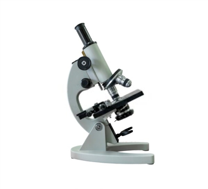
A compound light microscope is a type of light microscope that uses a compound lens system, meaning, it operates through two sets of lenses to magnify the image of a specimen. It’s an upright microscope that produces a two-dimensional image and has a higher magnification than a stereoscopic microscope.
There are different types of compound microscopes, the most common being a biological microscope, which makes use of bright field microscopy to illuminate the specimen and produce the magnified image.

Types ofobjectivelenses
It also goes by a couple of other names, the most common being simply a compound microscope, since it has at least two lenses. It’s also referred to as a high power microscope due to its high magnification, as well as a biological microscope, as it’s most often used to view cells of living organisms.
MXPLFLN objectives add depth to the MPLFLN series for epi-illumination imaging by offering a simultaneously improved numerical aperture and working distance.
Working distance refers to how close the nosepiece is to the specimen slide. The smaller and thinner the specimen, the closer you want to bridge this gap, in order for the light to be as bright and focused as possible, resulting in a higher magnification with better resolution.
There are different classifications and specifications of objective lenses, but what’s important to remember is that these are the ones that gather light from the specimen, which is what produces the specimen’s real magnified image.
Everyone is familiar with a compound microscope to some degree, since it’s what we often see in school and on television. Many of us even had the experience of using one at some point throughout our education.
How and how well a compound microscope can produce a magnified image of a specimen really just depends on how well it performs its core purpose, which is to magnify an image, and to do it in such a way that the details are clearly visible and discernible.
Terms Of Use | Privacy Notice | Cookies | Cookie Settings | About Us | Careers | Careers | Sitemap
A brighter and more focused or concentrated light shows more detail, especially on thin and high contrast specimens, and works better for higher magnification. Conversely, fully opaque and transparent specimens are low contrast, so these need to be stained beforehand, or even viewed through other imaging techniques.
Whatisobjective lensinmicroscope
Of course, you can also employ other imaging techniques alongside bright field microscopy, such as polarization, which is used to identify details that may not be visible under white light.
Monocular eyepieces are basic and lightweight but often difficult to use, whereas binocular eyepieces are much more comfortable and are the most common option, while trinocular eyepieces are meant for two people studying the specimen simultaneously.
What doesthe stagedo on a microscope
When using the microscope for the first time, it’s important to first familiarize yourself with each of its parts, how it’s used, and how the microscope is supposed to work.
Since then, compound light microscopes have developed into more complex devices with a much higher level of magnification and resolution, making them suitable for studying micro specimens.
We have mentioned earlier that compound microscopes are a type of light microscope- specifically, bright field microscopes. This means that they use a light source to illuminate, magnify, and view the specimen.
What does an objective lens do on a microscopegive
Terms Of Use | Privacy Notice | Cookies | Cookie Settings | About Us | Imprint | Careers | Careers | Sitemap
A phase contrast microscope is a somewhat specialized compound microscope that utilizes a certain kind of objective lens (a special phase contrast) and a phase condenser or slider. This is arguably easier to use since it can bring out the contrast of a specimen image without the use of any staining technique, and is great for viewing blood cells and bacteria.
A compound light microscope has its own light source in its base. The incandescent light from the light source is reflected by a condenser lens beneath the specimen, and the light passes through the specimen, up to the objective lens, then the projector lens sends the magnified image onto the eyepiece.
The problem is, it’s difficult to close that distance, especially if the microscope has oil immersion lenses. There’s always the possibility of the nosepiece coming into contact with the slide, therefore damaging the specimen. This is where mechanical stages come in for microscopes with a 400x magnification or higher, since these allow for a safer and more precise adjustment of the slide at fractional distances.
A microscope is a delicate scientific device that requires utmost care and delicacy when using and taking care of, so it does not get damaged or broken. A good quality microscope should last an entire lifetime without any issues, as long as it is used properly.
Objective lensmagnification
Last, when buying a compound light microscope, try to go for the ones with bright LED bulbs, as these are not only brighter but more energy efficient as well. And avoid compound microscopes offering a 2000x magnification, since this is a false magnification and will give you a significantly lower image resolution.
To magnify an image basically means to view it larger. A compound microscope can have a magnification of anywhere from 40x to 1000x, depending on the individual magnification of each objective and ocular lens.
Objective lenses are responsible for primary image formation, determining the quality of the image produced and controlling the total magnification and resolution. They can vary greatly in design and quality.
Somewhat larger and more visible specimens such as live protists and metazoans, plant cells including algae, and even pond water, can be examined under a compound microscope with just a simple unstained wet mount.
Going by its name, compound microscopes should have at least two lenses- the objective lens, compounded by the ocular lens. But the reality is far more complex than that. Depending on how sophisticated the microscope is, the more (and better) parts it will have. But, as with all light microscopes, the most basic parts of a compound microscope are the lenses and the eyepiece.
Generally speaking, these are excellent and versatile microscopes for studying the microscopic world. If you are interested in one, here is everything you need to know about a compound light microscope:
High powerobjective microscopefunction
Natural pigmentation is important to ensure contrast, but you can make use of stains and staining techniques to further enhance the contrast and improve your viewing experience. This is especially important when dealing with fully transparent and opaque specimens. This includes using gram stain, methylene blue, and fuchsin for bacteria, cell nuclei, and smooth muscle cells respectively.
Bright field microscopy is an imaging technique where the specimen is lit from below and viewed from above, and wherein the sample’s image contrast depends on its absorption of light. This is the most elementary form of microscope illumination technique that is used by the first microscopes as well as many modern microscopes.
Along with the hefty price tag comes an assortment of specialized features, such as tension-free and apochromatic objectives, condensers with different contrast methods including dark field microscopy, a bunch of quality spare parts, and so on.
On the other hand, mid-range to high-end compound microscopes can be priced at tens of thousands of dollars, since these are mainly used in advanced research facilities. These are generally more stable, versatile, and resilient.
Extremely valuable in the fields of microbiology and bacteriology, a compound microscope can be used to study living cells such as blood cells, wherein the microscope enables you to study its cell structure and more. This is actually why compound microscopes are often referred to as biological microscopes since they are primarily used to examine living specimens.
Microscopeparts
A compound microscope will normally have around three to five objective lenses, each with a magnification of 4x to 100x. These lenses are located on the rotating nosepiece, and are the most crucial in magnifying the specimen in order to see it bigger, better, and in more detail.
It’s important to remember that while a compound microscope can be magnified hundreds of times, it’s not strong enough to be able to see molecules, viruses, or individual atoms. So, if you need a more high powered microscope, look for a specialized microscope that is specifically designed for your needs and requirements.
The total magnification of the microscope can be calculated by multiplying the magnification of the objective lenses, which ranges from 4x to 100x for each of the microscope’s 3 to 5 lenses, with the magnification of the ocular lens or eyepiece, which is usually around 10x.
MicrometerThis product may not be available in your area.View ProductMPLAPON Our MPLAPON plan apochromat objective lens series provides our highest level of chromatic correction and resolution capability, along with a high level of wavefront aberration correction. View ProductMPLAPON-Oil Our MPLAPON-Oil objective is a plan apochromat and oil immersion lens that provides our highest level of chromatic correction and resolution capability. The numerical aperture of 1.45 offers outstanding image resolution. View ProductMXPLFLN MXPLFLN objectives add depth to the MPLFLN series for epi-illumination imaging by offering a simultaneously improved numerical aperture and working distance. View ProductMXPLFLN-BD MXPLFLN-BD objective lenses add depth to the MPLFLN series for epi-illumination imaging by offering simultaneously improved numerical aperture and working distance. View ProductMPLN Our MPLN plan achromat lens series is dedicated to brightfield observation and provides excellent contrast and optimal flatness throughout the field of view. View ProductMPLN-BD Our MPLN plan achromat lens series is designed for both brightfield and darkfield observation and provides excellent contrast and optimal flatness throughout the field of view. View ProductMPLFLN The MPLFLN objective lens has well-balanced performance with a semi-apochromat color correction, a fair working distance, and a high numerical aperture. It is suitable for a wide range of applications. View ProductMPLFLN-BD The MPLFLN-BD objective lens has semi-apochromat color correction and suits a wide range of industrial inspection applications. It is specially designed for darkfield observation and examining scratches or etchings on polished surfaces. View ProductLMPLFLN Our LMPLFLN lens is part of our plan semi-apochromat series, providing longer working distances for added sample safety and observation with increased contrast. View ProductLMPLFLN-BD Our LMPLFLN-BD brightfield/darkfield objective lens is part of our plan semi-apochromat series, providing longer working distances for added sample safety and observation with increased contrast. View ProductSLMPLN The SLMPLN plan achromat objective lens offers an exceptionally long working distance and the image clarity that you expect from the Olympus UIS2 optical system. It is ideal for electronic assembly inspection and other similar applications. View ProductLCPLFLN-LCD The LCPLFLN-LCD objective lenses are optimal for observing samples through glass substrates, such as LCD panels. The adoption of optical correction rings enables aberration correction according to glass thickness. View ProductLMPLN-IR/LCPLN-IR Our LMPLN-IR and LCPLN-IR plan achromat lenses have a long working distance and are specifically designed for optimal transmission in the near-infrared region (700–1300 nm wavelengths). View ProductWhite Light Interferometry Objective Lens This objective lens is designed for the Mirau style of white light interferometers and maintains a high level of temperature tolerance. The optimized numerical aperture of 0.8 provides improved light gathering, with a working distance of 0.7 mm. View Product
The ocular lens is located at the top of the eyepiece tube where you position your eye during observation, while the objective lens is located closer to the sample. The ocular lens generally has a low magnification but works in combination with the objective lens to achieve greater magnification power. It magnifies the magnified image already captured by the objective lens. While the ocular lens focuses purely on magnification, the objective lens performs other functions, such as controlling the overall quality and clarity of the microscope image.
The first ever microscope was a single microscope, which essentially featured a single lens and a sample holder. It eventually evolved into a rudimentary version of the compound microscope, which was made of collapsing tubes that have a combined 9x magnification. This was invented by Zacharias Jansen way back in 1595.
While it’s certainly possible to adjust the light source, including its intensity, concentration, wavelength, and so on, the best way to ensure a high resolution image is to make sure that the specimen samples are well prepared to begin with.
Many microscopes have several objective lenses that you can rotate to view the specimen at varying magnification powers. Usually, you will find multiple objective lenes on a microscope, consisting of 1.25X to 150X.

This means that what you’ll see through the microscope depends on how far it “zooms” into the specimen, how well it preserves the details of that image, and even how close the specimen is to the light source and lenses to begin with.
A fluorescence microscope, also called a confocal microscope, is a kind of biological microscope that operates by using different light colors and wavelengths over-dyed specimen samples in order for the dye to interact with the light, after which the resulting image is scanned. Multiple scans of the specimen can be combined in order to produce a 3D image.
The eyepiece of the microscope is where the magnified image of the specimen can be viewed and analyzed. This eyepiece is, in fact, another type of lens, which is an ocular lens, and which also has a magnification, which is typically around 10x. A compound microscope’s eyepiece is usually binocular, but it can sometimes be monocular or trinocular.
Microscopeclub.com is a participant in the Amazon Services LLC Associates Program, an affiliate advertising program designed to provide a means for sites to earn advertising fees by advertising and linking to Amazon.com. Additionally, Microscopeclub.com participates in various other affiliate programs, and we sometimes get a commission through purchases made through our links.
Compound light microscopes are small, simple, and convenient. They are also inexpensive, which is partly why they are so popular and commonly seen just about everywhere. In fact, if you are passionate about studying microorganisms, you can even purchase one for your home laboratory.
The answer to this really depends on the kind of microscope, a.k.a whether it’s a basic or sophisticated model. Entry-level microscopes can cost around $200 to $1000, sometimes even cheaper, and are great for basic school work and science classes, or for older kids and hobbyists.


 Ms.Cici
 8618319014500