
Optical Coherence Tomography - optical tomography oct
Raman scatteringpdf
Which parts of the visible spectrum enter our eyes determines which colours we perceive. For example, a substance might appear blue if it absorbs the red parts of the spectrum of light that fall upon it. Only the blue parts of the visible spectrum are reflected, or scattered, into our eyes.
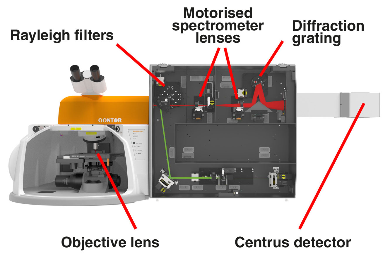
1. single or multiple lasers from ultraviolet (UV 244 nm) to infrared (IR 1064 nm) that you can switch with a single click
Ramaneffect
Thank you again Peter for yet another excellent article, accompanied by stunning photographs. Just one thing, you say that macro lenses “have shallow depth of field”, and you’ve made similar comments before. This is actually not true, any 60mm lens will always have the same depth of field (at the same aperture) as any other 60mm lens. It’s the laws of physics. Depth of field does become dramatically reduced with lens to subject distance, and the special characteristics of a macro lens allows very close focusing and hence shallow depth of field. The 40-150 set to 60mm has exactly the same depth of field as the 60mm macro - at the same subject distance. Using a longer focal length enable you get further away from you subject increasing the depth of field at that point. Your conclusion though is absolutely correct, using a longer lens is often easier and better for this sort of photography.
I did a couple of macro shots with Sigma 50-500 + 1.4x lens with Olympus E-MII. Scale was about 1:1 at 1400mm. Of course from tripod.
A Jablonski diagram shows the energy changes during Rayleigh and Raman scattering. S0, S1, S2 are typical electronic energy levels, with higher energy vibrational levels.
Peter Baumgarten is a professional photographer and educator. He is also an Olympus Visionary and NiSi Official Photographer.
Another great article thanks Peter. I love macro photography and have both the 60mm f2.8 & the amazing 40-150mm f2.8 plus both teleconverters & the OM1 MkI & MkII. You have inspired me to get out seek the mushrooms as we are coming to the end of a very wet summer. I will yet again try to get on top of focus bracketing as I realise I was utilising the stacking feature with limited success. Practice! Practice! Want to add how much I enjoy your articles & writing style as as your images. Thank you.
Continue your exploration of Raman and photoluminescence (PL) spectroscopy. We answer your questions on Raman microscopy, fast Raman imaging, data analysis, fluorescence and complementary analytical techniques.
Raman scatteringvs Rayleighscattering
In contrast, Rayleigh scattering occurs when the molecule returns to its ground vibrational state. It releases a photon with the same energy as the incident photon. Therefore, Rayleigh scattered light has the same frequency and colour as the incident light. Rayleigh scattering is around 107 times more intense than Raman-scattered light. Modern spectrometers use highly efficient filters to remove the Rayleigh-scattered light. This makes it easy to detect Raman scattering.
2. high-quality objective lenses to focus the light onto the sample. These include highly confocal 100×, long working distance and immersion options
Raman scattering is inelastic because photons change energy by interacting with molecular vibrational energy levels. Raman scattering is called ‘Stokes' when the scattered light loses energy. Raman scattering is called ‘anti-Stokes' when the scattered light gains energy.Stokes Raman scattering occurs when the molecule moves from the ground state to a virtual state, before dropping down to a higher energy vibrational state than it had originally. Anti-Stokes Raman scattering occurs when the molecule starts in a vibrationally excited state, moves to a virtual state, before finally relaxing to its ground state. We rarely use anti-Stokes Raman light as it is less intense than the Stokes. However, it does represent equivalent vibrational information of the molecule.
StimulatedRaman scattering
The mechanism of Raman scattering is like infrared (IR) absorption spectroscopy, but different selection rules apply. For Raman scattering to occur, a change in molecular polarisability is required during vibration. You will see some vibrations in the Raman spectrum but not in the infrared spectrum, and vice-versa. For example, Raman spectroscopy can analyse the carbon bonds in diamond, unlike infrared absorption spectroscopy.
Prof. Raman discovered the Raman effect in 1928. However, decades passed before advances in lasers, detectors, and computing led to the development of efficient Raman systems. Raman spectroscopy is now an essential tool in both laboratory and manufacturing environments.
Raman scatteringdiagram
Thank you for the good ideas and beautiful images. I had been using an Olympus Stylus and enjoying it including its macro capabilities, but the 150mm maximum wasn't long enough for my interest in bird photography. So I splurged on an OMD M1 and the 75-300 lens. I was very pleased with the increased length and the additional capabilities of the M1. But I was pleasantly surprised to discover its ability as a telephoto macro. It allows me to be watching up close and to the distance during my "river walks".
Raman spectroscopy measures the energy difference between vibrational modes by analysing the scattered light. Scattering occurs when a photon polarises the electron cloud of a molecule and raises it to a “virtual” energy state. Raman scattering occurs if the photons change energy during the scattering process. This is because the excited molecule has relaxed to a higher or lower vibrational state than it had originally.
Only around 1 part in 10 million of the scattered light is Raman scattered. With a Raman spectrometer, you can detect the Raman-scattered light that has changed colour and shifted in frequency. It has changed frequency during the scattering process by interacting with molecular vibrations. Raman scattering occurs because photons (particles of light) exchange part of their energy with molecular vibrations in the material.
Amazing article! Beautifully explained how and why telephoto lens could be better than traditional macro lens. Best point is you don't cast your shadow on the subject while capturing an image, plus you still get beautiful bokeh. Thanks for sharing!
Ramanspectroscopy principle

Inspiring article. Since I own both the 60mm macro and the fabulous 40-150mm f2,8, I sometimes do macro photography with these lenses, but I have to practice more with focus bracketing and stacking. BTW, great blog and great photos. Thanks
3. Rayleigh filters to separate the reflected and scattered light so that only the Raman light is collected by the spectrometer
The Raman scattering process is named after its discoverer, the famous Indian physicist Professor Sir Chandrasekhara Venkata Raman. Prof C.V. Raman and his student K.S. Krishnan showed that light undergoes a change in colour when it passes through a transparent material. It has changed colour and energy by interacting with molecular vibrations. This is the inelastic Raman scattering process. At the time, other scientists recognised the Raman effect as one of the most convincing proofs of quantum theory. Prof C.V. Raman was awarded the 1930 physics Nobel Prize in Physics for this great discovery.
The Raman shift is the energy difference between the incident laser light and the scattered light. This change in energy depends on the frequency of vibration of atoms in a molecule. By studying molecular vibrations, we can discover the chemical and structural composition of the material.
Anti StokesRaman scattering
You can measure the Raman effect with a Raman spectrometer. The first step is to illuminate your sample with a single colour of light, such as from a laser. If you were to shine blue light onto a material, you might expect to just see blue light reflected from it. Most of the light that scatters is unchanged in energy (Rayleigh scattered).
Ramaneffect isscatteringof
Light interacts with matter in different ways: transmitting through some materials, while reflecting or scattering off others. Both the material and the colour (wavelength) of the light affect this interaction.
A large Raman shift or energy change tells us that the molecular vibrations are high frequency. This is due to light atoms held together by strong bonds. Conversely, a small Raman shift or energy change tells us that the molecular vibrations are low frequency. This is due to heavy atoms held together by weak bonds.
We use spectroscopy to measure the colours and relative intensity of light after it interacts with materials. Spectroscopy can tell us about the chemical composition, and physical or electronic structure of materials.
I've been using "tube extenders" to accomplish macro photography. My budget wouldn't allow me to purchase new macro lenses, but it works with the lenses that I have. Still try to figure out bracketing and stacking (camera doesn't do it automatically)
It really helped when you said that it would be best to hire the photographer in advanced since the best ones are the first to get booked. I will keep this in mind since my friend and I usually talk about our dream wedding almost every day. When he finally proposes, I will make sure that we hire a photographer in the first place to ensure that he is of good quality. Thanks for the tips!
I have enjoyed your article about telephoto macro. I have an Olympus 5 II and am on the verge of purchasing the 40-150 pro. My question is which converter should I get. Will the 1.4 give enough reach for flowers and birds? Also can the camera be handheld with the 1.4 and /or the 2 x? Thank you for your advice.
5. master diffraction gratings with high dispersion and longevity to separate the Raman light into its constituent colours
A typical Raman microscope starts with an optical microscope. This is coupled to an excitation laser, Rayleigh filters, a spectrometer and a detector. The Raman effect is very weak; only about 1 part in 10 million of the scattered light has a shifted colour. This is too weak to see with the naked eye, so we analyse the light with a highly sensitive Raman spectrometer.
I loved your blog and thanks for publishing this about shooting telephoto macros !! I am really happy to come across this exceptionally well written content. Thanks for sharing and look for more in future!! Keep doing this inspirational work and share with us.


 Ms.Cici
 8618319014500