
Notch Filters | Stop-Band Optical Filters - band stop notch filter
OCT is not the same as a computed tomography (CT) scan. OCT uses light rays to create images, and CT scans use X-ray beams.

Read on to learn more about how OCT works. This article also explains the conditions that OCT can help with and what happens during the procedure.
The OCT device then uses these measurements to create images of a person’s retina that are cross-sectional and three-dimensional.
Lasersafetyglassesguide
The beam waist of a Laser beam is the location along the propagation direction where the Beam Radius has a minimum. The waist radius is the beam radius at this ...
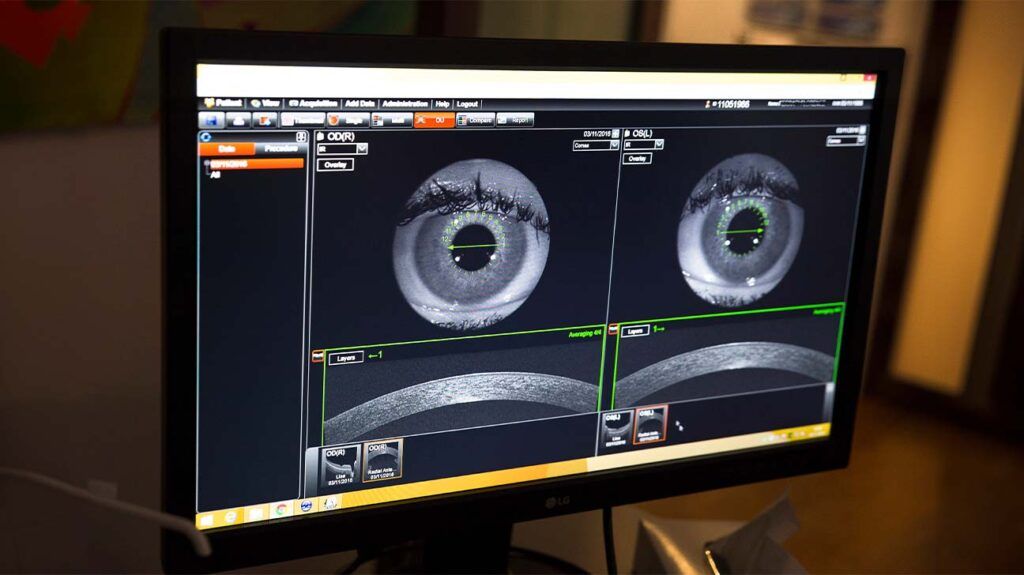
Optical coherence tomography (OCT) is a type of noninvasive imaging test. Ophthalmologists use OCT to create high-resolution, cross-sectional images of the inside of a person’s eye.
The focal length of a lens is the optical distance from the point where the light meets inside the lens to the camera's sensor. Learn more today.
Vitreous is the substance that fills the middle of the eye. Posterior vitreous detachment (PVD) occurs when the vitreous pulls away from the retina.
OCT can help an ophthalmologist see the different layers of the retina. This helps them map and measure the thickness of these layers.
When the intensity of coherent light rises above a certain level, it penetrates the half mirror to reach the outside. This is known as a laser beam. A variety ...
Nov 16, 2021 — Imaging with flat optics: metalenses or diffractive lenses? ... What is a metalens? Metalenses are flat surfaces that use nanostructures to focus ...
Laser protection glassesprice
OCT can help an ophthalmologist diagnose a number of medical conditions including problems with the macula, AMD, glaucoma, and different types of retinopathy.
Adjustable Bridge, Wrap Around Frame Glasses: feature a wrap around design with an adjustable bridge and comfort fit temples
An ophthalmologist may use OCT to help confirm a diagnosis if a person is displaying symptoms of certain eye conditions. Below are the symptoms that may lead an ophthalmologist to use OCT.
Aug 22, 2023 — USB CAMERA is a versatile and free Android app brought to you by US apps 2020. This handy tool, falling under the Utilities & Tools category, ...
A person may not experience any symptoms if they have early stage glaucoma. Over time, a person may slowly experience vision loss.
Prolonged high blood sugar levels can damage the retina. This occurs when diabetes damages the blood vessels that supply the retina with blood.
In some cases, an ophthalmologist may administer dilating eye drops before the procedure. This is to help widen the pupil to make imaging easier.
They must then remain still and follow the instructions of the person carrying out the procedure. These instructions will direct the person to look in certain directions throughout the scan.
Researchers say the drug aflibercept performed better in a clinical trial than bevacizumab in helping wean people off treatments for wet age-related…
Zinc Selenide is the best for that range from a transmittance perspective. Others have mentioned Germanium, which has ~45% transmittance over 7- ...
During the scan, a person may focus on a target within the scanner. They may also see a red line appear during the scan.
During a CT scan, a medical professional aims the X-rays at a part of the person’s body and quickly rotates them around the body. The CT scanner processes the signals that these X-rays produce. The CT scanner then creates cross-sectional images.
LaserSafetyGlassesCanada
Bestlaser protection glasses
If a person has received dilating eye drops during OCT, their eye may be sensitive to light for several hours after the procedure.
There is no research to prove that exercises can help with drooping eyelids. Learn more about this here and find out about other treatment options.
Without treatment, diabetic retinopathy can lead to blindness. OCT can help a medical professional diagnose diabetic retinopathy.
OCT can also help medical professionals diagnose optic nerve disorders. This is because an ophthalmologist can use OCT to spot changes to the fibers of the optic nerve.
Laser protection glassesamazon
A medical professional can use OCT to help diagnose AMD. It can be suitable for helping diagnose both early and advanced-stage AMD.
Measuring the thickness of the different layers of a person’s retina can help an ophthalmologist diagnose a number of medical conditions, such as age-related macular degeneration (AMD) and glaucoma.
Laser safety eyewear can be cleaned using water and neutral cleaning agents such as a mild, household glass cleaner. They should be dried gently with a soft cloth to prevent both scratches and water spots.
Central serous retinopathy is a condition that occurs when fluid builds up under the retina. Medical professionals may also refer to this condition as central serous chorioretinopathy.
Laser Safety Glasses and Goggles feature high optical density at their specified wavelength range to protect against laser radiation. The durable lenses are molded with laser-protective absorptive dyes, causing accidental scratches to have no effect on their performance. Designs that protect against ultraviolet, visible, and infrared lasers are available. All Laser Safety Eyewear products are CE certified and come with a hard case, neck cord, and microfiber cloth.
Laser Glasses
Optical coherence tomography (OCT) is a form of noninvasive imaging test that creates high-resolution, cross-sectional images of the inside of a person’s eye.
Glaucoma is a group of eye diseases that damage the optic nerve. Glaucoma can lead to a person experiencing vision loss and blindness.
LaserSafetyGlasses1064 nm
LasersafetyglassesClass 4
Dichroic filters create only the purest color in their spectrum and will last forever. Dichroics transmit only wanted frequencies, absorbing no unwanted ...
During OCT, an ophthalmologist uses a beam of light to scan an area of a person’s eye. The OCT device then measures the light that the structures within the eye have reflected back.
By allowing an ophthalmologist to do all the above, OCT can help them diagnose certain eye disorders and guide specific treatments.
Warning: Because of the potential for eye damage, the degree of protection required in each circumstance should be determined by the Laser Safety Officer, the industrial hygienist, or the individual responsible for the safety program.
The MTC (Micro Turning Center) series of machines was designed according to the needs of ultra-precision turning. By using diamond tools, optical surface ...
Diabetic retinopathy is a complication of diabetes that causes damage to the blood vessels in the retina. Learn about its causes, symptoms, and…
Lithium fluoride is exceptionally chemically stable and LiF/BeF2 mixtures (FLiBe) have low melting points (360 to 459 °C or 680 to 858 °F) and the best ...
Please select your shipping country to view the most accurate inventory information, and to determine the correct Edmund Optics sales office for your order.
PVD can cause a person to develop vitreous traction. This is a condition that occurs when part of the vitreous remains stuck to the macula during PVD. This part of the vitreous then pulls on the macula, causing symptoms of vitreous traction.
Share your videos with friends, family, and the world.
In its early stages, diabetic retinopathy usually does not cause symptoms. Some people may notice changes in their vision, such as problems seeing things that are far away.
The macula is a small area in the center of the retina. It plays a role in helping people clearly see the details of objects.


 Ms.Cici
 8618319014500