
Microscopy in the Electronics Manufacturing Industry - binocular vs trinocular
OCTeye test price
Apr 20, 2024 — Edmunds Scientific Catalog Spirit of 1974 W/ Original Envelope No Cover. $34.99. +$6.99 delivery & $5.31 fees. Add to cart. Make offer.
Figure 1: Using a pinhole to block out-of-focus light. Light from above (a) or below (b) the focal plane is rejected bythe pinhole, while light from the focal plane (c) passes the pinhole to the detector. Image obtained from Leica Science Lab.
OCTmachine
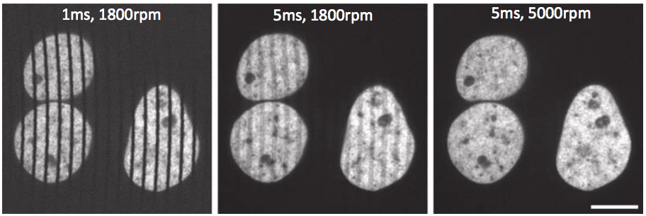
An important point to consider is that SDCMs excite/image multiple points at once and can spin at very high speeds, meaning that high-quality scientific cameras are needed in order to capture images from the sample. These cameras need to have high framerates due to the speed of the spinning disk, because longer exposure times or a slow disk rotation speed may result in a streaky image. By using cameras that can match high rotation speeds, high-quality images are produced, as seen in figure 4.
Optical coherence tomography (OCT) is an imaging technique that uses light to capture 2D and 3D images up to a resolution of a micrometer (μm). It has many uses in medical imaging and research.
Sam graduated from the University of Manchester with a B.Sc. (Hons) in Biomedical Sciences. He has experience in a wide range of life science topics, including; Biochemistry, Molecular Biology, Anatomy and Physiology, Developmental Biology, Cell Biology, Immunology, Neurology and Genetics.
Optical coherence tomography
OCT was invented by the addition of a transversal scanner to a low coherence interferometer to scan the beam over the object laterally. A cross-section image is obtained by taking adjacent scans of the object; this is also known as axial TD-OCT. Another method of performing TD-OCT is en face OCT, which involves taking one-dimensional reflectivity scans (T-scans), with the end image being comprised of many T-scans.
Mar 30, 2017 — Each coloured filter produces a different effect on the scene. Yellow Filter. A yellow filter has always been the classic first choice ...
There are several modes of OCT that can be used to image an object. They each have advantages and disadvantages that dictate which one is used in any given situation. These methods of OCT have been developed over many years and more research is going on to improve its efficiency. This will allow better images to be produced, which would be of great use to both researchers and the healthcare industry.
OCTinterpretation PDF
SB-OCT is based on the demodulation output of the optical spectrum of the low coherence interferometer. The spectrum has peaks and troughs, the period of the modulation being proportional to the OPD in the interferometer. This relationship means the larger the OPD, the larger number of peaks in the spectrum. The camera used to image needs pixels small enough to be able to sample the peaks and troughs in the spectrum. In SB-OCT, the cameras used are at 20-70 kHz.
OCTeye test results
Watch transgender mtf porn videos. Explore tons of XXX movies with shemale sex scenes in 2024 on xHamster!
She would probably be jealous/possesive/would seek alot of atteniton/fish for answers she want's to hear/act hostile/ passieve agressive/guilt ...
SD-OCT refers to the spectral interrogation of the spectrum at the interferometer output. This can be done in two ways. The first involves using a combination of broadband optical source and the processing unit which utilizes a spectrometer. The second involves using a swept source (SS) and the processing unit which employs a photodetector. This is like the TD-OCT procedure, but instead of scanning the OPD the charges on the array are read in the spectrometer in SB-OCT or by tuning the frequency of the laser in SS-OCT.
Professor Nancy Ip discusses her groundbreaking neuroscience research, focusing on neurotrophic factors and innovative Alzheimer's disease treatment approaches.
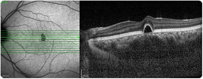
Figure 2: A standard Nipkow-Petran disk used in SDCMs. Hundreds of pinholes are arranged in Archimedeanspirals (left), which pass over the sample as the disk spins. The pinholes have diameter D and separation distance S, changing these values can optimize the resultant image received.
Leverage the power of Anthropologie's collection of mirrors, to amplify spaces, especially compact bedrooms. Elevate the illusion of expanded space using wall ...
Learn about the usage of process raman spectroscopy in the optimization of bioreactor monitoring and then improvement of cultivated meat production.
OCTppt
While we only use edited and approved content for Azthena answers, it may on occasions provide incorrect responses. Please confirm any data provided with the related suppliers or authors. We do not provide medical advice, if you search for medical information you must always consult a medical professional before acting on any information provided.
Mckenzie, Samuel. (2018, October 10). What is Optical Coherence Tomography?. News-Medical. Retrieved on November 17, 2024 from https://www.news-medical.net/health/What-is-Optical-Coherence-Tomography.aspx.
Mckenzie, Samuel. "What is Optical Coherence Tomography?". News-Medical. 17 November 2024. .
High-performance CMOS image sensors use active-pixel architectures invented at NASA's Jet Propulsion Laboratory in the mid 1990s. They can perform camera ...
OCTprinciple
SDCM offers a clear improvement on LSCM and conventional fluorescence microscopy, allowing for fast and efficient imaging of live samples, dynamic processes and optical sectioning of 3D samples in 2D slices. Multichannel images can be taken at multiple wavelengths, resulting in high-quality images dense with information, and deconvolution methods exist to further improve contrast and image quality.
With SS-OCT the laser line needs to be narrower than the spectral distance between adjacent peaks for the photodetector to detect it when tuning the SS. This allows the photo-detected signal to take the shape of the channeled spectrum. This method translates the periodicity of the spectrum into peaks of differing frequency in relation to the OPD, which gives an A-scan. The A-scan is then used to produce an image.
Registered members can chat with Azthena, request quotations, download pdf's, brochures and subscribe to our related newsletter content.
In this interview, News Medical speaks with Dr Martin Biggs from Synoptics about Syngene's G:Box, a high-quality imaging and gel documentation system.
The main disadvantage of SDCM is that only a small proportion of light is transmitted through the disk to the sample, but with customizable pinhole disks, secondary micro-lens disks and highly sensitive cameras these limitations can be easily overcome, while also improving imaging speed and field of view even further to produce high resolution images faster than ever.
2024813 — Festbrennweiten lassen durch ihre Offenblende viel Licht auf den Sensor fallen – und das nicht gerade wenig: Sie erlauben dir gegenüber einem ...
May 15, 2024 — All but two rare earth metals — scandium and yttrium — are part of a chemical group called lanthanides, and light rare earths are the ...
A TD-OCT setup uses an optical light source and an interferometer. A reference mirror and an optical splitter are used to produce a reference beam. A microscopy interface optics is used to convey light from the splitter to and from the object of interest and up to a processing unit. The processing unit performs the interference of light between the reference beam and the beam returned from the object of interest, and it also processes and analyzes the interference signal. The optical path difference (OPD) is obtained by subtracting the length of the reference path from the length of the object path. The OPD can be used to work out which layers within an object can be imaged.
OCT is a high-resolution optical imaging technique that is based on the interference between the signal from the object being investigated (the sample) and a local reference signal. OCT can produce a cross-sectional image of an object in real time. For OCT, the axial resolution of the image is dependent on the optical source, which is an advantage when compared to other methods such as confocal microscopy. OCT can be used to gain very high-resolution images of the retina even with optical aberrations and when narrow light beams are used.
OCTin Cardiology
Typical fluorescence microscopy involves illuminating the entire sample and detecting the resulting fluorescence. Illuminating and detecting from the entire sample includes collection of out-of-focus light above and below the focal plane, causing blurriness and image degradation. This effect is exacerbated when imaging 3D samples such as cells that contain liquid-filled areas that scatter light, causing information to be lost.
Mckenzie, Samuel. 2018. What is Optical Coherence Tomography?. News-Medical, viewed 17 November 2024, https://www.news-medical.net/health/What-is-Optical-Coherence-Tomography.aspx.
Mckenzie, Samuel. "What is Optical Coherence Tomography?". News-Medical. https://www.news-medical.net/health/What-is-Optical-Coherence-Tomography.aspx. (accessed November 17, 2024).
The pinholes in the disk are arranged so that every part of the image is scanned as the disk is spun. By altering the disk rotation speed, pinhole diameter and/or pinhole spacing; the image brightness, contrast and quality can be optimized, making SDCMs highly customizable. However, while larger pinhole diameters results in improved light transmittance through the disk it also reduces resolution, and while having big pinhole spacing eliminates any pinhole cross-talk, it also reduces light transmittance.
Figure 3: Using a secondary micro-lens disk to improve lighttransmittance through the primary pinhole disk. Laser illumination is focused through the pinholes by the micro-lenses, with both disks spinning in sync over the sample.Image obtained from Graf et al. 2005.
Find the largest offer in Spacers and Pilasters at Richelieu.com, the one stop shop for woodworking industry.
There are two main methods of OCT, time domain (TD)-OCT and spectral domain (SD)-OCT. Both have pros and cons and are varied in their procedure. SD-OCT is performed in two ways: spectrometer-based (SB), or using a tunable laser or a swept source (SS).
"Discover the 241-2003 25.4mm Dia. BG3 Color Glass Filter from EKSMA Optics, featuring 3mm thickness and UVFS Mal-1965 material.
News-Medical.Net provides this medical information service in accordance with these terms and conditions. Please note that medical information found on this website is designed to support, not to replace the relationship between patient and physician/doctor and the medical advice they may provide.
Confocal microscopy improves on standard fluorescence microscopy by using pinholes to reject out-of-focus light (figure 1), which results in greater resolution, greater contrast and reduced noise. However, this pinhole only images a tiny area of a sample (approx. 100 nm) and consequently needs to be scanned across the whole sample, which takes time and can cause photo damage. This format is known as laser scanning confocal microscopy (LSCM).
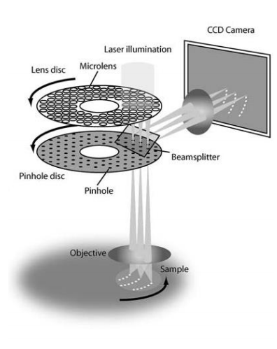
Figure 4: The effects of disk rotation speed and exposure time on the resultant image. From left to right, images show increasing exposure times and/or disk rotation speeds, which results in a cleaner image without streaks. Scale bar 10 µm.Image obtained from Stehbens et al. 2012.
Spinning disk confocal microscopy (SDCM) represents an alternative to LSCM. Rather than a single pinhole, a SDCM has hundreds of pinholes arranged in spirals on an opaque disk (figure 2), which rotates at high speeds. When spun, the pinholes scan across the sample in rows, building up an image. Using a spinning disk vastly improves the speed of image acquisition (allowing for imaging of fast dynamic processes and live specimens), and considerably reduces photo damage.
Having small pinholes with large spacing would result in higher resolution but the lowest transmittance of light through the disk. Transmittance can be improved with the addition of a second disk which contains micrometer-scale lenses in the place of pinholes. The micro-lens disk focuses light through each pinhole of the primary disk, with considerably improves light transmittance to the sample, as seen in figure 3.
Your questions, but not your email details will be shared with OpenAI and retained for 30 days in accordance with their privacy principles.


 Ms.Cici
 8618319014500