
Microscope Resolution: Concepts, Factors and Calculation - diffraction limit of light
Functionof condenserin microscope
To make it easier for you to find which Leica objectives work best for your microscope and application, you can take advantage of the Objective Finder
What isobjective lens in microscope
Leica semi-apochromats are objectives for applications in the visual spectral range with higher specifications, offering field flatness up to 25 mm. The absolute values of the focus differences for the red wavelength and the blue wavelength to green wavelength (3 colors) are ≤ 2.5x depth of field of the objective.
It is easy to think that more microscope magnification will result in seeing more detail and a better image. However, keep in mind that more magnification means you will see a much smaller area on your sample. Additionally if you get too much magnification (anything over 1000x), you will end up with empty magnification and poor resolution. So no, more microscope magnification is not necessarily better.
Objective lens function in microscopeand their functions
The objective lens of a microscope forms a magnified, real, intermediate image of the sample or specimen which is then magnified further by the eyepieces or oculars and observed by the user as a virtual image. When a camera is used to observe the sample, then a phototube lens is installed after the objective in addition to, or even in place of, the eyepieces. The phototube lens forms a real image of the sample onto the camera sensor. The objective’s numerical aperture (NA), its ability to gather light, largely determines the microscope’s resolution or resolving power to distinguish fine details of the sample. Also, the working distance, the distance between the sample and objective, and the depth of field, the depth of the space in the field of view within which the sample can be moved without noticeable loss of image sharpness, both greatly depend on the properties of the objective lens. For more information, refer to: Collecting Light: The Importance of Numerical Aperture in Microscopy, How Sharp Images Are Formed, & Optical Microscopes – Some Basics & Labeling of Objectives
Ocularlens microscope function
All Leica objectives are marked with codes and labels. These identify the objective, its most important optical performance properties, and the main applications it can be used for. For more information, refer to: Labeling of Objectives
An important point to understand when working with compound microscopes and their objective lenses is that the field of view changes as the magnification changes. Typically a lower magnification objective lens will have a larger field of view, and a higher magnification objective lens will have a smaller field of view. For more information on how magnification affects the field of view read this article on microscope magnification and field of view.
Compoundobjective lens function in microscope
For standard applications, Leica Microsystems offers an extensive range of top-class microscope objectives. There are also Leica objectives which have been optimized for special applications. The highest-performance Leica objectives feature maximum correction and optical efficiency and have won several awards. All over the world, scientists are relying on Leica microscope objectives to gain insights into their area of research.
Objective lens function in microscopeand itsfunction
Leica apochromats are objectives for applications with highest specifications in the visual range and beyond, offering field flatness up to 25 mm. The absolute values of the focus differences for the red wavelength and the blue wavelength to green wavelength (3 colors) are ≤ 1.0 x depth of field of the objective.
Stagemicroscope function
Leica achromats are powerful objectives for standard applications in the visual spectral range, offering field flatness (OFN) up to 25 mm. The absolute value of the focus differences between red wavelength and blue wavelength (2 colors) is ≤ 2x depth of field of the objective.
Not all products or services are approved or offered in every market, and approved labelling and instructions may vary between countries. Please contact your local representative for further information.
The optics of the most basic microscope includes an objective lens and ocular or eyepiece. The objective lens is closest to the sample, specimen, or object being observed with the microscope (see the schematic diagram below). For more information, refer to the article: Optical Microscopes – Some Basics Show schematic diagram

Do you need an individual objective for your application? Then contact our Leica OEM Optic Center so that we can offer you a customized solution.
Leica microscope objective lenses are designed and made by our optics specialists to have the highest performance with a minimum of aberrations. The objectives help to deliver superior microscope image quality for many applications, such as life science and materials research, industrial quality control and failure analysis, and medical and surgical imaging.
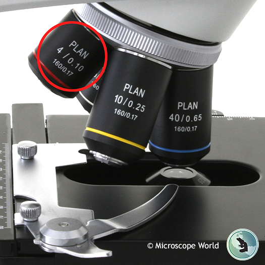
Since the 4x objective lens has the least magnification, but a larger field of view, it allows for more of the specimen to be seen, as well as locating the part of the sample you wish to view. This in turn makes it easier to focus on the sample. Most microscope objective lenses are parfocal, meaning once you have one objective lens in focus, all other objectives should be in focus as well as you move from 4x to 10x, etc. Occasionally, when switching to the highest magnification lens you may need to make a slight adjustment to the fine focus, but this is not required when you start out with the 4x lens. So starting your microscopy viewing at the lowest objective is usually the most simple way to start. You can certainly use another magnification to begin, but it will likely take more time and can be a bit discouraging if you just can't seem to locate the area of your specimen you wish to view, especially if you are a microscopy novice.


 Ms.Cici
 8618319014500