
Make the distortion of image, effect of the curved mirror online - picture of distortion
ChemicalsSHOP ALL CHEMICALSBuffersElectrophoresisHiFliQ® FPLC ColumnsProtein Ark ResinsStains for Electron MicroscopyStains for Light MicroscopyCryogenicCutting Wheels & Blades
Slide & Block StorageSlide StainingSHOP ALL SLIDE STAININGStains for Light MicroscopyStage MicrometersTissue EmbeddingTissue Processing Consumables
Replication MaterialsSpecimen Stub Storage BoxesSpecimen Stubs & MountsSpecimen Stubs - ModularScintillatorsSputter Targets
Diamond Saws & CuttingDry IceFibre Optic IlluminatorsFlowmetersGlow Discharge UnitsHotplates & StirrersIncubators & OvensKnifemakersLam Plan - Sample PreparationLiquid Nitrogen Dewars
Calibration StandardsSHOP ALL CALIBRATION STANDARDSGeller Reference StandardsCoverglasses / CoverslipsDiamond Knives - Histo DissectionEyepiece GraticulesFinder Grids
The eyepieces (also known as ‘ocular lenses’ or ‘oculars’) are where the final image of your specimen ends up and is viewed. The eyepieces add a final additional magnification factor on top of that from the chosen objective (this is typically a 10X magnification). Eyepieces may look like relatively simple components of the microscope- indeed, some are simply a metal tube with lenses top and bottom, but in reality (and in many research grade microscopes) these tubes contain groups of lenses which work together to give a corrected view of your specimen as well as complimenting the optical set-up and properties of the objectives.
For optimal viewing and imaging of specimens down the microscope, the eyepieces and objectives must work harmoniously with each other. Microscope manufacturers design these optical components to work together, so this is something to bear in mind if you are changing eyepieces or objectives between instruments. If you are buying an ‘off-the-shelf’ instrument, then the optics fitted will be designed and matched in such a way as to complement each other. If, on the other hand, you are building your own research grade instrument, then the choice of objectives will ultimately determine the matching eyepieces and vice versa.
TweezersSHOP ALL TWEEZERSCeramic TweezersHigh Precision TweezersOther TweezersPlastic TweezersVacuum TweezersWafer Tweezers
AdhesivesBags & LabelsBeakers, Tubes & ContainersCleaning ProductsSHOP ALL CLEANING PRODUCTSAir DustersCleaners, Solvents & CreamsCloths & WipesPolishesTools
Microscopeparts and functions
AperturesCalibration & Test SpecimensSHOP ALL CALIBRATION & TEST SPECIMENSGeller Reference StandardsCertified Particle Size StandardsCritical Dimension StandardsMagnification CalibrationResolution & Grey Level Test SpecimensConsumables KitsFilaments
MicroscopesSHOP ALL MICROSCOPESAsbestos MicroscopesBiological MicroscopesIndustrial MicroscopesMounting MediaSectioningSlides & Accessories
Adam Equipment Balances & ScalesAirflow & Ventilation TestingSHOP ALL AIRFLOW & VENTILATION TESTINGInstruments & AnemometersProbesCapture HoodsFlowmetersAccessoriesCell Manipulation InstrumentationSHOP ALL CELL MANIPULATION INSTRUMENTATIONElectroporatorMicromanipulatorsMicroinjectorsMicrocapillariesVibration ProtectionAccessories
Whatisthefunction of arm inmicroscope
Slide & Block StorageSlide StainingSHOP ALL SLIDE STAININGStains for Light MicroscopyStage MicrometersTissue EmbeddingTissue Processing Consumables
Cell Manipulation by Calibre ScientificSHOP ALL CELL MANIPULATION BY CALIBRE SCIENTIFICElectroporatorMicromanipulatorsMicroinjectorsMicrocapillariesVibration ProtectionAccessories for Cell Manipulation
Calibration StandardsSHOP ALL CALIBRATION STANDARDSGeller Reference StandardsCoverglasses / CoverslipsDiamond Knives - Histo DissectionEyepiece GraticulesFinder Grids
Whatiseyepieceinmicroscope
Replication MaterialsSpecimen Stub Storage BoxesSpecimen Stubs & MountsSpecimen Stubs - ModularScintillatorsSputter Targets
Grids - SEM FinderGrinding & PolishingMaterials EmbeddingSHOP ALL MATERIALS EMBEDDINGCold Mounting ResinsHot Mounting ResinsMounting Tabs & AdhesivesPreparation
Diamond Saws & CuttingDry IceFibre Optic IlluminatorsFlowmetersGlow Discharge UnitsHotplates & StirrersIncubators & OvensKnifemakersLam Plan - Sample PreparationLiquid Nitrogen Dewars
AperturesCalibration StandardsCalibration Standard - Lattice PlaneCirclip Injector and CirclipsCoated GridsCryo PreparationDiamond Knives - DiATOMEFilamentsGrid Boxes & StorageGrids - FinderGrids - Omniprobe
MicroscopesSHOP ALL MICROSCOPESAsbestos MicroscopesBiological MicroscopesIndustrial MicroscopesMounting MediaSectioningSlides & Accessories
Sample HoldersSectioningStainingSupport Films - Carbon Support Films - Forming MaterialsSupport Films - Formvar / PioloformSupport Films - Formvar CarbonSupport Films - GrapheneSupport Films - Holey Carbon
TweezersSHOP ALL TWEEZERSCeramic TweezersHigh Precision TweezersOther TweezersPlastic TweezersVacuum TweezersWafer Tweezers
Polishing & Grinding MaterialsSHOP ALL POLISHING & GRINDING MATERIALSAbrasive DiscsDiamond DiscsDiamond Polishing CompoundsDiamond Suspensions & SpraysPolishing Cloths & PadsPolishing CompoundsAccessories
ChemicalsSHOP ALL CHEMICALSBuffersElectrophoresisHiFliQ® FPLC ColumnsProtein Ark ResinsStains for Electron MicroscopyStains for Light MicroscopyCryogenicCutting Wheels & Blades
Support Films - Lacey Carbon Support Films - QuantifoilSupport Films - SiliconTissue Processing ChemicalsTissue Processing ConsumablesVacuum Coating MaterialsVacuum Oils & GreasesX-ray Microanalysis Standards
Objectivelensfunction
Objectivelens microscopefunction
Adjusting eyepieces to suit your vision is a relatively simple step. This is known as ‘diopter adjustment’ and is used to adjust the focus and vision differences between eyes which many people have (unless you have perfect normal visual acuity or ‘20/20 vision’). The first simple step is to adjust the distance between the eyepieces to suit the anatomy of your own head. The eyepieces on a binocular set-up are sometimes mounted on a horizontal ‘slider’ and can be moved to suit the distance between the eyes. Alternatively, these may be mounted in separate housing which can be revolved.
Grids - Athene by Agar ScientificSHOP ALL GRIDS - ATHENE BY AGAR SCIENTIFICStandard Square PatternThin BarThick Bar/Thin BarSlot and Multiple SlotThick SlotHexagonalRound Hole PatternSingle HoleFoldingOtherK-kits for Liquid TEMLight Element Support GridsMaterial ProcessingPhotographic Films & Papers
Sample HoldersSectioningStainingSupport Films - Carbon Support Films - Forming MaterialsSupport Films - Formvar / PioloformSupport Films - Formvar CarbonSupport Films - GrapheneSupport Films - Holey Carbon
Magnetic Field CancellingMicroscopesSHOP ALL MICROSCOPESMic-Fi Digital MicroscopesMicrowave ProcessorsNanoparticle DepositionOhaus Analytical & Precision BalancesPelco EquipmentpH MeasurementPlatform Rockers
The eye lenses are usually surrounded by rubber eyecups. These serve two purposes- if you wear glasses, then these allow you to view your specimens down the microscope without bumping or damaging your glasses on the metal body of the eyepiece. Secondly, they block out some of the ambient light giving you a clearer view of what’s on the stage. They can be rolled back or removed if you would prefer not to use them.
Magnetic Field CancellingMicroscopesSHOP ALL MICROSCOPESMic-Fi Digital MicroscopesMicrowave ProcessorsNanoparticle DepositionOhaus Analytical & Precision BalancesPelco EquipmentpH MeasurementPlatform Rockers
Function of body tube inmicroscope
If you use microscopes within a shared facility, then cleanliness and hygiene should be important factors. If you have any sort of eye infection, then you should refrain from using shared microscopes until this has cleared up. Many eye infections are highly contagious, so don’t go spreading them around your lab! After using a shared microscope you should also wipe clean the eyepieces and cup (as well as the other cleaning of the microscope before you finish). I’ve nothing against mascara and eyeliner, but some people really don’t suit it!
Basemicroscopefunction
Polishing & Grinding MaterialsSHOP ALL POLISHING & GRINDING MATERIALSAbrasive DiscsDiamond DiscsDiamond Polishing CompoundsDiamond Suspensions & SpraysPolishing Cloths & PadsPolishing CompoundsAccessories
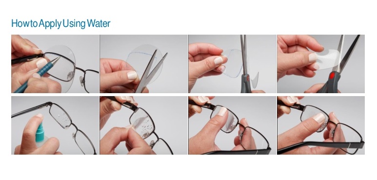
• 3M™ Press-On™ Optics, also known as Fresnel Prisms, and Press-On Aspherics, provide a system for creating prismatic and spherical corrections in a cost effective and temporary manner.• 3M™ Press-On™ Optics offer the eye care professional and the patient several benefits:• A simple, therapeutic, inexpensive, way to correct several visual disorders.• Provide an immediate correction.• They are more comfortable, more cosmetically appealing treatment for strabismus and diplopia than conventional prisms.• They add NO noticeable weight or thickness to the spectacle.• Press-on prism material is flexible static vinyl which can easily be cut to shape with scissors to determine the acceptance of a proposed corrective prescription.• Adheres with just water to existing lenses, yet it can be easily repositioned.• Apply a Press-On sphere or prism to the back surface of one or both lenses of the patient`s eyeglasses with just water.• It can be applied to the entire lens or to any region of the lens.• A full range of seventeen powers from 1.00 to 40.00 prism diopters allows you to broaden therapeutic uses and provides additional treatment options in your practice.Made of polyvinyl chloride1mm thick63.5 mm diameter
Grids - SEM FinderGrinding & PolishingMaterials EmbeddingSHOP ALL MATERIALS EMBEDDINGCold Mounting ResinsHot Mounting ResinsMounting Tabs & AdhesivesPreparation
Although the modern microscope is an array of optics, mirrors and lenses, in general, there are only two set of optics which are adjusted and changed by the user when viewing samples- the eyepieces and the objectives.
AperturesCalibration & Test SpecimensSHOP ALL CALIBRATION & TEST SPECIMENSGeller Reference StandardsCertified Particle Size StandardsCritical Dimension StandardsMagnification CalibrationResolution & Grey Level Test SpecimensConsumables KitsFilaments
Nosepiecemicroscopefunction
Support Films - Lacey Carbon Support Films - QuantifoilSupport Films - SiliconTissue Processing ChemicalsTissue Processing ConsumablesVacuum Coating MaterialsVacuum Oils & GreasesX-ray Microanalysis Standards
Adam Equipment Balances & ScalesAirflow & Ventilation TestingSHOP ALL AIRFLOW & VENTILATION TESTINGInstruments & AnemometersProbesCapture HoodsFlowmetersAccessoriesCell Manipulation InstrumentationSHOP ALL CELL MANIPULATION INSTRUMENTATIONElectroporatorMicromanipulatorsMicroinjectorsMicrocapillariesVibration ProtectionAccessories
Cell Manipulation by Calibre ScientificSHOP ALL CELL MANIPULATION BY CALIBRE SCIENTIFICElectroporatorMicromanipulatorsMicroinjectorsMicrocapillariesVibration ProtectionAccessories for Cell Manipulation
Grids - Athene by Agar ScientificSHOP ALL GRIDS - ATHENE BY AGAR SCIENTIFICStandard Square PatternThin BarThick Bar/Thin BarSlot and Multiple SlotThick SlotHexagonalRound Hole PatternSingle HoleFoldingOtherK-kits for Liquid TEMLight Element Support GridsMaterial ProcessingPhotographic Films & Papers
Whether the eyepieces are made up of two or more lenses, the only visible lenses are at either end of the eyepiece. The lenses closest to your eyes are known as the ‘Eye Lens’, whilst those which face into the body of the microscope are known as the ‘Field Lens’.
Grids - AgarSHOP ALL GRIDS - AGARSquare MeshRectangular Mesh Parallel BarFoldingHexagonal MeshThin BarVery Fine MeshSingle & Triple SlotSingle HoleTabbedResin Embedding - AcrylicResin Embedding - EpoxyResin Embedding - London ResinResin Embedding Consumables
Oils & GreasesSafety GlovesSafety ProductsSpecimen PreparationStorage BoxesSHOP ALL STORAGE BOXESGel-Pak BoxesMembrane Boxes
Grids - AgarSHOP ALL GRIDS - AGARSquare MeshRectangular Mesh Parallel BarFoldingHexagonal MeshThin BarVery Fine MeshSingle & Triple SlotSingle HoleTabbedResin Embedding - AcrylicResin Embedding - EpoxyResin Embedding - London ResinResin Embedding Consumables
AperturesCalibration StandardsCalibration Standard - Lattice PlaneCirclip Injector and CirclipsCoated GridsCryo PreparationDiamond Knives - DiATOMEFilamentsGrid Boxes & StorageGrids - FinderGrids - Omniprobe
JavaScript seems to be disabled in your browser. For the best experience on our site, be sure to turn on Javascript in your browser.
AdhesivesBags & LabelsBeakers, Tubes & ContainersCleaning ProductsSHOP ALL CLEANING PRODUCTSAir DustersCleaners, Solvents & CreamsCloths & WipesPolishesTools
Oils & GreasesSafety GlovesSafety ProductsSpecimen PreparationStorage BoxesSHOP ALL STORAGE BOXESGel-Pak BoxesMembrane Boxes
The next step is to make the diopter adjustment. If you have a look at the two eyepieces in a binocular set-up, then you’ll see that one of them has a ring adjuster around the eyepiece tube. Firstly, look at your specimen with the fixed eyepiece only. Adjust the focus of the specimen using the focus on the microscope body. Without adjusting the microscope focus, look down the adjustable eyepiece and slowly turn the diopter ring until your specimen comes into sharp focus. Once this is set, then this adjustment stays the same no matter which objective you use (unlike Kohler Illumination which I’ll cover in a future article).


 Ms.Cici
 8618319014500