
LWIR Transparent Thermosets | New polymeric materials ... - ir transparent materials
The key issue is this: high NA objectives bring a large portion of their light in at a high angle. This high angle results in longer paths for the excitation light to take, and this results in more scattering events. The end result is that excitation intensity decreases. This has been shown theoretically and empirically. So if you’ll be imaging deep, consider moderate NA objectives.
Stagemicroscopefunction
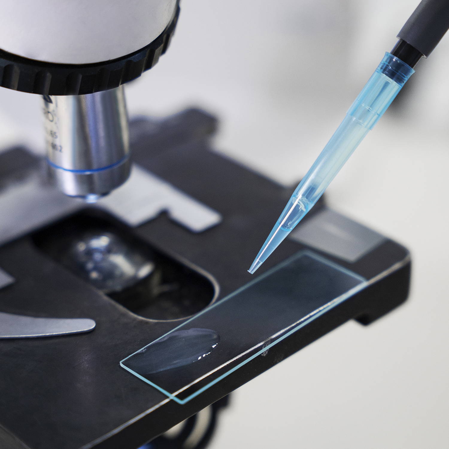
A specimen is a sample or example used for scientific study. It can be anything from biological tissues to materials, examined under a microscope or other instruments for analysis.
Types ofobjectivelenses
Even many commercially available scopes fail to overfill the large back apertures of today’s low magnification/high NA objectives. The major microscope manufacturers need their objectives to fit onto their existing microscope bodies and systems, and this is a major engineering constraint in their design for new imaging systems.
Magnification is the process of enlarging the appearance of an object, making it look bigger than its actual size. In optics, it is the ratio of the size of the image produced by a lens or microscope to the actual size of the object being viewed.
A microscope is a scientific instrument used to magnify and observe objects that are too small to be seen with the naked eye. It works by focusing light or electrons to create an enlarged image of the specimen.
Capable of high magnification, which is achieved through the combination of the objective lens (typically 4x, 10x, 40x, and 100x) and the eyepiece (usually 10x).
Recently, microscope manufacturers have been releasing ever higher NA objectives for multiphoton imaging. Although higher NA objectives should give better axial resolution, they might not be ideal for imaging deep into the brain compared to more moderate NAs.
Objectivelens magnification
A Compound Microscope is a type of optical microscope that uses multiple lenses to magnify small objects. It consists of two sets of lenses: the objective lens, which is closer to the specimen and provides the initial magnification, and the eyepiece lens, which further magnifies the image for the viewer's eye. Light passes through the specimen and is magnified by the objective lens, then further magnified by the eyepiece lens, resulting in a highly magnified image visible to the observer. Compound microscopes are commonly used in biology, medicine, and other scientific fields for viewing cells, tissues, and other small structures.
A monocular microscope head is a basic type of microscope head with a single eyepiece, ideal for cost-effective and straightforward applications. It is particularly useful in educational settings and for beginners, but it can lead to eye strain over long periods and lacks the depth perception provided by more advanced binocular and trinocular heads.
A binocular microscope head utilizes two eyepieces for simultaneous viewing with both eyes, providing enhanced comfort, depth perception, and superior image quality. Ideal for professional and research settings requiring detailed observation, its design minimizes eye strain and enhances ergonomic support compared to monocular microscopes.
Objective microscopefunction
For glass, the range of refractive indices is usually between 1.4 and 1.7. Different types of glass have different refractive indices, so this property can be ...

Microscope objectives are vital lenses that determine the magnification, resolution, and quality of the images produced by a microscope. They come in various types and magnifications, each suited for different applications and levels of detail, making them indispensable in scientific research, medical diagnostics, and educational settings.
4.5 mW 632.8nm (RED) HELIUM NEON LASER - NOTE: Lasers and power supplies are sold separately. A power supply is NOT provided with this unit.
Microscopeparts
Im trying to make a dobsonian for my birthday , and im wondering if a concave mirror would work as the primary mirror.
Commonly used in biological research, medical diagnostics, and educational settings for detailed examination of specimens.
explorē 8 is a handheld portable magnifier that is ideal for anyone with vision loss who needs an electronic reading aid for magnifying documents.
Flexible Fiber Optic Light Guide · Free bending, flexible use · A variety of HECHO standard sizes · Standard input accepts HECHO LED and Halogen series · Custom ...
Navigate effortlessly through magnification levels and focus adjustments. Our microscopes feature intuitive controls, allowing you to concentrate on your research without the hassle of complicated settings.
The terms monocular, binocular, and trinocular refer to the different types of microscope heads, each offering a distinct way of viewing the specimen.
These are also called USB serial converters and they provide full RS-232 / 422 / 485 signals with LED diagnostics and activity indicators.
I think the perception that higher NAs always improve images arises when people try out new, high NA objectives that have smaller back apertures than their old objectives (e.g., an Olympus 20x/0.95 NA or a Nikon 16x/0.8 NA). If the back aperature on the 25x, 1.0+ NA objective they’re trying is smaller, then suddenly they’re overfilling more than before and their axial resolution and S:N are improved. They chalk it up to the NA and swear never to go back to 0.8 NA objectives. However, their old objective might actually be better, and what they really need to work on is their scanning optics.
Stagemicroscope definition
Used in fields like biology, geology, entomology, electronics assembly, and manufacturing for tasks requiring manipulation and examination of objects in three dimensions.
Provides high magnification (up to 1000x or more) and high resolution for viewing fine details of cells, tissues, and microorganisms.
Compound Magnification is calculated by multiplying the magnification of the objective lens by the magnification of the eyepiece.
Magnification works by bending light through lenses or using digital technology to enlarge the appearance of an object, allowing for detailed observation and analysis.
Fluocinonide 0.05%, Cream, 60gm Tube · Triamcinolone Acetonide, 0.10%, Cream, 454gm (16oz) Jar Each · Carisoprodol, (C-IV) 350mg, 100 Tablets/Bottle.
Witness the microscopic world in stunning detail with our high-quality optics. Every slide comes to life with crystal-clear clarity, allowing you to delve into the intricacies of biology, chemistry, and beyond.
Objective microscope definitionand function
Compound microscopes are suited for detailed examination of microscopic structures, while stereo microscopes are more appropriate for observing larger objects in three dimensions and for tasks that involve manipulation and dissection.
A phase contrast microscope is an optical microscope designed to enhance the contrast of transparent and colorless specimens without the need for staining. It works by exploiting differences in the refractive index of different parts of the specimen, transforming these differences into variations in light intensity.
Mar 26, 2024 — At the fast end of the maximum aperture spectrum (f/1 to f/2.8), the implication is that you can hold a 1/60 second shutter speed into dimmer ...
By contrast, underfilling the back aperture is a great way to destroy one’s axial resolution. Since the lateral resolution is relatively unaffected, this problem often goes unnoticed (see figure below, its link, and this review). If the excitation beam is less than half of the diameter of the back aperture of a 20x/0.95 NA, then the axial FWHM could be 3x what it should be, or roughly the equivilant of a 0.60 NA objective (theoretical FWHM 5.6 microns), or worse.
Objectivelens telescope
A darkfield microscope is a type of optical microscope that provides high contrast images of unstained specimens by using scattered light. The specimen appears bright against a dark background
AmScope exclusive ALL-IN-ONE 3D DIGITAL INSPECTION MICROSCOPE. View different angles and perspectives of objects with ease.
Compound zero order waveplates. If two identical multiple order plates are placed together so that one's fast axis is along the other's slow axis, they cancel ...
Illuminate your subjects with brilliance. Our microscopes feature advanced lighting technologies, providing the perfect balance for optimal observation, even in low-light conditions.

Prisms that deviate the ray path, rotate the image, or simply displace the image from its original axis are helpful in many imaging systems. Ray deviations are ...
A trinocular microscope head combines the benefits of binocular viewing with the capability to capture digital images or videos of specimens. It is particularly suited for advanced research, educational purposes, and industrial applications where precise imaging and documentation are essential.
A stereo microscope, also known as a stereoscopic or dissecting microscope, provides three-dimensional viewing of larger, opaque specimens through dual optical paths with objective lenses. It offers lower magnification (typically 5x to 40x) than compound microscopes but enhances depth perception. Ideal for tasks in biology, geology, and manufacturing, it allows comfortable, extended viewing with ergonomic adjustments.
Uses two separate optical paths with two objective lenses to provide a stereoscopic (3D) view of larger, opaque specimens.


 Ms.Cici
 8618319014500