
Liquid Light Guides - liquid light guide
Whatdoes a microscopedo
![]()
This page titled 3.2A: Microscopy is shared under a CC BY-SA 4.0 license and was authored, remixed, and/or curated by Boundless via source content that was edited to the style and standards of the LibreTexts platform.
How does a microscopework step by step
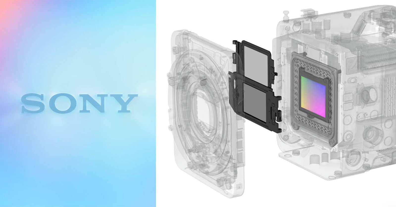
Instead, it’s also possible to use a filter at the sensor level, which is how Sony’s electronic variable ND filter system works. Sony’s optical filter system lives inside the camera, in front of the image sensor, and it responds to user input in the camera’s menu system.
Cells vary in size. With few exceptions, individual cells cannot be seen with the naked eye, so scientists use microscopes (micro- = “small”; -scope = “to look at”) to study them. A microscope is an instrument that magnifies an object. Most photographs of cells are taken with a microscope; these images can also be called micrographs.
Microscope
Most student microscopes are classified as light microscopes. Visible light passes and is bent through the lens system to enable the user to see the specimen. Light microscopes are advantageous for viewing living organisms, but since individual cells are generally transparent, their components are not distinguishable unless they are colored with special stains. Staining, however, usually kills the cells.
While it is relatively straightforward to use a filter like this on the lens itself, this comes with some usability concerns and a potential impact on image quality. Plus, getting a variety of ND filter strengths in different filter thread sizes is tedious and costly.
In contrast to light microscopes, electron microscopes use a beam of electrons instead of a beam of light. Not only does this allow for higher magnification and, thus, more detail, it also provides higher resolving power. The method used to prepare the specimen for viewing with an electron microscope kills the specimen. Electrons have short wavelengths (shorter than photons) that move best in a vacuum, so living cells cannot be viewed with an electron microscope.
To give you a sense of cell size, a typical human red blood cell is about eight millionths of a meter or eight micrometers (abbreviated as eight μm) in diameter; the head of a pin of is about two thousandths of a meter (two mm) in diameter. That means about 250 red blood cells could fit on the head of a pin.
For example, the user can adjust the ND filter’s strength from 1/4 to 1/128, enabling fine-tuned control. However, since the system is electronic and fully integrated into the camera, it can also be adjusted automatically based on a user’s selected settings and the metered light.
How Does a microscopeWork simple
ND filters are a common tool for filmmakers, enabling them to adjust the amount of light that hits the image sensor. This is important because filmmakers often want to shoot at specific shutter speeds, like 1/48s when shooting 24p footage. While that shutter speed may be easy to achieve with a slow lens or in dim conditions, bright light, and fast lenses can demand faster-than-desired shutter speeds. Using an ND filter makes it possible to cut the amount of light and comfortably shoot at the desired shutter speed, even with a fast aperture.
Whatdoes astage do ina microscope
How does a microscopemagnify
In a scanning electron microscope, a beam of electrons moves back and forth across a cell’s surface, creating details of cell surface characteristics. In a transmission electron microscope, the electron beam penetrates the cell and provides details of a cell’s internal structures. As you might imagine, electron microscopes are significantly more bulky and expensive than light microscopes.

It is really impressive technology and is available in Sony’s FX6, FX9, FS5, FS5 II, FS7 II, and Burano cameras, among others.
The optics of a microscope’s lenses change the orientation of the image that the user sees. A specimen that is right-side up and facing right on the microscope slide will appear upside-down and facing left when viewed through a microscope, and vice versa. Similarly, if the slide is moved left while looking through the microscope, it will appear to move right, and if moved down, it will seem to move up. This occurs because microscopes use two sets of lenses to magnify the image. Because of the manner by which light travels through the lenses, this system of two lenses produces an inverted image (binocular, or dissecting microscopes, work in a similar manner, but they include an additional magnification system that makes the final image appear to be upright).
How Does a microscopeWork for Kids
The electronic variable ND filter has another added benefit. Not every lens, including third-party E-mount lenses, has seamless iris control. When recording video, cinematographers don’t want to deal with rough transitions from one aperture to the next, nor do they want to adjust their shutter speed during a shot. Electronic variable ND filters help the camera compensate for changing light values without changing the aperture or shutter speed.
The LibreTexts libraries are Powered by NICE CXone Expert and are supported by the Department of Education Open Textbook Pilot Project, the UC Davis Office of the Provost, the UC Davis Library, the California State University Affordable Learning Solutions Program, and Merlot. We also acknowledge previous National Science Foundation support under grant numbers 1246120, 1525057, and 1413739. Legal. Accessibility Statement For more information contact us at info@libretexts.org.
The system relies upon a crystal LCD through which light travels from the lens to the image sensor. What makes this “variable” is electric voltage. The camera precisely controls the voltage that goes into the liquid crystal panel, which changes how much light can travel through it and reach the image sensor. There is no color shift or other negative impact on image quality.
Light microscopes, commonly used in undergraduate college laboratories, magnify up to approximately 400 times. Two parameters that are important in microscopy are magnification and resolving power. Magnification is the process of enlarging an object in appearance. Resolving power is the ability of a microscope to distinguish two adjacent structures as separate: the higher the resolution, the better the clarity and detail of the image. When oil immersion lenses are used for the study of small objects, magnification is usually increased to 1,000 times. In order to gain a better understanding of cellular structure and function, scientists typically use electron microscopes.
At its core, an electronic variable ND system enables users to control their camera’s exposure settings and depth of field, even when ambient light conditions are excessively bright or constantly changing.
Changing the ISO is not always an optimal option, either. If it’s bright enough, there may not be an ISO speed slow sufficient to nail the shot at the required shutter speed. Further, many videographers want to maintain a consistent look and image quality across multiple shots, so constant ISO adjustments may not be the best choice.
Numerous Sony cinema cameras include electronic variable neutral density (ND) filters, including the popular full-frame FX9.


 Ms.Cici
 8618319014500