LED lighting helps Koehler Paper Group to deliver its ... - koehler lighting
Understanding each part's function ensures proper usage of the microscope, leading to clearer images and accurate observations.

The angle they actually care about when calculating the amount of reflected light (and possibly refracted light, for transparent objects) is between the vector (or ray , if you prefer) created from the viewer to the spot you’re looking at on the object, and the surface normal of the object.

The illumination system provides a consistent and controlled light source, ensuring that specimens are properly illuminated for clear visualization under the microscope.
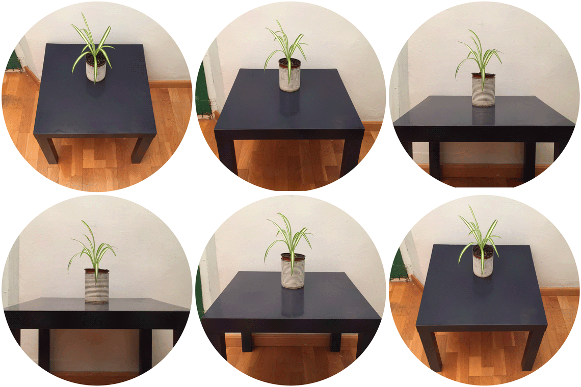
Take a look around the space you are in. Can you find an example of the Fresnel Effect? Look at shiny floors and plastic surfaces. Crouch down as I did and see how the intensity of the reflection changes.
And here is what we see from the viewpoint of the person. Use the arrows to the left and right of the image below to switch between versions with Fresnel Effect and without Fresnel Effect.
To understand the Fresnel Effect, you have to understand the basics of reflections.We’ll keep this minimal – the key is the Angle of Incidence.The Angle of Incidence is the angle between your line of sight and the surface of the object you are looking at.
Hi Vaughn! Yes, usually the surface normal is used to explain the Fresnel effect. My goal with this article was to make the concept of the Fresnel effect as simple to grasp as possible. Adding the concept of surface normals would have increased complexity and cognitive load unnecessarily. I included a link to the Wikipedia entry for those readers that want to get the full technical explanation. 🙂
Fresnellens
I thought the angle of incidence is taken from the normal to the surface, why is yours taken from parallel to the surface?
I was blind to the Fresnel Effect until someone pointed it out to me — now I can see that it is everywhere! If you’re looking for it, you’ll find it.
Here is another example: reflections change across distance – because the angle of incidence changes. As you look down to the ground close to your feet, the angle of incidence is very steep. If you look at a point on the ground that’s further away from you, the angle gets more shallow – and the reflection becomes more visible.
Fresnellight
Microscopes come in various types, including compound microscopes, scanning electron microscopes, transmission electron microscopes, etc. Each type of microscope has unique capabilities and magnification levels.
Microscope parts, like the eyepiece, objective lens, stage, condenser, diaphragm, and light source, work together to magnify and illuminate specimens for detailed observation.
The objective lens collects light from the specimen, which is further magnified by the eyepiece. The condenser and illuminator work to provide adequate illumination for clear visualization.
Thank you Kevin! I wanted to use as few technical terms as possible in order to keep the article short and easy to understand. The surface normal is a very useful concept and I like your way of using it to describe the principle behind the Fresnel Effect.
Fresnelscreen
fresnellens中文
Hey John! The two are related. Highlights are “specular reflections” and the fresnel effect describes how the intensity of specular reflection changes depending on your angle of view. This video might clarify it: https://www.dorian-iten.com/see-more/
A microscope is an optical instrument used to magnify small objects or specimens that are too small to be seen by the naked eye. It works by using lenses or a combination of lenses and mirrors to focus light on the specimen and magnify its image. Microscopes are essential tools in scientific disciplines, including biology, chemistry, and forensic science, allowing researchers to study the complex details of cells, tissues, microorganisms, etc.
Fresnelequation
The diagram in a microscope serves as a visual guide, illustrating the different components and their functions, helping users in operating the microscope effectively for observation and analysis of specimens.
In other words, if you look directly down at the table, the angle is close to zero, and as you get closer to eye level with the table, the angle increases. This makes it easier to explain that “as the angle increases, the amount of reflection increases.”
Fresneldiffraction
In Class 9th, a microscope is introduced as a scientific instrument used for magnifying tiny objects to observe details not visible to the naked eye.
Excellent explanation, making simple the (apparently) complex. Indeed, your description demonstrates that “complexity is n the eye of the beholder.’
On curved surfaces, the angle of incidence gets steeper towards the edges of the form. On a cylinder, the fresnel effect leads to the specular reflections being most visible in the red areas:
The diagram of microscope class 9 is an important topic in the biology syllabus. The following is a diagram of a miscroscope with full labelling:

A diagram of a microscope is a useful visual aid for understanding its complex structure and functioning. Microscopes have long been essential tools in research, and industry, allowing us to study the microscopic world. The diagram of a microscope with labels provides an easy way to understand its various parts. From the base to the eyepiece, each component of a microscope plays an important role in magnifying and demonstrating specimens. In this article, we will learn a simple diagram of a microscope along with the parts of a microscope and their function.
Yes, different types include compound microscopes, stereo microscopes, electron microscopes, each with unique features made for specific applications and magnification levels.
fresnel中文
The objective lens gathers light from the specimen and magnifies it, while the eyepiece further magnifies the image for observation.
Fresnelreflection
I had a hard time understanding this concept till I found this explanation, very simple and easy to grasp, thanks for keeping things simple.
In conclusion, a detailed diagram of a microscope provides lots of information on the internal components of a microscope. Understanding its components and their functions with the help of microscope diagram with labels shows its important role in various fields. The microscope is still an essential instrument that helps us explore new areas of research and expand our understanding of the world around us. It allows us to see into the tiny world and demonstrate it. The well-labeled compound microscope diagram is given above in the article.
PS. If you find this interesting, you might enjoy learning about the 11 Modeling Factors – light effects that create the sensation of form.
A compound microscope, typically studied with a diagram, is a type of microscope consisting of multiple lenses to magnify objects by passing light through them, enabling detailed examination of microscopic specimens.
The main components usually include the eyepiece, objective lens, stage, condenser, diaphragm, illuminator, and various adjustment knobs.
Definition of Microscope: A microscope is an instrument that magnifies small objects or specimens to make them visible for detailed examination. It uses lenses or a combination of lenses and mirrors to focus light on the specimen and produce an enlarged image, enabling observation of structures that are not visible to the naked eye.


 Ms.Cici
 8618319014500