
How to Calculate Microscope On-Screen Magnification - how to find magnification

The Prime series of 95% quantum efficient, back-illuminated sCMOS cameras are designed to support the most demanding, low-light research applications
This leads to the obvious question, why use 100x or 60x magnification if 40x magnification achieves the same resolution? When using higher magnification objectives, field of view is being restricted for no reason.
Figure 2 highlights how increasing the numerical aperture of the objective lens reduces the size of the Airy disk and therefore increases the amount of resolvable detail.
See what others are doing. Stories and images from scientists using our high-performance sCMOS, EMCCD and CCD cameras to advance their research.
Supplying custom cameras to instrument designers for most of our 40 year history, we will work with you every step of the way.
All cameras are controllable with the PVCAM driver and supported in Ocular software. The PVCAM driver SDK can also be used integrate into other software packages.
Laser Safety glassesClass 4
High content imaging is primarily concerned with the automated analysis of large cell populations where the goal is to process as many cells as possible in the fastest time with the highest resolution.
Biochip, genomics and microarray detection represent a large mix of applications with varying needs of a scientific camera.
Good news! You have already signed up to our mailing list. If you would like to amend your preferences, please look out for one of our emails- don’t forget to check your junk folder just in case.
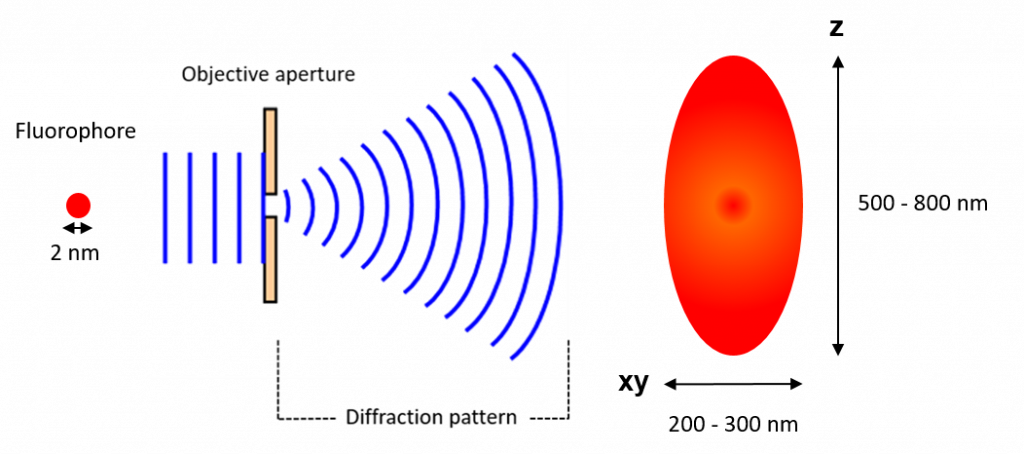
In the case of GFP, which emits at ~510 nm, and a high (NA 1.4) numerical aperture objective, the size of the fluorophore as resolved by the microscope would be 182 nm. This is much larger than the actual fluorophore which may be just 2 nm.
Laser safety glassesnear me
The issue with resolving adjacent fluorophores is that as Airy disks move closer together they merge with one another and become unresolvable (Figure 3). Thus, in 1896, Lord Rayleigh refined the Abbe equation to take into account how far apart two fluorophores need to be to differentiate them:
The Iris family of sCMOS cameras deliver up to a 15 megapixel sensor with a 25 millimetre field of view for high-resolution imaging over a large imaging area.
See what others are doing. Stories and images from scientists using our high-performance sCMOS, EMCCD and CCD cameras to advance their research.
CMOS made scientific. The Moment is a true global shutter CMOS camera with an ultra-compact form factor, powered through USB 3.2 Gen 2.
In microscopy, it is vital to have some form of contrast or stain that gives areas of the sample color and makes it possible to image. Advanced fluorescence microscopy techniques take advantage of this.
The pixel size required to achieve Nyquist sampling at different magnifications is illustrated in Tables 2 and 3. These tables provide the answer to the question posed before: Why use 100x or 60x magnification if 40x magnification achieves the same resolution?
Biochip, genomics and microarray detection represent a large mix of applications with varying needs of a scientific camera.
There are two types of resolution; microscope resolution and camera resolution. Microscope resolution is determined by the numerical aperture of the objective and the emission wavelength whereas camera resolution is determined by pixel size.
The maximum resolution of the microscope is a function of the numerical aperture of the objective lens and the emission wavelength of the sample, whereas camera resolution is determined entirely by pixel size.
Light travels as a wave so when it is focused to a small spot with a lens, no matter how good the objective lens is, the focal spot will have a larger size than the actual fluorophore.
Laser safety glassesguide
CMOS made scientific. The Moment is a true global shutter CMOS camera with an ultra-compact form factor, powered through USB 3.2 Gen 2.
The size of the diffraction limited spot is approximately half the size of the wavelength of light emitted but the full equation, determined by Ernst Abbe in 1873, is:
So why use higher magnification objectives at all? This is where camera resolution comes in. Magnification plays a big part in the resolving power of a scientific camera.
The diffraction limited spot takes the shape of an Airy disk (Figure 2), named after George Biddell Airy. It consists of a bright central spot with a series of diffraction rings surrounding it. The size of the central spot is determined by the wavelength of light being emitted and the numerical aperture of the objective.
The QImaging CCD family of scientific cameras are designed with solutions for electrophysiology, long stare, color imaging, documentation and live cell imaging.
A smaller pixel camera allows for a lower magnification to be used which has the advantage of seeing more of the sample in the field of view.
Laser safety glassesamazon
Physics and biophysics imaging encompasses a wide range of techniques used to interrogate physical phenomena using high tech imaging systems.
The Evolve family of cameras are high-resolution, back-illuminated EMCCD providing high sensitivity for the lowest light applications.
A smaller pixel is therefore required to achieve Nyquist sampling at 40x. Table 3 shows that a smaller pixel size of 4.25 μm reaches a camera resolution of 0.24 μm with 40x magnification which satisfies Nyquist.
Cooled, low-noise CMOS cameras designed for integration. With unprecedented thermal control, Retiga E cameras are capable of exposures over an hour!
With this refinement, the distance required to differentiate two GFP fluorophores with a high (NA 1.4) objective would be 222 nm.
It’s important to note that objective magnification has no impact on resolution, numerical aperture is the only important value. A 100x, 1.40 NA objective has the same resolving power as a 60x, 1.40 NA objective. Equally, a 100x, 1.30 NA objective has the same resolving power as a 40x, 1.30 NA objective.
The most obvious way to match camera resolution to microscope resolution would appear to be simply matching the diffraction-limited resolution given in Table 1 to the size of a single pixel. However, this is not the case. The goal isn’t just to match the microscope resolution but to distinguish adjacent objects.
All cameras are controllable with the PVCAM driver and supported in Ocular software. The PVCAM driver SDK can also be used integrate into other software packages.
Browse our LaserShield® filters using the Wavelength Selection Tool in the left side navigation. If you need to calculate the OD for your required level of protection, please use our OD Calculator tool or call us at 800-521-9746 to assist you with choosing the right filter for your application.
The data presented in this section illustrates that the lowest magnification that can be used is limited by the pixel size of the camera, which needs to be small enough to match the microscope resolution.
The QImaging CCD family of scientific cameras are designed with solutions for electrophysiology, long stare, color imaging, documentation and live cell imaging.
The Evolve family of cameras are high-resolution, back-illuminated EMCCD providing high sensitivity for the lowest light applications.
Laser safety glasseshome depot
By using lower magnification objectives, changing from a 100x, 1.30 NA objective to a 40x, 1.30 NA objective, field of view is increased by 450%. That represents a 450% increase in sample area with no loss in resolution.
It’s possible to reach diffraction-limited resolution with lower (40x) magnification objectives but pixel size becomes limiting. To remedy this, cameras have been developed with smaller pixels to allow researchers to move to lower magnification objectives. This has the advantage of increasing the field of view without sacrificing resolution. Researchers can expect to increase throughput 200% just by switching from 60x to 40x.
Laser Safety Glasses1064 nm
High content imaging is primarily concerned with the automated analysis of large cell populations where the goal is to process as many cells as possible in the fastest time with the highest resolution.
The brand new Kinetix family of back-illuminated sCMOS cameras delivers a framerate and field of view unmatched by any other sCMOS camera.
Laser Safety GlassesCanada
In microscopy, it is vital to have some form of contrast or stain that gives areas of the sample color and makes it possible to image. Advanced fluorescence microscopy techniques take advantage of this.
450nmlaser Safety Glasses
Where d is the size of the diffraction limited spot, λ is the wavelength of light used and 2NA is 2 times the numerical aperture of the objective.
Cooled, low-noise CMOS cameras designed for integration. With unprecedented thermal control, Retiga E cameras are capable of exposures over an hour!
JavaScript seems to be disabled in your browser. For the best experience on our site, be sure to turn on Javascript in your browser.
Separating adjacent features requires the presence of at least one intervening pixel with a different intensity value. For this reason, the best spatial resolution that can be achieved requires matching the diffraction-limited resolution of the microscope to two pixels on the sensor. This is called Nyquist sampling and it can be calculated using the equation:
The brand new Kinetix family of back-illuminated sCMOS cameras delivers a framerate and field of view unmatched by any other sCMOS camera.
Resolution in fluorescence microscopy is defined as the shortest distance between two points on a specimen that can still be distinguished. This is primarily determined by two factors; microscope resolution, which is the smallest object the microscope can resolve, and camera resolution, which is the ability of the camera to detect what the microscope can resolve.
Physics and biophysics imaging encompasses a wide range of techniques used to interrogate physical phenomena using high tech imaging systems.
As discussed previously, to achieve Nyquist sampling and match the microscope resolution using GFP (510 nm), the camera resolution should reach 0.22 μm. Table 2 shows that for a 40x objective, a 6.5 μm pixel is too large as the resulting camera resolution is 0.37 μm. A 6.5 μm pixel is considered sufficient for 60x magnification, however, as the resulting camera resolution reaches 0.25 μm.
This happens because the wavefront of the fluorescence emission becomes diffracted at the edges of the objective aperture. This effectively spreads the wavefront out, widening the fluorescence emission into a diffraction pattern which has a central spot larger than the original fluorophore (Figure 1).
Camera resolution is defined as the ability of the camera sensor to sample the image and the resolving power of a scientific camera is entirely dependent on the size of the pixel and by how much it is magnified. Figure 4 highlights how scientific camera pixel size is changed by objective magnification.
The information in Table 1 highlights the resolution limit possible with a variety of different magnification objectives and numerical apertures using GFP (510 nm) emission.
Laser safety glasses are designed to block certain wavelengths of light while allowing others through, to enable you to perform your job safely and prevent exposure to direct, reflected, or scattered laser radiation that could harm your eyes or even blind you. NoIR offers a wide selection of carefully formulated filters for various applications and laser types.
As Figure 5 illustrates, if microscope resolution is matched to the size of a single pixel, then it is possible that two adjacent objects could be imaged onto adjacent pixels. In this case, there would be no way of discerning them as two separate objects in the resulting image.
However, the resolving power of a fluorescence microscope is ultimately restricted by the diffraction limit of light which, when using green light (510 nm) for example, would be around 220 nm. This sets a lower limit on what can be resolved. It is therefore common practice in standard fluorescence microscopy to use a microscope setup capable of reaching this lower limit to detect the smallest resolvable object. Attempting to resolve lower than this isn’t possible using conventional microscopy, it’s only possible to break the diffraction limit of light using super-resolution techniques.
The Iris family of sCMOS cameras deliver up to a 15 megapixel sensor with a 25 millimetre field of view for high-resolution imaging over a large imaging area.
The Prime series of 95% quantum efficient, back-illuminated sCMOS cameras are designed to support the most demanding, low-light research applications
Supplying custom cameras to instrument designers for most of our 40 year history, we will work with you every step of the way.


 Ms.Cici
 8618319014500