
How Lens Compression and Perspective Distortion Work - lens distortion
While a conventional microscope uses visible light (400-700 nm) as the light source to irradiate the sample, a fluorescent microscope uses higher-intensity light to excite the fluorophores in the specimen. Furthermore, fluorophores emit light of longer wavelengths during fluorescence, producing a magnified image with a color different from that of the original light source.
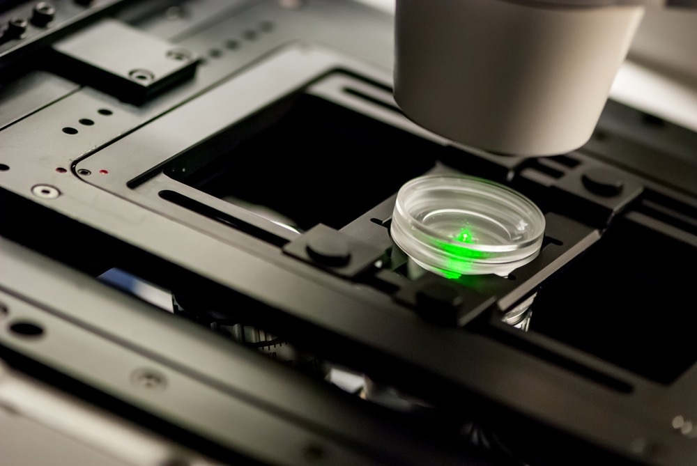
A fluorescent microscope is a powerful tool that selectively reveals the object of interest (signal) on a black background. Various fluorescent indicators, also called fluorescent probes or fluorophores, are tailor-modified to label specific targets such as lipids, proteins, or ions.
3400usd to cad

Although fluorescence microscopy has significantly helped in understanding the dynamics and physiology of biological materials, further investigation into combating existing limitations can provide a wider scope for applying this type of microscope.
340USD to CAD
Sanderson, M. J., Smith, I., Parker, I., Bootman, M. D. 2014. Fluorescence microscopy. Cold Spring Harbor Protocols. https://www.ncbi.nlm.nih.gov/pmc/articles/PMC4711767/
Fluorescent microscopes have several advantages, including high sensitivity, high resolution, multiplexing ability to simultaneously visualize multiple pathways in a sample, high specificity, non-destructive analysis of samples, ability to image live cells, and ability to trace the exact location of proteins.
Kaveti, Bhavna. "How Do Fluorescent Microscopes Work?". AZoOptics. 25 November 2024. .
34000 cad toinr
Disclaimer: The views expressed here are those of the author expressed in their private capacity and do not necessarily represent the views of AZoM.com Limited T/A AZoNetwork the owner and operator of this website. This disclaimer forms part of the Terms and conditions of use of this website.
Kaveti, Bhavna. 2023. How Do Fluorescent Microscopes Work?. AZoOptics, viewed 25 November 2024, https://www.azooptics.com/Article.aspx?ArticleID=2385.
34000 usd toinr
The idea of studying biological materials using fluorescence was conceived in the early 19th century. However, its practical application was not demonstrated until 1938, when Richard Manly and Robert Goldstein first developed the fluorescent microscope.
30,000USD to CAD
George Rice. Fluorescent Microscopy. https://serc.carleton.edu/microbelife/research_methods/microscopy/fluromic.html#:~:text=The%20conventional%20microscope%20uses%20visible,in%20a%20sample%20of%20interest.
Another piece published in PhotoniX devised a two-channel attention network (TCAN) that showed superior performance over STED in generating high-resolution images from their low-resolution counterparts, enabling generalization to different modalities of fluorescence microscopy. The improved resolution helped unravel the fine structures and dynamic instability of the microtubules.
The VINCI series of ultrafast fiber lasers has a central emission wavelength of 1064 nm and features a unique combination of short pulse durations.
Bhavna Kaveti is a science writer based in Hyderabad, India. She has a Masters in Pharmaceutical Chemistry from Vellore Institute of Technology, India, and a Ph.D. in Organic and Medicinal Chemistry from Universidad de Guanajuato, Mexico. Her research work involved designing and synthesizing heterocycle-based bioactive molecules, where she had exposure to both multistep and multicomponent synthesis. During her doctoral studies, she worked on synthesizing various linked and fused heterocycle-based peptidomimetic molecules that are anticipated to have a bioactive potential for further functionalization. While working on her thesis and research papers, she explored her passion for scientific writing and communications.
34000 usd to cadtoday
Kaveti, Bhavna. "How Do Fluorescent Microscopes Work?". AZoOptics. https://www.azooptics.com/Article.aspx?ArticleID=2385. (accessed November 25, 2024).
Registered members can chat with Azthena, request quotations, download pdf's, brochures and subscribe to our related newsletter content.
To understand the workings of a fluorescent microscope, it is important to become familiar with the underlying phenomenon of fluorescence. When light of a specific wavelength is irradiated on a fluorophore-labeled specimen, the electrons in the specimen are excited to higher energy levels. The subsequent de-excitation of electrons to attain stability causes the emission of light with longer wavelengths, which is called fluorescence.
These F-theta lenses by Avantier are designed for consistent spot size and uniform field curvature correction, ideal for high-resolution imaging applications.
35000USD to CAD
However, the spectral emission during this phenomenon also includes weaker fluorescence, which is separated using a spectral emission filter to ensure the distribution of a single wavelength of light during the imaging process. Because the color of the light depends on its wavelength, the emitted light has a different color than the absorbed light.
Your questions, but not your email details will be shared with OpenAI and retained for 30 days in accordance with their privacy principles.
Fluorescence-labeled biomolecules stand out against a black background under a fluorescence microscope, providing an understanding of the role of various biomolecules in cell physiology and disease development.
Combs, C. A. 2010. Fluorescence microscopy: a concise guide to current imaging methods. Current protocols in neuroscience. https://currentprotocols.onlinelibrary.wiley.com/doi/abs/10.1002/0471142301.ns0201s50
Reuven Silverman of Ophir discusses the critical role of M2 measurements in laser technology for optimization and quality control in various industries.
A recent article published in the journal Nature Communications introduced optical astigmatism-based volumetric wide-field fluorescence microscopy with the localization of a fluorescence source and demonstrated its application in the mapping of murine cortical microcirculation. Through this work, the authors performed real-3D brain vascular network imaging with high resolution to uncover information on the velocity and direction of cerebral blood flow.
33000usd to cad
Although transmitted light microscopy techniques, including differential interference contrast (DIC), phase contrast, and polarized microscopy have improved the visualization of living specimens by enhancing their intrinsic contrast, live imaging using fluorescence microscopy has allowed life science enthusiasts to visualize subcellular structures at higher resolution.
Zhou, Q. et al. 2022. Three-dimensional wide-field fluorescence microscopy for transcranial mapping of cortical microcirculation. Nature Communications. https://doi.org/10.1038/s41467-022-35733-0
Kaveti, Bhavna. (2023, January 23). How Do Fluorescent Microscopes Work?. AZoOptics. Retrieved on November 25, 2024 from https://www.azooptics.com/Article.aspx?ArticleID=2385.
LIS Technologies is on the road to transforming nuclear fuel enrichment through advanced laser techniques, ensuring a sustainable and cost-effective approach to energy production.
While we only use edited and approved content for Azthena answers, it may on occasions provide incorrect responses. Please confirm any data provided with the related suppliers or authors. We do not provide medical advice, if you search for medical information you must always consult a medical professional before acting on any information provided.

However, there are limitations in fluorescence microscopy, including photobleaching of fluorophores due to chemical damage during fluorescence, phototoxicity of cells due to the generation of reactive species, excitation and emission overlap, leading to false results, limited sample thickness, limited multiple labeling, and observation of structures that are only fluorescence-labeled.
Huang, B. et al. 2023. Enhancing image resolution of confocal fluorescence microscopy with deep learning. PhotoniX. https://doi.org/10.1186/s43074-022-00077-x


 Ms.Cici
 8618319014500