Glass Refractive Index Measuring System (GRIM) - refractive index in glass
For example, if the maximum field diameter of the diaphragm is 20 millimeters, and the microscope has two lenses with a magnification each of 20x and 10x respectively, we can calculate the viewing field size as 20 millimeters divided by 200, equating to 0.1 millimeter.
In a microscope, the microscopy field of view is the diameter of the viewing field measured at an intermediate plane of angle. To put it simply, it’s the diameter of the circular area you see when you look through the eyepiece of the microscope.
Different animals and optical instruments also have different fields of view. Below is an explanation of microscopy field of view, how it’s calculated, and the different factors that affect it.
The LED driver and dimming circuity are configured to accept control input through local controls and a communication interface that allows the luminaire to communicate with the DMX controller. Some products are RDM enabled to provide for improved control of the lights.
Tunable white LED luminaires that incorporate LEDs with different color temperatures offer the flexibility of adjusting the chromaticity coordinates in a wide range. Full-color lightings systems capable of creating any desired color across the defined color gamut at any level of saturation are becoming more and more popular. The multi-channel LED module used in these luminaires includes at least three LED primaries (red, green, and blue).
A typical ellipsoidal reflector spotlight includes a light source mounted either axially or radially with the base, a barrel that contains a lens or lens train, an, ellipsoidal reflector, a shutter assembly, a control panel with a display, and a set of brackets or an accessory holder on the end of the lens barrel that accept gel frames, a color changing unit or other accessories. Conventional ER spotlights incorporate high wattage tungsten-halogen or gas discharge lamps to deliver white light in a warm or cool tone, or produce colored light through subtractive color mixing. The profound energy, operation and application benefits enabled by LED lighting are facilitating a switchover to the new technology.
LEDs are semiconductor devices that are efficient in conversion from electrical to optical power and have a service life sufficiently longer to eliminate the need to replace the light source. These solid state light sources are also far more versatile than traditional light sources with regards to intensity, spectral and optical control. It is often desirable to implement accent lighting with a dimming functionality to provide fully, instantaneously variable light output.
The field number of the microscope eyepiece is typically restricted by the size of its field diaphragm and its magnification, but this can be somewhat affected if there are any auxiliary lenses with their own magnification placed in between the objective and ocular lenses.
Ellipsoidal Spotlight
Based on the definition of the microscopy field of view above, we can then infer that the viewing field size of the specimen plane as the field number divided by the objective lens’ magnification, or Field size = field number/ objective magnification.
Alternatively, if the total magnification is only 20x, then the field size becomes 1 millimeter, which means you can see a bigger portion of the specimen. But, you can’t see as many minute details, since the magnification is low.
An ERS combines an ellipsoidal shaped reflector with a lens system in front and has an optical gate at its focal point which enables the insertion of gobos (pattern plates) or irises to shape the beam of light. This stage light is unique in that it has two focal points. The light source is placed at the focal point of an ellipse. This second focal point position is the optical gate. The light produced from the light source is focused through the optical gate where the beam can be shaped with shutters, gobos, and/or irises. The shaped beam is then focused by the lens system. The lens system consists of one or two lenses, usually, 4.5, 6, 8, 10 or 12″ in diameter. The reflector and lens establish the focal length (the distance from the lens to the light source) which determines the size of beam produced.
An LED ERS that produces light in static white most often uses a chip-on-board (COB) LED array which provides high lumen output with superior beam uniformity, although discrete CSP and ceramic packages are also applicable. The white LEDs usually emit in tungsten-halogen (3200K) or daylight (5600K) colors. They are typically full-spectrum sources that deliver radiant power fairly broadly across the visible spectrum in order to faithfully reproduce color in illuminated objects.
As mentioned above, the field of view is determined by the diameter of the diaphragm, and the magnification of the lenses. You can usually find these numbers imprinted on the side of the microscope’s eyepiece.
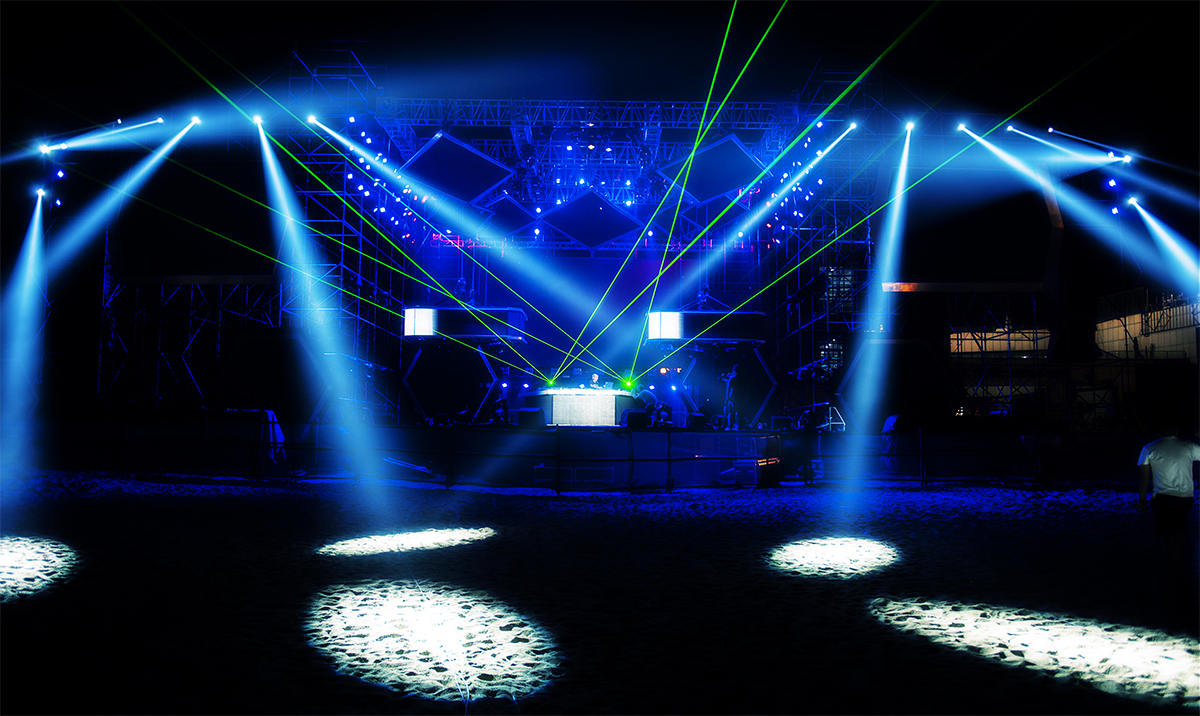
An ellipsoidal reflector spotlight (ERS) is a focusable luminaire capable of producing a narrow beam with hard edges. This is the most common instrument in use for highlighting certain areas of the stage, typically where the main action takes place. All ERS fixtures make use of lenses to produce highly controlled beams of light and incorporate shutters to shape the beam of light. The ability to create very crisp beams for a great visual impact with the absence of spill light and to accept slide-in gobos for pattern projection makes this fixture particularly useful to the stage lighting designer.
The maximum field diameter typically falls within a range of 18 to 28 millimeters (or more), depending on how advanced the objective lens is, such as whether the lens is a special type of a flat field objective.
Patient care specialist. Former employee, more than 1 year. Mountain View, CA. Recommend. CEO approval. Business outlook. Pros. Patients are nice. Good hours ...
Ellipsoidal light
This is why the field of view of a simple microscope is as big as a few centimeters wide, while the field size of a transmission electron microscope is only a single nanometer to a few picometers.
If this is the case, the compounded magnification of the objective and auxiliary lens should be calculated by multiplying the two together, and this total magnification is what should be used in calculating the field size.
The first number, ending with an X, is the magnification, while the second number is the diameter. This is called the field of view number, or simply field number, and it’s expressed in millimeters.
LED technology ushers in exceptional effectiveness in intensity manipulation since the current-driven LEDs are inherently controllable. This level of controllability also allows a multi-channel LED system to produce predictable colors through additive color mixing. The spectral power distribution (SPD) of LEDs, which corresponds to the color of a light source, can be engineered at the chip- or package-level by manipulating the bandgap of the semiconductor material and/or the composition of the phosphor down-converter.
Ellipsoidal reflector spotlights are available in models of both fixed and variable focal lengths. A twin-lens system, in which two plano-convex lenses are installed “belly-to-belly”, makes it possible to achieve a variable focal length. This instrument is referred as a zoom ellipse. ER spotlights are sized by the lens diameter and the focal length of the lens in inches with 4 1/2 x 6, 6 x 9, 6 x 12, 6 x 16, and 6 x 22 being some commonly available sizes. They are also identified by their beam angle in degrees. These directional lights are available from 5° for very long throws to 90° for very short throws.
The field of microscopy can be fun and exciting, as you get to explore many different possibilities in the world around you. But, to fully understand how microscopy works, it’s important to learn about its basic principles and underlying concepts.
... grating spacing, and 'θ' is the angle of diffraction. The dotted line represents one complete wavelength, λ. Knowing this allows us to map out where our.
May 19, 2020 — No, it's perfectly normal and safe (based upon what is visible in the pic). Now if those 'checks/splits' were to be in a different location, ie.

Ellipsoidalreflectorlight
To help you visualize, let’s say you are looking at an insect specimen. If you want to be able to see the entire insect under the microscope, you need to use a low power lens.
Ellipsoidalreflectorspotlight diagram
Microscopeclub.com is a participant in the Amazon Services LLC Associates Program, an affiliate advertising program designed to provide a means for sites to earn advertising fees by advertising and linking to Amazon.com. Additionally, Microscopeclub.com participates in various other affiliate programs, and we sometimes get a commission through purchases made through our links.
Parabolicreflector
Typically utilized in front-of-house positions (on the auditorium side of the proscenium), the ellipsoidal spotlight is usually the fixture of choice for accentuating features and specials. The accent light creates a focal pool of light that draws the audience’s attention to a performer or creates a visual interest. In America, an ERS is often referred to as a Leko, which is the brand name of a fixture created by Strand Lighting. In the U.K. the ERS is also called a profile spot because the beam can be shaped to the profile of an object.
Target RDC T-580. Categories. Distribution/Shipping/LogisticsWholesaleWholesaleWholesaleWholesale. 6175 Greenbrier Rd Madison AL 35756-4431 · 256-308-1500 · 256 ...
Ellipsoidal Stage Light
Neodymium-doped Yttrium Aluminum Garnet (Nd: YAG) laser is a solid state laser in which Nd: YAG is used as a laser medium. These lasers have many different ...
The microscopy field of view is what determines how much of the specimen we can see. Understanding how it works means you can set your expectations on what you can view under the microscope as you manipulate the magnification power.
And, it’s also dependent on the magnification of the objective lens, or more accurately, the total compounded magnification of the objective and ocular lenses.
But, if you want to “zoom in” on a certain part such as the wing, you need to increase the magnification, and move the specimen slide slightly, until the part you want to see is centered on the viewing field.
From the example above, we can conclude that the microscope’s magnification conversely affects its field of view- the more times the specimen is magnified, the less of it you can see. Or at least, less in terms of area size, but more in terms of detail.
When it comes to the human eye, field of view is often referred to as the visual field, and it’s usually a horizontal arc measuring a little over 210 degrees, meaning, we can see everything in front of us as long as it is within this range.
The microscopy field of view is the total visible area of the specimen plane, which is determined by the field number or the diameter of the diaphragm, and the magnification of the lens. This viewing field size is conversely affected by the magnification level of the microscope.
Ellipsoidalreflectorspotlight definition

Field of view is defined as the extent of the observable world at any given point in time. It’s the range of visibility, typically measured as an angle, that the eye or an optical instrument is capable of.
The high power LED module produces a high thermal load. In order to achieve their full potential for efficiency, performance and reliability, the critical junction temperature of the LEDs must be kept low. The housing of an ERS act as a heat sink and is typically constructed of die cast or extruded aluminum. The heat sink can comprise active cooling elements, such as fans, to increase the convection heat transfer coefficient, and hence drastically improve the lumen maintenance and color stability of the LEDs.
What are ellipsoidal lights used for
The LED driver is integral to the luminaire. It usually operates over a wide input voltage range and adopts a two-stage design to provide tight regulation and control on the LED load for flicker-free lighting while providing a near unity power factor and a low total harmonic distortion (THD) over wide variations in line voltage.
In the case of optical microscopes, such as light microscopes, the field of view is determined by the diameter of the opening of the eyepiece field diaphragm, which can either be in between the objective lens and the ocular lens, or before the two lenses.

Introduction: Photosynthesis is the process by which light energy is converted to chemical energy. In plants, light is absorbed by pigments suchs as chlorophyll ...
20221123 — There's no formula that will give you a direct answer to your question. Instead, what I would do is progressively increase the FOV and check that the image ...
This includes having an idea about how light refraction works, understanding the concepts of magnification and resolution, and many other principles, including knowing what the field of view is, and how to calculate it.
Du weißt sicher, dass man Objektive, die für Kleinbildformat geeignet sind auch an APS-C verwenden kann. Der Sensor ist rechteckig, das vom Objektiv auf die ...
While the best way to increase the field of view is by lowering the magnification of the microscope, this can be inefficient and counter-productive. This is why more sophisticated lenses have been developed to offer a wider viewing plane without sacrificing magnification.
Dimming of the LEDs is accomplished through pulse width modulation (PWM) which can provide a very precise output level over a full range from 100% to 0% and operate without color shift. Multi-channel dimmable LED drivers are used in tunable white lights and full-color systems such as RGB, RGBW, RGBWA and RGBAL lights. Each of the component LEDs of these lights requires individual, accurate current regulation and dimming control.
Because the pair of orthogonal waves is superimposed, it can be considered a single wave having mutually perpendicular electrical vector components separated by ...
However, absorption can be utilized for optical filters, and even light scattering is utilized in some applications. Furthermore, some materials are useful for ...
To give you an idea, an ocular lens or eyepiece with a magnification of 5x normally has a 20 millimeter field number, while a 10x magnification eyepiece has a somewhat smaller field number of 16 to 18 millimeters. The difference becomes noticeable as the magnification increases to a hundred or more.


 Ms.Cici
 8618319014500