
GB2 Fresnel Lens Magnifier - fresnel lens magnifying glass
Microscope objective lenses are a crucial part of a microscope, responsible for magnifying the specimen being observed. They are used to gather light from the object being observed and focus the light rays to produce a real image. The objective lens is one of the most important parts of a microscope, as it determines the microscope’s basic performance and function [3].
Dimensions: Display - 11.0" x 7.0" x 0.5"; Camera - 2.0" x 2.0"; Combined Wt. - 3.0lbs. Shipping dimensions: 13.5" x 9.5" x 4.5".
The included K2View software provides for excellent color rendering and fast image processing especially when capturing multiple images. Simple, intuitive and easy-to-use controls offer image capture, notation, geometric measuring and storage in JPG, BMP, TIFF, RAW format. The software also includes auto white balance, color enhancement, anti-flicker and other benefits. TWAIN and DirectX plug-ins are also included on the installation CD and are independent of the main driver, allowing discreet control of the camera for these applications.
The Summit K2 series camera bring affordable digital microscopy to a wide range of documentation demands of routine life science, industrial and educational environments. Built around the proven Aptina MT9T001 CMOS color sensor, they share a broad dynamic range, progressive scan and 12-bit parallel resolution for accurate color reproduction and optimal image quality.
In cases where the objective is not meant to be used in infinity corrected microscopes, there will be a number, usually 160) referring to the length of the microscope tube. Some microscope objectives will show the letters “DIN” which stands for “Deutsche Industrial Normen.” that sets a length of 160 mm.
TrinocularMicroscope price
In previous entries, we have talked about the design of scanning microscopes, infinity corrected microscopes, confocal microscope design, and Koehler illumination systems-a common illumination system in microscopes. The most essential microscope element in a borescope design is the objective lens.
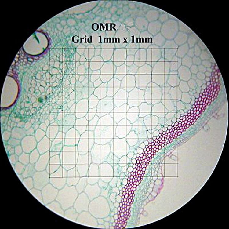
For accurate and convenient microscopic calibration and measurements we offer our 20mm linear reticle. Linear Scale: 100 divisions = 1mmGrid Scale: 10 divisions per side = 1mm, 100 cellsCross (Pinwheel) Scale: 0-6-0, 10 divisons = 1mm
Trinocularmicroscope drawing
All Summit K2 Series microscope cameras share a rugged aluminum alloy body fitted with different size Aptina CMOS sensors depending on the model. They stand out for remarkable color reproduction as well as a large color palette for improved resolution and sensitivity. In addition, the included 0.5x reducing lens makes for an image that is significantly closer to that seen through the eyepieces with the naked eye. Robust documentation and measuring software is included with all the OptixCam K2 cameras along with a c-mount adapter with 23.2mm smooth bore to fit most microscopes.
The simplest designs are usually called ‘achromat objectives’ and contain only a front lens and a couple of achromatic doublets to correct for aberrations. On the other hand, we have Apochromat microscope objectives in which several apochromatic doublets are used, in addition to some achromats for a better image quality. For a better explanation of the difference between achromatic and apochromatic lenses, please read the linked articles.
Where R is the resolution, ? is the light wavelength, n is the refractive index, and θ is the half angle of the acceptance light cone (NA is the numerical angle defined as sin(θ)). For example, a microscope objective that works with visible light, with air surrounding the sample, and an acceptance half-angle cone of 72-degrees, will have a minimum resolution of 256 nm. If we surround the sample in a liquid with a refractive index of 1.5, our resolution will improve to 171 nm.
TrinocularMicroscope with Camera
The new OptixCam Summit K2 Series 1.3MP digital microscope camera is a terrific way to quickly turn any modern microscope into a digital teaching tool. Designed for students and hobbyists, this sturdy and affordable camera will maximize a student's ability to discover, analyze and learn from their microscopy experiences. It is well-suited to both PC and Mac operating systems with software designed to get you up and running with both live and still imaging. The camera includes a C-mount adapter with 0.5X reducing lens and 23.2mm smooth bore for mounting on the eyetube or trinocular port of most microscopes. We highly recommend this camera for the hobbyist, coin-collector, educator and all general microscopy use.
Trinocularmicroscope vs binocular
With over 15 years of experience and 500+ unique optical systems designed, Optics for Hire specializes in advanced optical engineering. If it uses light, we've worked on it.
Other features include fast refresh rates, auto/ manual exposure and white balance control, full screen live imaging, time-lapse photography and user-friendly image capture. In short, these microscope cameras offer a well-designed package at an affordable price. We recommend them for any standard photomicrography application where the primary requirements are image capture and documentation.

The new OptixCam Summit K2 Series 10.0MP digital microscope camera is specifically designed as a cost-effective and versatile solution for the wide-ranging imaging requirements of life science researchers, healthcare professionals, industrial inspectors and other disciplines. 10.0MP resolution offers excellent details for images projected on large screens or when printing with large-format applications. PC and MAC users benefit from easy-to-use feature-rich software, accurate color, clarity and image detail, while low noise USB 2.0 data transfer maximizes workflow and productivity.
JavaScript seems to be disabled in your browser. For the best experience on our site, be sure to turn on Javascript in your browser.
TrinocularMicroscope function

With over 20 years of experience and 800+ unique optical systems designed, Optics for Hire specializes in advanced optical engineering. If it uses light, we've worked on it.
Most off the shelf microscope objectives have several body markings to better identify them. Typical markings can be seen in Figure 2.
For accurate and convenient microscopic calibration and measurements we offer our 20mm Grid reticle - Grid Scale: 100 divisions = 1mmWe also offer the following optional reticles:Linear Scale: 100 divisions = 1mmCross (Pinwheel) Scale: 0-6-0, 10 divisons = 1mm
TrinocularMicroscope
The brand new OptixCam Summit K2 Series cameras represent the latest innovation in digital microscopy, built to meet the documentation demands of routine life science, industrial and educational applications at very competitive price-points. Designed around the proven Aptina MT9T001 CMOS color sensor, they share a broad dynamic range, progressive scan and 12-bit parallel resolution for accurate color replication and optimal image quality.
Another specification can be “Plan Fluor” for fluorite and “APO” for apochromatic. Next we have the magnification, numerical aperture, and the immersion medium. As mentioned before, dry objective lenses usually have a NA no larger than 0.95, but that number can be considerably higher in immerse objectives. We next have an infinity symbol, meaning that the lens is infinity corrected.
The CX3 display finally brings a useful sized monitor to the world of industrial inspection, laboratories and schools. At 11.6", the display is a pleasure to use with good on screen controls and a remote control. A standard SD card slot allows you to conveniently transfer image files via SD card for later analysis and file-sharing on your PC. A particularly useful feature is a cross-hair generator that enables up to eight on-screen, cross-hairs in five different color options. Other features include auto white balance, RGB adjustment, auto and manual white balance and auto exposure.
Three high quality Plan Achromatic objective lenses include PL4x, PL10x, and PL60xS for six different levels of magnification - 40x, 64x, 100x, 160x, 600x, and 1,008x.
Trinocularmeaning
The camera has a standard C-Mount fitment, and a C-mount adapter (not included) will be necessary to mount the camera to a microscope.
The magnification of the objective lens can vary, depending on the intended use of the microscope. For example, objective lenses for biological applications typically range from 4x to 100x, while those used for metallurgical applications can range up to 200x or more [1].
There are three design variables that can help us calculate the microscope objective resolution: the system wavelength, the light cone captured by the objective (also known as numerical aperture), and the refractive index between the first lens of the objective and the sample. This can be expressed by the following formula:
The included K2View software creates a complete imaging solution with intuitive camera controls, image capture and advanced options like annotation, geometric measuring and storage in JPG, BMP, TIFF, PNG, PCX AND TARGA format. The software also includes auto white balance, color enhancement, anti-flicker and other benefits. TWAIN and DirectX plug-ins are also included on the installation CD and are independent of the main driver, allowing discreet control of the camera for these applications. PLEASE NOTE: MAC capability is limited to image capture only.
Two condensers are included: a 1.25 N.A brightfield with aperture and field diaphragm and included blue, yellow and frosted filters. The darkfield condenser (0.91) includes blue, green, yellow and metal filter disks. Illumination includes a halogen EPI-Illuminator with variable controls with brightfield illumination powered by 12V 20W in-base transmitted light. Both sources of illumination include variable controls.
The OM200 is a trinocular brightfield/darkfield metallurgical microscope that has proven its value over many years. It features four Plan Achromatic objective lenses and both brightfield and darkfield condensers for a good quality, affordable solution for metallurgical microscopy. There are two sets of wide field eyepieces (WF10x and WF16x) for maximum magnification of 1,008x. Universal 110v-220V voltage powers a halogen EPI-Illuminator in addition to integrated 12V 20W transmitted halogen. Available in trinocular version with included camera adapter. Lifetime Limited Warranty.
The microscope objective will show the manufacturer (not shown in the figure), followed by the type of aberration correction; in our image, we have a “Plan Achromat” which produces a flat surface at the image plane and achromat for the type of chromatic aberration.
The graduated mechanical stage is glass covered and measures a generous 90mm x 140mm with dual gradations under the glass and along the stage body. The range of movement is 30mm x 70mm. Rack and pinion focusing includes graduated coarse/fine focus controls on both sides with adjustment down to 0.002mm.
Trinocularcamera
The microscope head is inclined 45° for comfortable viewing and includes two sets of eyepieces, WF10x and WF16x, as standard equipment. Dual diopters enable convenient, individual eye adjustment while the interpupillary adjustment range is 55mm-75mm. The head rotates through 360 degrees for flexible viewing positions. A smooth 23mm trinocular adapter is included for ease of documentation with eyepiece cameras.
Objective lenses can have just a couple of lens elements, (an achromat and simple lens, for example) or multiple groups of elements. Even two microscope objectives with the same magnification can have a completely different design, as shown in Figure 1.
All Summit K2 microscope cameras share a rugged aluminum alloy body fitted with different size Aptina CMOS sensors depending on the model. Advanced documentation and measuring software is included along with a C-mount adapter with 23mm smooth bore and 0.5X reducing lens to more closely match the captured image with the eyepiece view. Optional focusing lenses, calibration slide and 30mm and 30.5mm collars are available.
Objective lenses for microscopes typically have several components, including the front lens, the rear lens, the aperture, the lens barrel, and the thread. Each component plays an important role in determining the objective’s performance. For example, the aperture determines the resolution and depth of field of the objective lens, while the thread allows the objective to be attached to the microscope.
In conclusion, microscope objective lenses are an essential part of a microscope and are used to magnify the specimen being observed. They consist of several components that work together to produce a clear image, and their magnification can vary depending on the intended use of the microscope.
An upgrade to the old CX-3 microscope camera, the new CX-3-116 sports a larger 11.6" monitor with improved connections and use interface. It is better quality than the old HDMI 1080 in every respect with double the functional resolution, an expansive, 11.6"-inch display and an improved on screen menu system, all at the same price. The TFT-LCD display offers vivid resolution and exceptional off-angle viewing capability and with a standard C-mount, the new camera attaches to any standard trinocular microscope fitted with a C-mount adapter.
In the previous calculation, I assumed an angle of acceptance of 72-degrees with a reasonable upper limit when working with air (that angle gives us a NA of 0.95). However, by immersing the sample and microscope in oil or another liquid, it is possible to have a larger NA. This affects not only the resolution of our image but also its brightness (the brightness is calculated as the square of its NA).


 Ms.Cici
 8618319014500