
FLIR Chameleon3 USB3 CM3-U3-31S4C-CS - flir cs
Bennett, B. D., T. L. Jetton, G. Ying, M. A. Magnuson, and D. W. Piston. Quantitative sub-cellular imaging of glucose metabolism within intact pancreatic islets. J. Biol. Chem. 271: 3647–3651, 1996.
Mainen, Z. E, R. Malinow, and K. Svoboda. Synaptic calcium transients in single spines indicate that NMDA receptors are not saturated. Nature 399: 151–155, 1999.
Hi Tim, I think that is “stick remover” and yes it possible to buy here in Portugal. Which by the way is a good country to born if you want to talk Portuguese.
UVlaserpointer
Svoboda, K., F. Helmchen, W. Denk, and D. W. Tank. Spread of dendritic excitation in layer 2/3 pyramidal neurons in rat barrel cortex in vivo. Nat. Neurosci. 2: 65–73, 1999.
Bhawalkar, J. D., N. D. Kumar, C. F. Zhao, and P. N. Prasad. Two-photon photodynamic therapy. J. Clin. Laser Med. Surg. 15: 201–204, 1997.
Helmchen, F., K. Svoboda, W. Denk, and D. W. Tank. In vivo dendritic calcium dynamics in deep-layer cortical pyramidal neurons. Nat. Neurosci. 2:989–996, 1999.
Piston, D. W., B. R. Masters, and W. W. Webb. Three-dimensionally resolved NAD(P)H cellular metabolic redox imaging of the in situ cornea with two-photon excitation laser scanning microscopy. J. Microsc. 178: 20–27, 1995.
Masters, B. R., P. T. C. So, and E. Gratton. Optical biopsy of in vivo human skin: Multi-photon excitation microscopy. Laser Med. Sci. 13: 196–203, 1998.
Bodaness, R. S., D. R Heller, J. Krasinski, and D. S. King. The two-photon laser-induced fluorescence of the tumor-localizing photosensitizer hematoporphyrin derivative. Resonance-enhanced 750 nm two-photon excitation into the near-UV Soret band. J. Biol. Chem. 261: 12098–12101, 1986.
Hello Tim, thank you for your reply. That’s an option I’ve been thinking about. My concern is if it left any residues of tape on acrylic. That´s not suitable for what I intend to do.
Up to 2/3", C-Mount Lens; Up to 7.5 MegaPixels, 2.8µm Pixel Size Sensors; Compact (C), FA Lens for Machine Vision; 3.5mm to 100mm Focal Length ...
I found a recent thread mentioning that you can order it from Amazon in Portugal. https://www.reddit.com/r/MonsterHigh/comments/12zrvcu/anyone_know_of_an_alternate_to_goo_gone_available/
Kierdaszuk, B., H. Malak, I. Gryczynski, R. Callis, and J. R. Lakowicz. Fluorescence of reduced nicotinamides using one-and two-photon excitation. Biophys. Chem. 62: 1–13, 1996.
Shear, J.B., C. Xu, and W. W. Webb. Multiphoton-excited visible emission by serotonin solutions. Photochem. Photobiol. 65: 931–936, 1997.
Masters, B. R., and B. Chance. Redox confocal imaging: Intrinsic fluorescent probes of cellular metabolism. In: Fluorescent and Luminescent Probes for Biological Activity, edited by W. T. Mason. London: Academic Press, 1999, pp. 361–374.
Gu, M., and C. J. R. Sheppard. 1995. Comparison of three-dimensional imaging properties between two-photon and single-photon fluorescence microscopy. J. Microsc. 177: 128–137, 1995.
Mohler, W. A., and J. G. White. Stereo-4-D reconstruction and animation from living fluorescent specimens. Biotechniques 24: 1006–1010, 1012, 1998.
Denk, W. Two-photon scanning photochemical microscopy: Mapping ligand-gated ion channel distributions. Proc. Natl. Acad. Sci. U.S.A. 91: 6629–6633, 1994.
SM, S. H., Y. Hayashi, R. S. Petralia, S. H. Zaman, R. J. Wenthold, K. Svoboda, and R. Malinow. Rapid spine delivery and redistribution of AMPA receptors after synaptic NMDA receptor activation. Science 284: 1811–1816, 1999.
Centonze, V. E., and J. G. White. Multiphoton excitation provides optical sections from deeper within scattering specimens than confocal imaging. Biophys. J. 75: 2015–2024, 1998.
Fisher, W. G., W. P. Partridge, Jr., C. Dees, and E. A. Wachter. Simultaneous two-photon activation of type-I photodynamic therapy agents. Photochem. Photobiol. 66: 141–155, 1997.
Ultraviolet light laserprice
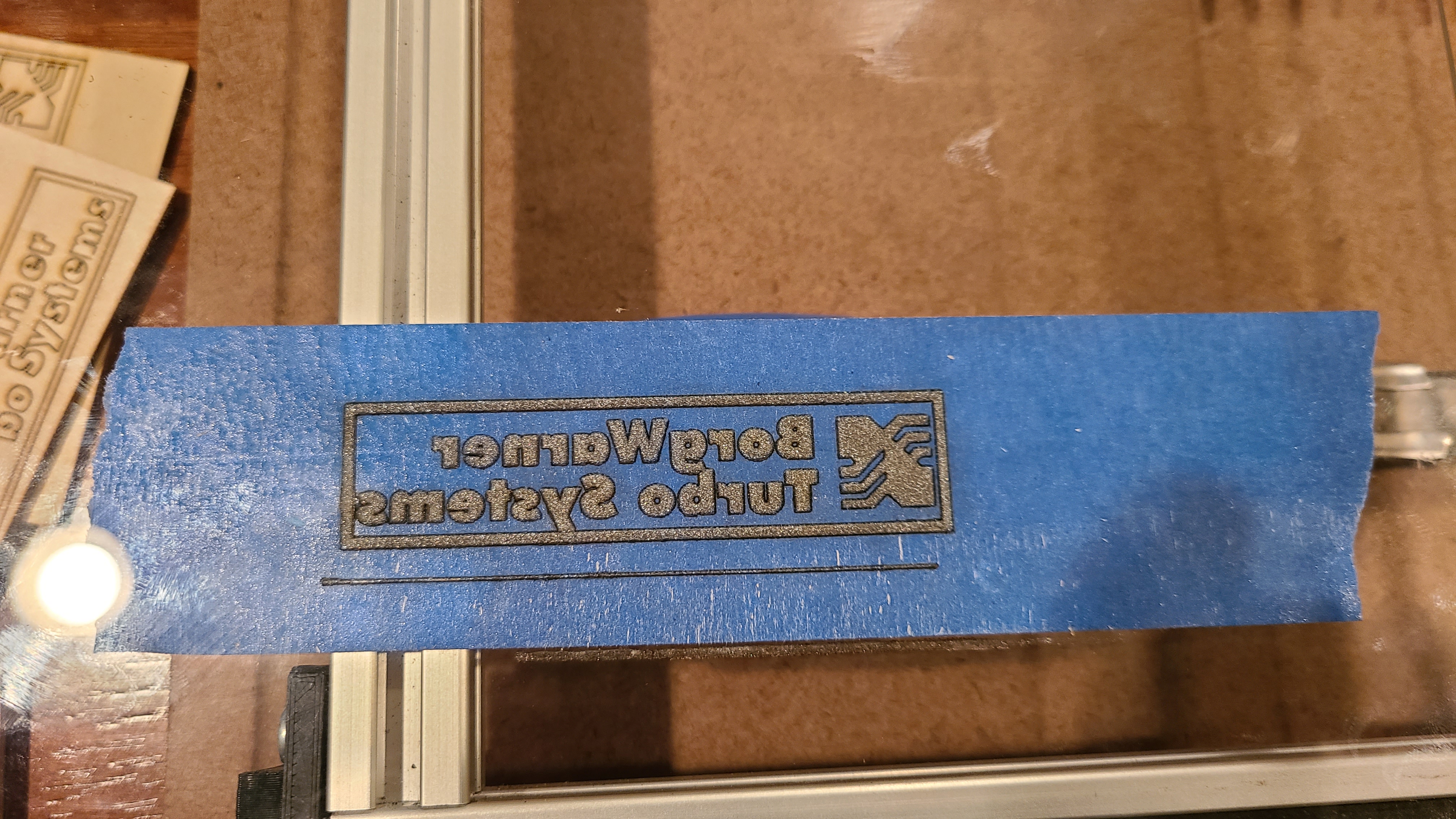
EDIT: Just tried with blue painters tape on an old CD case. I know I have some real acrylic around, but I just moved and cannot find it.
Pettit, D. L., S. S. Wang, K. R. Gee, and G. J. Augustine. Chemical two-photon uncaging: A novel approach to mapping glutamate receptors. Neuron 19: 465–471, 1997.
Squirrell, J. M., D. L. Wokosin, J. G. White, and B. D. Bavister. Long-term two-photon fluorescence imaging of mammalian embryos without compromising viability. Nat. Biotechnol. 17: 763–767, 1999.
Lakowicz, J. R., B. Kierdaszuk, R. Callis, H. Malak, and I. Gryczynski. 1995. Fluorescence anisotropy of tyrosine using one-and two-photon excitation. Biophys. Chem. 56: 263–271, 1995.
If it’s not in the visible range you can’t see it… that’s a lot of bandwidth of frequencies that can be used… There are a few…
Furuta, T., S. S. Wang, J. L. Dantzker, T. M. Dore, W. J. Bybee, E. M. Callaway, W. Denk, and R. Y. Tsien. Brominated 7-hydroxycoumarin-4-ylmethyls: Photolabile protecting groups with biologically useful cross-sections for two photon photolysis. Proc. Natl. Acad. Sci. U.S.A. 96: 1193–1200, 1999.
Ultraviolet light lasernear me
Denk, W., K. R. Delaney, A. Gelperin, D. Kleinfeld, B. W. Strowbridge, D. W. Tank, and R. Yuste. Anatomical and functional imaging of neurons using 2-photon laser scanning microscopy. J. Neurosci. Methods 54: 151–162, 1994.
I understand they work for acrylic but I don’t have one of those, mine are led, co2 and fiber… they have a different to no effect, depending on the material.
Piston, D. W., S. M. Knobel, C. Postic, K. D. Shelton, and M. A. Magnuson. Adenovirusmediated knockout of a conditional glucokinase gene in isolated pancreatic islets reveals an essential role for proximal metabolic coupling events in glucose-stimulated insulin secretion. J. Biol. Chem. 274: 1000–1004, 1999.
So, P. T. C., H. Kim, and I. E. Kochevar. Two-photon deep tissue ex vivo imaging of mouse dermal and subcutaneous structures. Opt. Exp. 3: 339–350, 1998.
Made from premium multi-coated glass, the SANDMARC Telephoto 60mm lens gives you 2x optical magnification. The Telephoto lens is ideal for documenting the ...
Jones, K. T., C. Soeller, and M. B. Cannell. The passage of Cat+ and fluorescent markers between the sperm and egg after fusion in the mouse. Development 125: 4627–4635, 1998.
Albota, M. A., C. Xu, and W. W. Webb. Two-photon fluorescence excitation cross sections of biomolecular probes from 690 to 960 nm. Appl. Opt. 37: 7352–7356, 1998b.
Tanks Jack. For now I am clear about the doubt that originated this topic. Of course now I’m more curious about UV lasers but that will be a subject for debate later.
by C He · 2019 · Cited by 121 — Graded index (GRIN) lenses are commonly used for compact imaging systems. It is not widely appreciated that the ion-exchange process that ...
Farrer, R. A., M. J. R. Previte, C. E. Olson, L. A. Peyser, J. T. Fourkas, and P. T. C. So. Single-molecule detection with a two-photon fluorescence microscope with fast-scanning capabilities and polarization sensitivity. Opt. Lett. 24: 1832–1834, 1999.
20230508_2225311920×1080 177 KB 20230508_2228251920×1080 219 KB 20230508_2232501920×1080 257 KB 20230508_2234511920×1080 217 KB
Bestultraviolet light laser
Lakowicz, J. R., and I. Gryczynski. Tryptophan fluorescence intensity and anisotropy decays of human serum albumin resulting from one-photon and two-photon excitation. Biophys. Chem. 45: 1–6, 1992.
What are UV lasers used for
If the laser is in the visible spectrum of frequencies then anything that is clear will not be able to damage (cut or engrave) that material…
I was wondering: my laser has 455 nm wavelength too. It is beyond UV, of course, in the blue range: that’s the visible color when it works. Isn’t blue tape (or anything blue) inefficient? My understanding of physics (which might be wrong) is that blue material is blue because it strongly reflects this color. Isn’t something red or orange, or just plain black, more efficient for such masking?
Photonic integrated circuit products on the market now · Applied Nanotools is an integrated photonics foundry that specialises in rapid turnaround prototyping ...
Multiphoton microscopy is one of the fastest growing areas in biomedical imaging. The potential of multiphoton excitation was first theorized by Maria GöppertMayer in 1931. Generating three-dimensionally resolved microscopic images based on nonlinear optical excitation was postulated in the 1970s (Gannaway and Sheppard, 1978; Wilson and Sheppard, 1984). The definitive experiment was done by Denk, Webb, and co-workers (1990), who accomplished two-photon, three-dimensional (3D) imaging of biological specimens. Furthermore, they demonstrated 3D localized uncaging and photobleaching using two-photon excitation to trigger a photochemical reaction in a subfemtoliter volume.
I’m working with Lightburn and a diode laser GRBL generic machine. I’ve been experimenting a lot with different materials (as ideas come up). Right now I’m trying to engrave clear acrylic and from what I’ve read UV technology would be the most suitable for what I want. Knowing some of the risks of diode laser machines due to the emission of UV rays, I had a doubt whether a diode laser is the same thing as a UV laser.
UVlaserwavelength
Summers, R. G., D. W. Piston, K. M. Harris, and J. B. Morrill. The orientation of first cleavage in the sea urchin embryo, Lytechinus variegatus, does not specify the axes of bilateral symmetry. Dev. Biol. 175: 177–183, 1996.
Third test, as i do not have any solvent at my disposal I try to invert the acrylic sample and and run a material test. Until here all tests were made with painted surface face up, this test was made with painted surface face down. I run a test like this because I intend to evaluate the reflection effect on acrylic. The result was interesting,… cracks everywhere, down side (painted side) melted on the most power and lower speeds hits, and curios is at 100% power between 2000 and 1500mm/min it burns face up side. Testing acrilyc011040×780 182 KB Testing acrilyc021920×1440 150 KB
Here at Newnex, we focus on creating top-quality USB cables and have crafted this detailed guide to help you navigate through the complex world of USB cable ...
Hell, S. W., M. Booth, and S. Wilms. Two-photon near-and far-field fluorescence microscopy with continuous-wave excitation. Opt. Lett. 23: 1238–1240, 1998.
Svoboda, K., W. Denk, D. Kleinfeld, and D. W. Tank. In vivo dendritic calcium dynamics in neocortical pyramidal neurons. Nature 385:161–165, 1997.
First test i try to remove the paint with thinner. (I am convinced that I already clean acrylic with thinner but,… maybe not. )
Chalfie, M., Y. Tu, G. Euskirchen, W. W. Ward, and D. C. Prasher. Green fluorescent protein as a marker for gene expression. Science 263: 802–805, 1994.
Mohler, W. A., J. S. Simske, E. M. Williams-Masson, J. D. Hardin, and J. G. White. Dynamics and ultrastructure of developmental cell fusions in the Caenorhabditis elegans hypodermis. Curr. Biol. 8: 1087–1090, 1998.
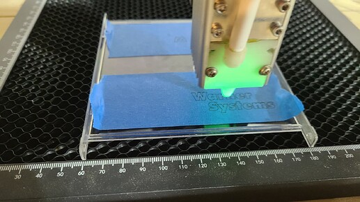
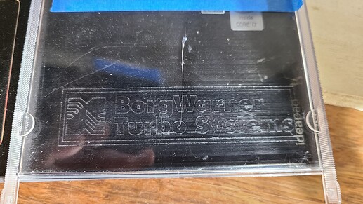
Here’s some more glass lasering. I mirrored the cut and put the tape on the bottom of the glass. I figured that way it’s burning the tape closest to the glass first, instead of burning thorough the tape. 200mm/min, 100% power, “Fill” in Lightburn...
Ammasi Periasamy Ph.D. (Director, Professor of Biology and Biomedical Engineering) (Director, Professor of Biology and Biomedical Engineering)
Hello everyone! By the topic you can guess how noob I am. I am portuguese and my english is far from perfect, so sorry in advance. (Any misunderstood is google translator fault ) So, i’ve tried to clarify my doubt on the internet but I haven’t found anything that clarifies me properly. I resort to your help hoping this is not already a debated subject (at least I didn’t find anything enlightening on the forum)
Oct 24, 2014 — Magnifying power is defined as the ratio between the dimensions of the image and the object. The process of magnification can occur in ...
If I understood correctly, then from what you mention and the image below, only diodes that work at a frequency below 400nm are considered UV, right? Wavelength723×377 25 KB
Gauderon, R., R. B. Lukins, and C. J. R. Sheppard. Effects of a confocal pinhole in two-photon microscopy. Microsc. Res. Tech. 47: 210–214, 1999.
So, P.T.C., Kim, K.H., Buehler, C., Masters, B.R., Hsu, L., Dong, CY. (2001). Basic Principles of Multiphoton Excitation Microscopy. In: Periasamy, A. (eds) Methods in Cellular Imaging. Methods in Physiology. Springer, New York, NY. https://doi.org/10.1007/978-1-4614-7513-2_9
Dec 27, 2020 — View> Toggle Axis cross. I think it is on after that until you toggle it off. And in Edit>Preferences>Display 3D View tab there is a checkbox ...
It does leave some residue on the plastic, but was easily removed with a product called “Goo Gone”. I’m sure it is sold under different names, also called citrus cleaner, or something like that. The cleaner does not harm the plastic at all.
Ultraviolet light laseramazon
Gannaway, J. N., and C. J. R. Sheppard. Second harmonic imaging in the scanning optical microscope. Opt. Quantum Electronics 10: 435–439, 1978.
Ultraviolet light laserfor sale
Hi Jack, thanks for the quick response. If I understand correctly, summarizing the technology (if that’s what it can be called) a generic laser diode (in my case 40W) is not considered UV because, quite simply speaking, you can see the beam of light and from what I saw in a video of a UV laser the ash is invisible, only the engraving is visible.
Kim, K. H., R. T. C. So, I. E. Kochevar, B. R. Masters, and E. Gratton. Two-photon fluroescence and confocal reflected light imaging of thick tissue structures. SPIE Proc. 3260: 46–57, 1998.
You are correct. If we were sipping amps in some mission-critical application, or using a much lower power laser, efficiency might be a concern. At this level, unless its white tape, I don’t think it’s factor worth worrying about.
Denk, W., M. Sugimori, and R. Llinas. Two types of calcium response limited to single spines in cerebellar Purkinje cells. Proc. Natl. Acad. Sci. U.S.A. 92: 8279–8282, 1995.
Electromagnetic waves are polarized if their electric field vectors are all in a single plane (linear polarization), or in 2 plans perpendicular to each ...
Maletic-Savatic, M., R. Malinow, and K. Svoboda. Rapid dendritic morphogenesis in CAl hippocampal dendrites induced by synaptic activity. Science 283: 1923–1927, 1999.
What you can do is mask the clear acrylic with something like tape or paint. My guess is that when the laser burns the mask, it will melt the acrylic underneath and make it seem engraved. I have a 450nm 7watt diode laser and have done this with glass achiving pretty good results.
Kleinfeld, D., R. R. Mitra, F. Helmchen, and W. Denk. Fluctuations and stimulus-induced changes in blood flow observed in individual capillaries in layers 2 through 4 of rat neocortex. Proc. Natl. Acad. Sci. U.S.A. 95: 15741–15746, 1998.
Engert, F., and T. Bonhoeffer. Dendritic spine changes associated with hippocampal longterm synaptic plasticity. Nature 399: 66–70, 1999.
Anything that damages material will produce some type of debris… the laser doesn’t destroy things out of existence. Could be gas if it vaporizes the material or any size of debris depending on how the laser damages the material…
Xu, C., J. Guild, W. W. Webb, and W. Denk. Determination of absolute two-photon excitation cross sections by in situ second-order autocorrelation. Opt. Lett. 20: 2372–2374, 1995.
Wokosin, D. L., V. E. Centonze, J. White, D. Armstrong, G. Robertson, and A. I. Ferguson. All-solid-state ultrafast lasers facilitate multiphoton excitation fluorescence imaging. IEEE J. Selected Top. Quantum Electronics 2: 1051–1065, 1996.
NIT designs and manufactures SWIR InGaAs sensors & cameras. About us ; Full HD & High Sensitivity SWIR camera SenS 1920 Ideal for ISR and semiconductor ...
Masters, B. R., P. T. So, and E. Gratton. Multiphoton excitation fluorescence microscopy and spectroscopy of in vivo human skin. Biophys. J. 72: 2405–2412, 1997.
Potter, S. M., C. M. Wang, R. A. Garrity, and S. E. Fraser. Intravital imaging of green fluorescent protein using two-photon laser-scanning microscopy. Gene 173: 25–31, 1996.
Straub, M., and S. W. Hell. Multifocal multiphoton microscopy: A fast and efficient tool for 3-D fluorescence imaging. Bioimaging 6: 177–184, 1998.
XYZ Linear Translation Stage, MPositioning T60XYZ-25L10A Precision 3 Axis Positioning System 25 mm Travel in XY and 10 mm Travel in Z: Amazon.com: ...
Buehler, C., K. H. Kim, C. Y. Dong, B. R. Masters, and P. T. C. So. Innovations in two-photon deep tissue microscopy. IEEE Eng. Med. Biol. Mag. 18: 23–30, 1999.
Yuste, R., A. Majewska, S. S. Cash, and W. Denk. Mechanisms of calcium influx into hippocampal spines: Heterogeneity among spines, coincidence detection by NMDA receptors, and optical quantal analysis. J. Neurosci. 19: 1976–1987, 1999.
Albota, M., D. Beljonne, J.-L. Bredas, J. E. Ehrlich, J.-Y. Fu, A. A. Heikal, S. E. Hess, T. Kogej, M. D. Levin, S. R. Marder, D. McCord-Maughon, J. W. Perry, H. Rockel, M. Rumi, G. Subramaniam, W. Webb, X.-L. Wu, and C. Xu. Design of organic molecules with large two-photon absorption cross sections. Science 281: 1653–1656, 1998a.
Niswender, K. D., S. M. Blackman, L. Rohde, M. A. Magnuson, and D. W. Piston. Quantitative imaging of green fluorescent protein in cultured cells: Comparison of microscopic techniques, use in fusion proteins and detection limits. J. Microsc. 180: 109–116, 1995.
Meanwhile,… Some material test i have made with acrylic. Power from 10 to 100%, speed from 500 to 2000mm/min, 40W diode laser 450nm and air assist. Spayed with black acrylic paint on one side.
Callis, P. R. The theory of two-photon induced fluorescence anisotropy. In: Nonlinear and Two-Photon-Induced Fluorescence, edited by J. Lakowicz. New York: Plenum Press, 1997, pp. 1–42.


 Ms.Cici
 8618319014500