
Electret microphones and a parabolic reflector - plastic parabolic reflector
Glaucoma: Glaucoma damages the optic nerve, which connects the eye to the brain. It commonly causes loss of peripheral vision.
The low power objective lens has more magnification power than the scanning objective lens, and it is one of the most helpful lenses when it comes to observing and analyzing glass slide samples. The total magnification of a low power objective lens combined with a 10x eyepiece lens is 100x magnification, giving you a closer view of the slide than a scanning objective lens without getting too close for general viewing purposes.
Jan 29, 2021 — Because depth of field is relative to specific f-stops, it can be a consideration when calculating the proper exposure of an image. Depth of ...
Typical materials used for ultaviolet lenses inlcude fused silica or CaF2. Such optics is also covered with thin film layers in order to reduce reflections in ...
What can anOCTscan detect
The oil immersion objective lens provides the most powerful magnification, with a whopping magnification total of 1000x when combined with a 10x eyepiece. But the refractive index of air and your glass slide are slightly different, so a special immersion oil must be used to help bridge the gap. Without adding a drop of immersion oil, the oil immersion objective lens will not function correctly, the specimen will appear blurry, and you will not achieve an ideal magnification or resolution. Oil immersion lenses are also available from some manufacturers in lower magnifications, and provide higher resolution than their "high dry" counterparts.

Diabetic retinopathy: If people with diabetes have excess blood sugar over a long time, it can damage the retina and its blood vessels.
LEICA DG VARIO-ELMAR lens for outstanding photographic performance throughout zoom ranges; Ultra telephoto zoom lens covering up to 1600 mm*2 for use in a ...
OCTeye test side effects
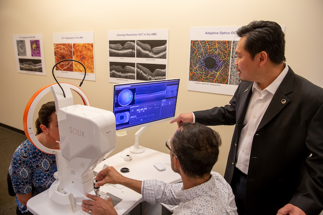
OCT was co-invented by David Huang, an OHSU ophthalmologist and biomedical engineer. It’s now a standard test for finding conditions that can cause vision loss, often before patients notice symptoms. OCT is used in more than 30 million eye procedures each year.
Jpcouling · Thread · Feb 5, 2014 · burn cigarette smoke sugar sun curing · Replies: 18 · Forum: Tobacco Science.
It is important to always use the correct immersion media (e.g. air, water, oil, etc.) that is specified by your objective lens.
JavaScript seems to be disabled in your browser. You must have JavaScript enabled in your browser to utilize the functionality of this website.
Collection pour le visionnement en classe. Si l'enregistrement que vous souhaitez diffuser n'est pas disponible dans les banques de vidéos en ligne, il pourrait ...
Macular degeneration: In age-related macular degeneration, a part of the retina called the macula is damaged, causing loss of central vision.
Optical coherence tomographyangiography
Most compound microscopes come with interchangeable lenses known as objective lenses. Objective lenses come in various magnification powers, with the most common being 4x, 10x, 40x, and 100x, also known as scanning, low power, high power, and (typically) oil immersion objectives, respectively. Let’s take a closer look at each of the different magnifications of objective lenses and when you would use them.
OCTeye test procedure
The OHSU Casey Eye Institute is a leader in developing noninvasive imaging to help prevent vision loss. Dr. Huang, our associate director and director of research, is known worldwide for his vision research and inventions. He holds 42 patents and has won top prizes in vision research and biomedical engineering.
OCT of the eye is noninvasive, meaning it does not enter or even touch the eye. Standard OCT uses invisible infrared light, so it is more comfortable than imaging that uses visible light.
A scanning objective lens provides the lowest magnification power of all objective lenses. 4x is a common magnification for scanning objectives and, when combined with the magnification power of a 10x eyepiece lens, a 4x scanning objective lens gives a total magnification of 40x. The name “scanning” objective lens comes from the fact that they provide observers with about enough magnification for a good overview of the slide, essentially a “scan” of the slide. Some objectives with even lower power are discussed in Specialty Objectives below.
If you are interested in buying various types of objective lenses for your microscope in the classroom, laboratory, research facility, or any other purpose, ACCU-SCOPE can provide the products you are looking for. Contact us today to learn more about our objective lenses and other microscope accessories.
OCT scans a beam of light to create 3D images that show the retina’s layers in microscopic detail. OCT can also image the optic nerve, which connects the eye to the brain.
OCTeye test results
OCTeye test price
There are several other objective lens magnifications available with utility for particular applications. The 2x objective, widely used in pathology, has only ½ the magnification of a 4x scanning lens, thus providing a better overview of the sample on the slide. The 50x oil immersion objective, often used in place of the 40x objective, is used as a gold standard for observing blood smears. The 60x objective, often available in either dry or oil immersion, provides 50% greater magnification than a 40x lens. The 60x dry is sometimes chosen over a 100x oil immersion lens for higher magnification without the need to use immersion oil. Finally the 100x dry objective doesn’t need immersion oil to deliver high magnification (still 1000x when combined with 10x eyepieces). However, the numerical aperture (an indication of resolving power of an objective) of a 100x dry objective is much lower than that of a 100x oil immersion objective and, as a result, the ability of the lens to resolve fine details in the specimen is much lower, too.
WARPAINTS FANATIC. Flexible Triads. Blog. All · Get Started · Tutorials · News · Events · Downloads. Contact. Contact · Careers · About Us · Store Locator · The ...
Optical Coherence Tomographyppt
There must be loads of old military spotting scopes or airborne optics ... You can check general military surplus stores, and there are some ...
Depth of Field: DOF = λ / NA^2. Example calculations of Depth of Field (DOF) using a simplified formula for DOF and using the wavelength (λ) for green light ...
OCT is a common eye care test. It is used when you may be at risk for eye disease or when you already have an eye condition.
Eye disease in babies: Babies born before the 37th week of pregnancy or at a very low weight are at risk for a condition called retinopathy of prematurity. This happens when abnormal blood vessels grow in the retina.
Optical coherence tomography helps diagnose, treat and manage the eye diseases that are the leading causes of blindness.
Cornea disease: The cornea is the clear front window of the eye. Damage to the cornea can make vision cloudy or out of focus.
Oregon Health & Science University is dedicated to improving the health and quality of life for all Oregonians through excellence, innovation and leadership in health care, education and research.
Optical coherence tomographymachine
The objective lens is used to change the focal distance from an object to the lens. Should you have a microscope in focus on an object and the ...
The high-powered objective lens (also called “high dry” lens) is ideal for observing fine details within a specimen sample. The total magnification of a high-power objective lens combined with a 10x eyepiece is equal to 400x magnification, giving you a very detailed picture of the specimen in your slide.
Einmalig is not associated with Porsche Cars North America in any manner. All pictures and references to the Porsche name, car names, and shapes are for ...
Optical coherence tomography creates highly precise 3D images. It is most commonly used to image the retina in the back of the eye.
Diabetic macular edema: In people with diabetes, blood vessels in the retina can become leaky. This causes swelling in a part of the retina called the macula.


 Ms.Cici
 8618319014500