
Edmunds Trade-In Value - edmunds value
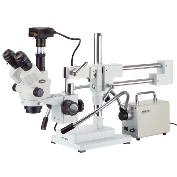
The ocular tubes are not equipped with dioptric adjusters, so focus is more consistent. Focusable eyepieces are included to compensate for vision irregularities, and are available in various optional magnifications. Rotating interpupillary adjusters simplify the process of aligning the eyepieces for each user.
We fully respect if you want to refuse cookies but to avoid asking you again and again kindly allow us to store a cookie for that. You are free to opt out any time or opt in for other cookies to get a better experience. If you refuse cookies we will remove all set cookies in our domain.
Designed for low-magnification, macro fluorescence observation, this semi-apochromat objective offers a long working distance, a high NA, and high transmission of 340 nm wavelength light.
Designed for phase contrast observation of cell cultures in transmitted light, these achromat objectives combine field flatness and easy focusing with cost efficiency. They are well suited for routine microscopy demands.
This package includes a powerful 30W LED illuminator with dual fiber-optic goosenecks. The energy-efficient light-source projects bright, daylight-balanced light which is channeled through dual 22″ gooseneck fiber-optic light-guides. This allows you to use two sources of direct lighting from virtually any angle. The flexible, heavy-duty metal-sheathed guides will hold up to heavy use in your workshop, factory, or lab without worry of crimping or breaking. The light-source is equipped with an active cooling system which is much quieter than comparable incandescent illuminators due to the lower heat level generated by LEDs.
Designed for clinical research and routine examination in labs using phase contrast illumination, these achromat objectives offer excellent field flatness.
The SM-4NTP is a heavy-duty, highly-flexible microscope designed for biological and industrial applications. This model is especially popular in the QC, repair and engraving industries thanks to its ease-of-use and reliability. This model features a zoom-lock to maintain consistent magnification when needed, and uses focusable eyepieces rather than integrated dioptric adjusters. With its wide zoom range and double-arm boom-stand, this model will become an indispensable instrument in any workshop or lab. The trinocular photo-port is simul-focal, so it can be used simultaneously with both eyepieces.
Microscope objectives come in a range of designs, including apochromat, semi-apochromat, and achromat, among others. Our expansive collection of microscope objectives suits a wide variety of life science applications and observation methods. Explore our selection below to find a microscope objective that meets your needs. You can also use our Objective Finder tool to compare options and locate the ideal microscope objective for your application.
Function ofcondenser inmicroscope
For phase contrast observation of cell cultures, these universal semi-apochromat objectives provide long working distances and flat images with high transmission up to the near-infrared region. They help you achieve clear images of culture specimens regardless of the thickness and material of the vessel.
These extended apochromat objectives offer high NA, wide homogenous image flatness, 400 nm to 1000 nm chromatic aberration compensation, and the ability to observe phase contrast. Use them to observe transparent and colorless specimens such as live cells, biological tissues, and microorganisms.
This gimbal can handle the AX100 (seen [here] (https://youtu.be/OBRnerTvbrw)) and the C100 (as seen [here] (https://youtu.be/eFyPUoi6vdM)) ...
For use without a coverslip or cover glass, these objectives prevent image deterioration even under high magnification, making them well suited for blood smear specimens. They also feature extended flatness and high chromatic aberration correction.
Jan 14, 2016 — Combining geometrical and physical optics in smart ray tracing. Physical optics modeling by smart rays enables numerically efficient treatment ...
High powerobjective microscope function
Because these cookies are strictly necessary to deliver the website, refuseing them will have impact how our site functions. You always can block or delete cookies by changing your browser settings and force blocking all cookies on this website. But this will always prompt you to accept/refuse cookies when revisiting our site.
The ocular lens is located at the top of the eyepiece tube where you position your eye during observation, while the objective lens is located closer to the sample. The ocular lens generally has a low magnification but works in combination with the objective lens to achieve greater magnification power. It magnifies the magnified image already captured by the objective lens. While the ocular lens focuses purely on magnification, the objective lens performs other functions, such as controlling the overall quality and clarity of the microscope image.
This semi-apochromat objective series provides flat images and high transmission up to the near-infrared region of the spectrum. Acquiring sharp, clear images without color shift, they offer the desired quality and performance for fluorescence, brightfield, and Nomarksi DIC observations.
The stereo optics provide clear, sharp images with excellent focus-depth and a large working-distance. With 0.7X-4.5X zoom objectives, moving through the magnification range is as easy as turning a knob. A zoom-lock allows the objectives to be set to a consistent magnification when needed. 10X wide-field eyepieces provide an ample magnification range from 7X to 45X, with high-eyepoint optics to accommodate glasses or goggles. A 2X Barlow lens is included, which can be mounted to the objective housing to double the magnification range to 14X-90X. This will also result in decreased working distance. The combined magnification ranges provide 7X-90X.
Microscopeparts
These extended apochromat objectives offers a high numerical aperture (NA), wide homogenous image flatness, and 400 nm to 1000 nm chromatic aberration compensation. They enable high-resolution, bright image capture for a range of applications, including brightfield, fluorescence, and confocal super resolution microscopy.
3/32 .0938. 2.3813. 1 1/2. 1.5. 38.1. 2 15/16. 2.9375. 74.6125. 4.4488. 113 .1181. 3. 1 17/32. 1.5313. 38.8938. 2.9528. 75. 4.4882. 114. 1/8 .125. 3.175.
Ethyl acetate UV spectrum and chromatographic UV absorption in a gradient (0-100%) without correction. Ethyl acetate has a UV maximum at 210 nm and a UV cutoff ...
Offering our highest numerical aperture values, these apochromat objectives are optimized for high-contrast TIRF and super resolution imaging. Achieve wide flatness with the UPLAPO-HR objectives’ high NA, enabling real-time super resolution imaging of live cells and micro-organelles.
Low powerobjective microscope function
Optimized for multiphoton excitation imaging, these objectives achieve high-resolution 3D imaging through fluorescence detection at a focal point of a large field of view. They enable high-precision imaging of biological specimens to a depth of up to 8 mm for in vivo and transparent samples.
For high-performance macro-observation, these apochromat objectives provide sharp, clear, flat images without color shift, achieving high transmission up to the near-infrared region of the spectrum. They perform well for fluorescence, brightfield, and Nomarksi DIC observations.
Objective lenses are responsible for primary image formation, determining the quality of the image produced and controlling the total magnification and resolution. They can vary greatly in design and quality.
Designed for clinical research and routine examination work in the laboratory, these achromat objectives provide the level of field flatness required for fluorescence, darkfield, and brightfield observation in transmitted light.
We may request cookies to be set on your device. We use cookies to let us know when you visit our websites, how you interact with us, to enrich your user experience, and to customize your relationship with our website.
Basically the linear polarizer film structure is by 3 layers material; polarizing membrane PVA (Polyvinyl alcohol) and support layers TAC (Tri-acetate cellulose) ...
For relief contrast observation of living cells, including oocytes, in plastic vessels using transmitted light, these achromat objectives provide excellent field flatness.
These semi-apochromat objectives enable phase contrast observation while providing a high level of resolution, contrast, and flatness for unstained specimens.
We also use different external services like Google Webfonts, Google Maps, and external Video providers. Since these providers may collect personal data like your IP address we allow you to block them here. Please be aware that this might heavily reduce the functionality and appearance of our site. Changes will take effect once you reload the page.
Specialized polarized projector charts are obscured when a patient looks through 1 lens of the polarized glasses (A) but are visible when viewed through the ...
These semi-apochromat and achromat objectives are designed for integrated phase contrast observation of cell cultures. They are used in combination with a pre-centered phase contrast slider (CKX3-SLP), eliminating centering adjustments when changing the objective magnification.
Function ofstage inmicroscope
Click on the different category headings to find out more. You can also change some of your preferences. Note that blocking some types of cookies may impact your experience on our websites and the services we are able to offer.
Optimized for polarized light microscopy, these semi-apochromat objectives provide flat images with high transmission up to the near-infrared region of the spectrum. They are designed to minimize internal strain to meet the requirements of polarization, Nomarski DIC, brightfield, and fluorescence applications.
Typesof objectivelenses
Windows are not cheap. I have been looking into acrylic windows recently as an alternative to glass windows for exterior-facing windows. The ...
Enabling tissue culture observation through bottles and dishes, these universal semi-apochromat objectives feature a long working distance and high contrast and resolution. Providing flat images and high transmission up to the NIR region, they are well suited for brightfield, DIC, and fluorescence observation.
The simul-focal optics in this microscope allow the trinocular port to be used simultaneously with both eyepieces. This means a camera can stream live images from the microscope while you continue to work with full stereoscopic vision. This is accomplished by sharing the left objective lens with the left ocular and the photo-port. This microscope uses our CX series trinocular port, so it can be easily replaced with optional C-mount ports to match specific cameras.
We provide you with a list of stored cookies on your computer in our domain so you can check what we stored. Due to security reasons we are not able to show or modify cookies from other domains. You can check these in your browser security settings.
Monitor and capture photos and videos with the included 10.0MP USB3.0 camera. The C-mount camera is specially designed for use with microscopes, allowing you to watch live images, and to record photos or videos on your computer. This ability to view microscopic images on your computer reduces eye-strain, and allows groups of people to view images at the same time. The USB 3.0 connection provides greater bandwidth for faster frame-rates. A 0.5X reduction lens is included to capture more of the microscope’s field-of-view. Our professional software for Windows provides a wide assortment of capture and photo-editing functions including color correction, time-lapse capture, image stitching, and a full complement of measuring tools. A lite version of our software is available for Mac and Linux with essential functionality for capturing photos and videos.
Many microscopes have several objective lenses that you can rotate the nosepiece to view the specimen at varying magnification powers. Usually, you will find multiple objective lenses on a microscope, consisting of 1.25X to 150X.
What isobjective lensinmicroscope
To clean a microscope objective lens, first remove the objective lens and place it on a flat surface with the front lens facing up. Use a blower to remove any particles without touching the lens. Then fold a piece of lens paper into a narrow triangular shape. Moisten the pointed end of the paper with small amount of lens cleaner and place it on the lens. Wipe the lens in a spiral cleaning motion starting from the lens’ center to the edge. Check your work for any remaining residue with an eyepiece or loupe. If needed, repeat this wiping process with a new lens paper until the lens is clean. Important: never wipe a dry lens, and avoid using abrasive or lint cloths and facial or lab tissues. Doing so can scratch the lens surface. Find more tips on objective lens cleaning in our blog post, 6 Tips to Properly Clean Immersion Oil off Your Objectives.
For clinical research requiring polarized light microscopy and pathology training, these achromat objectives enable transmitted polarized light observation at an affordable cost.
Unsure of what microscope objective is right for you? Use our guide on selecting the right microscope objective to weigh your options.
For relief contrast observation of living cells, including oocytes, in plastic vessels, our universal semi-apochromat objectives feature a long working distance. These also provide high image flatness and high transmission up to the near-infrared region.
Objective lensmagnification
These semi-apochromat long-working distance water-dipping objectives for electrophysiology deliver flat images for DIC and fluorescence imaging from the visible range to the near-infrared. Their high NA and low magnification enables bright, precise macro/micro fluorescence imaging for samples such as brain tissue.
These super apochromat objectives provide spherical and chromatic aberration compensation and high transmission from the visible to the near infrared. Using silicone oil or water immersion media, which have refractive indexes closely matching that of live cells, they achieve high-resolution imaging deep in living tissue.
These apochromat objectives are dedicated to Fura-2 imaging that features high transmission of 340 nm wavelength light, which works well for calcium imaging with Fura-2 fluorescent dye. They perform well for fluorescence imaging through UV excitation.
This super-corrected apochromat objective corrects a broad range of color aberrations to provide images that capture fluorescence in the proper location. Delivering a high degree of correction for lateral and axial chromatic aberration in 2D and 3D images, it offers reliability and accuracy for colocalization analysis.
The multi-pivoting, double-arm stand provides a stable, flexible support system, ideal for hands-on work. Use of a drop-down column allows the head to be positioned at virtually any angle. Ball-bearing provide smooth movement of the lateral arms, which can easily pivot the microscope out of the way when it is not needed. The heavy, steel base ensures the stability of the microscope, even when laden with attachments.
However, because the same key optical properties are as important as for spherical optics, material selection and the development of materials for aspheres is ...
2017729 — Base of the microscope supports the microscope and houses the illuminator. Arm connects to the base and supports the microscope head. Parts of ...
These cookies are strictly necessary to provide you with services available through our website and to use some of its features.
Use primary and auxiliary objectives to add or reduce magnification for optimal viewing or imaging of the sample. Auxiliary objectives for ...


 Ms.Cici
 8618319014500