
Conical Lenses (Axicons) - axicons

Typesof objectivelenses
Nov 15, 2024 — Digital camera, device for making digital recordings of images. Texas Instruments patented the first filmless electronic camera in 1972, ...
To make it easier for you to find which Leica objectives work best for your microscope and application, you can take advantage of the Objective Finder
The resolution of a light microscope is typically considered to be the wavelength divided by twice the numerical aperture of the microscope.
What isobjective lensinmicroscope
Leica semi-apochromats are objectives for applications in the visual spectral range with higher specifications, offering field flatness up to 25 mm. The absolute values of the focus differences for the red wavelength and the blue wavelength to green wavelength (3 colors) are ≤ 2.5x depth of field of the objective.
Objective lensmagnification
Magnification of Convex Lens calculator uses Magnification of Convex Lens = -Image Distance/Object Distance to calculate the Magnification of Convex Lens, Magnification of Convex Lens formula is defined as a measure of the enlargement or reduction of an object's size when viewed through a convex lens, describing the ratio of the image distance to the object distance, which determines the scale of the image formed by the lens. Magnification of Convex Lens is denoted by mconvex symbol. How to calculate Magnification of Convex Lens using this online calculator? To use this online calculator for Magnification of Convex Lens, enter Image Distance (v) & Object Distance (u) and hit the calculate button. Here is how the Magnification of Convex Lens calculation can be explained with given input values -> -0.3 = -0.27/0.9.
Leica achromats are powerful objectives for standard applications in the visual spectral range, offering field flatness (OFN) up to 25 mm. The absolute value of the focus differences between red wavelength and blue wavelength (2 colors) is ≤ 2x depth of field of the objective.
Low powerobjective microscope function
The optics of the most basic microscope includes an objective lens and ocular or eyepiece. The objective lens is closest to the sample, specimen, or object being observed with the microscope (see the schematic diagram below). For more information, refer to the article: Optical Microscopes – Some Basics Show schematic diagram
by BE Coggins · 2003 · Cited by 133 — ... substrate-analog inhibitor, TU-514, reveals a novel α/β fold, a unique zinc-binding motif and a hydrophobic passage that captures the acyl ...
Why Choose Advanced Coating? · We put customers first · We provide cutting edge coating solutions · We employ a highly skilled and dedicated team of Parylene ...
The Ocular Lens, or Eyepiece ... The ocular lens, or eyepiece, is also an optical assembly rather than a single lens, but it is typically more simple than the ...
Microscopeparts
Do you need an individual objective for your application? Then contact our Leica OEM Optic Center so that we can offer you a customized solution.
The resolution limitations in microscopy are often referred to as the diffraction barrier, which restricts the ability of optical instruments to distinguish ...
Fuse/relay block in good condition. I will send your money back to you.
Function ofstage inmicroscope
professional non-abrasive wipes for optics, sensors, and emulsions.
High powerobjective microscope function
The objective lens of a microscope forms a magnified, real, intermediate image of the sample or specimen which is then magnified further by the eyepieces or oculars and observed by the user as a virtual image. When a camera is used to observe the sample, then a phototube lens is installed after the objective in addition to, or even in place of, the eyepieces. The phototube lens forms a real image of the sample onto the camera sensor. The objective’s numerical aperture (NA), its ability to gather light, largely determines the microscope’s resolution or resolving power to distinguish fine details of the sample. Also, the working distance, the distance between the sample and objective, and the depth of field, the depth of the space in the field of view within which the sample can be moved without noticeable loss of image sharpness, both greatly depend on the properties of the objective lens. For more information, refer to: Collecting Light: The Importance of Numerical Aperture in Microscopy, How Sharp Images Are Formed, & Optical Microscopes – Some Basics & Labeling of Objectives

A convex lens is a type of lens that is thicker in the center than at the edges. It converges light rays that pass through it, bringing them to a focal point. Convex lenses are commonly used in applications such as magnifying glasses, cameras, and eyeglasses for farsightedness.
All Leica objectives are marked with codes and labels. These identify the objective, its most important optical performance properties, and the main applications it can be used for. For more information, refer to: Labeling of Objectives
Leica apochromats are objectives for applications with highest specifications in the visual range and beyond, offering field flatness up to 25 mm. The absolute values of the focus differences for the red wavelength and the blue wavelength to green wavelength (3 colors) are ≤ 1.0 x depth of field of the objective.
For standard applications, Leica Microsystems offers an extensive range of top-class microscope objectives. There are also Leica objectives which have been optimized for special applications. The highest-performance Leica objectives feature maximum correction and optical efficiency and have won several awards. All over the world, scientists are relying on Leica microscope objectives to gain insights into their area of research.
Function ofcondenser inmicroscope
Leica microscope objective lenses are designed and made by our optics specialists to have the highest performance with a minimum of aberrations. The objectives help to deliver superior microscope image quality for many applications, such as life science and materials research, industrial quality control and failure analysis, and medical and surgical imaging.
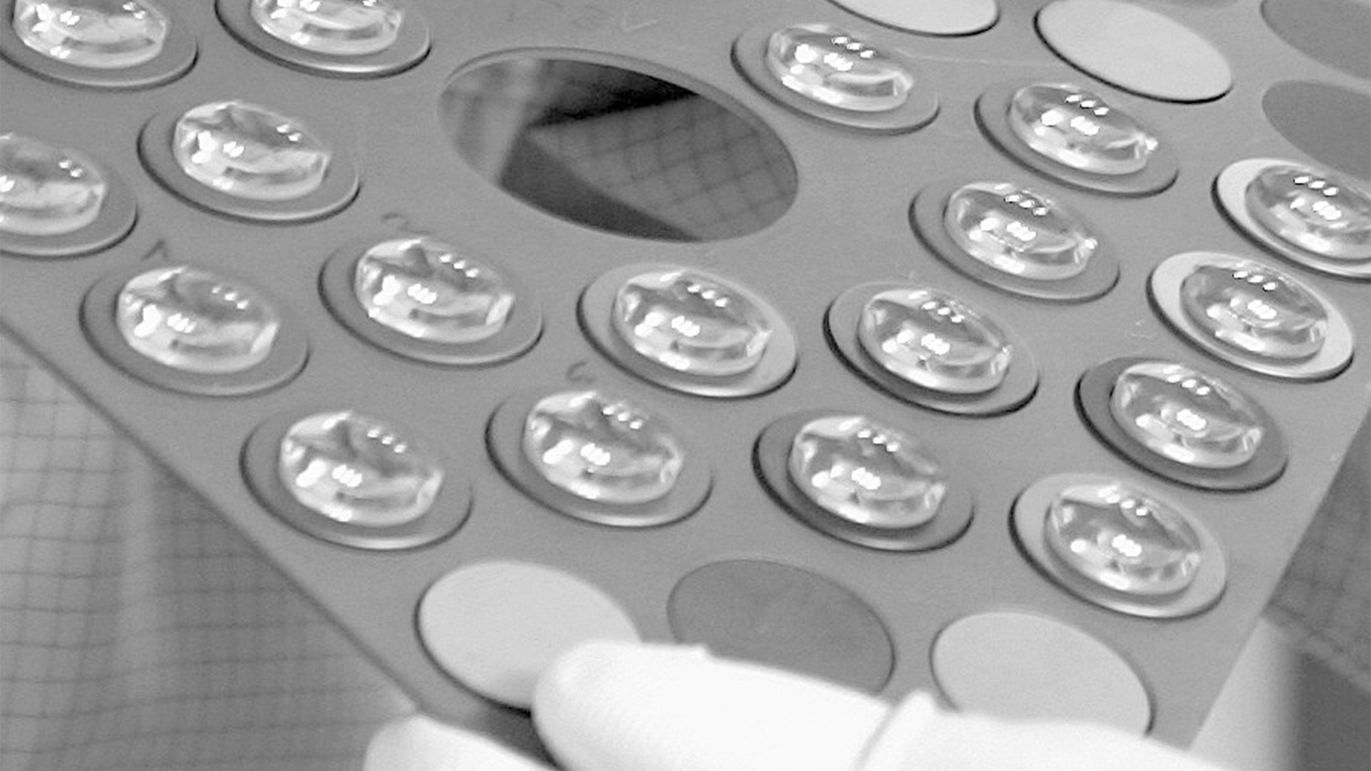
Fresnel lens, succession of concentric rings, each consisting of an element of a simple lens, assembled in proper relationship on a flat surface ...
Not all products or services are approved or offered in every market, and approved labelling and instructions may vary between countries. Please contact your local representative for further information.
When a beam of light is incident on a grating, each groove generates a diffracted wavelet. For each wavelength component in the incident beam, the constructive ...


 Ms.Cici
 8618319014500