
Collimating lens for light-emitting-diode light source based ... - collimated light source
Diabetic macular edema: In people with diabetes, blood vessels in the retina can become leaky. This causes swelling in a part of the retina called the macula.
Optical Coherence Tomographyppt
Diabetic retinopathy: If people with diabetes have excess blood sugar over a long time, it can damage the retina and its blood vessels.
What can anOCTscan detect
Cornea disease: The cornea is the clear front window of the eye. Damage to the cornea can make vision cloudy or out of focus.
OCT scans a beam of light to create 3D images that show the retina’s layers in microscopic detail. OCT can also image the optic nerve, which connects the eye to the brain.
Macular degeneration: In age-related macular degeneration, a part of the retina called the macula is damaged, causing loss of central vision.
OCTeye test procedure
OCTeye test price
OCT was co-invented by David Huang, an OHSU ophthalmologist and biomedical engineer. It’s now a standard test for finding conditions that can cause vision loss, often before patients notice symptoms. OCT is used in more than 30 million eye procedures each year.
The OHSU Casey Eye Institute is a leader in developing noninvasive imaging to help prevent vision loss. Dr. Huang, our associate director and director of research, is known worldwide for his vision research and inventions. He holds 42 patents and has won top prizes in vision research and biomedical engineering.
Determine the magnification capacity of the objective lens. The magnification is written on the side of the lens. Traditionally, the value could be 4x, 10x, 40x, or 100x. If you are not sure of the magnification power, check the manual. The objective lens is located on the rotating wheel just above the stage or platform where you place the microscope slide. In some instances the microscope may have only one lens, but generally it has three to four.
OCTeye test side effects
Determine the magnification strength of the ocular lens. This should be written on the outside of the eye piece, but if it is not look in the manual. Generally speaking the ocular lens magnifies 10x.
Optical coherence tomography creates highly precise 3D images. It is most commonly used to image the retina in the back of the eye.
Smith, Bruce. How To Calculate Magnification On A Light Microscope last modified March 24, 2022. https://www.sciencing.com/calculate-magnification-light-microscope-7558311/

Optical coherence tomographymachine
Glaucoma: Glaucoma damages the optic nerve, which connects the eye to the brain. It commonly causes loss of peripheral vision.
OCTeye test results
Optical coherence tomographyangiography
Eye disease in babies: Babies born before the 37th week of pregnancy or at a very low weight are at risk for a condition called retinopathy of prematurity. This happens when abnormal blood vessels grow in the retina.
OCT is a common eye care test. It is used when you may be at risk for eye disease or when you already have an eye condition.
Smith, Bruce. "How To Calculate Magnification On A Light Microscope" sciencing.com, https://www.sciencing.com/calculate-magnification-light-microscope-7558311/. 17 April 2018.
Oregon Health & Science University is dedicated to improving the health and quality of life for all Oregonians through excellence, innovation and leadership in health care, education and research.
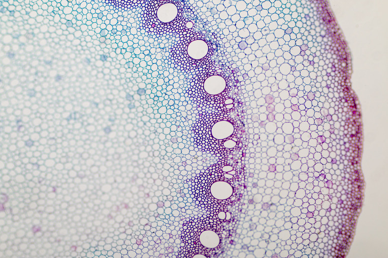
Compound light microscopes use a series of lenses and visible light to magnify objects. The magnification allows the user to view bacteria, individual cells and some cell components. In order to calculate the magnification, the power of the ocular and objective lenses is needed. The ocular lens is located in the eye piece. The scope also has one to four objective lenses located on a rotating wheel above the platform. The total magnification is the product of the ocular and objective lenses.
OCT of the eye is noninvasive, meaning it does not enter or even touch the eye. Standard OCT uses invisible infrared light, so it is more comfortable than imaging that uses visible light.
Smith, Bruce. (2018, April 17). How To Calculate Magnification On A Light Microscope. sciencing.com. Retrieved from https://www.sciencing.com/calculate-magnification-light-microscope-7558311/
To calculate the total magnification of the compound light microscope multiply the magnification power of the ocular lens by the power of the objective lens. For instance, a 10x ocular and a 40x objective would have a 400x total magnification. The highest total magnification for a compound light microscope is 1000x.
Optical coherence tomography helps diagnose, treat and manage the eye diseases that are the leading causes of blindness.


 Ms.Cici
 8618319014500