
CGI Office Locations - tech solutions gloucester
Typesof objectivelenses
There are different types of microscopes and each of these has different purposes of use. Some are suitable for biological applications, while others are used in educational institutions. There are also microscope types that find application in metallurgy and studying three-dimensional samples.
In addition to these core components, some objectives include a spring-loaded retractable assembly to protect the front lens elements and specimen from collision damage.
The depth of field in a microscope is defined as the distance from the nearest object plane in focus to the farthest plane in the same focus. In microscopes, the depth of field is very short and is measured in units of microns.
Rebecca holds a bachelor's degree in journalism from Endicott College and writes about trends and technologies in science and industry. She works closely with Evident engineers and scientists to write pieces about the latest laser scanning, super-resolution, multiphoton, upright, stereo, and inverted microscope systems, as well as leading-edge optics, cameras, and software. Follow her work to learn about Evident's latest for numerous applications, including cytology, pathology, education, and more.
The basic difference between low-powered and high-powered microscopes is that a high power microscope is used for resolving smaller features as the objective lenses have great magnification. However, the depth of focus is greatest for low powered objectives. As the power is switched to higher, the depth of focus reduces.
Knowledge is power when it comes to objective lenses. Many objective designs require you to make a tradeoff in one area of performance when you improve another. However, advances in objective technology enable our latest optical designs to overcome this common limitation.
Function ofstage inmicroscope
A compound microscope is defined as the type of microscope that has more than one lens. It has a combination of lenses and two optical parts known as an objective lens and an eyepiece or ocular lens. The magnifying power of the compound microscope is given as:
Low powerobjective microscope function
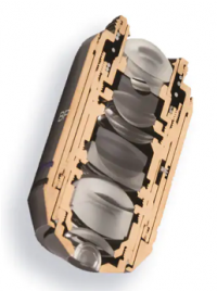
The objective lens is one of the most important parts of a microscope, since it determines its basic performance and function. Yet, these precision pieces of optical equipment are often not well understood.
Here, we break down the anatomy of an objective lens into easy-to-understand terms and discuss the common parts that make up an objective.
The scanning probe microscope has a probe tip that is mounted on the end of a cantilever. The tip is so sharp that it can move precisely and accurately across the surface of the sample scanning every atom. The tip is placed close to the surface of the sample, such that the cantilever experiences a deflection due to forces. This deflection distance is measured by the laser. The final image after scanning is obtained on the computer.
Keep in mind, objectives with more optical corrections for aberrations and flatness typically contain many lenses. For instance, sophisticated plan-apochromatic objectives have about 15 lens elements—while common achromatic objectives contain significantly fewer lenses.
For instance, Olympus X Line objectives are packed with many ultra-thin convex and concave lenses to offer exceptional flatness, aberration correction, and numerical aperture in one lens system. The result? Bright, high-quality images throughout the field of view.
Function ofcondenser inmicroscope
The field of view in a microscope is defined as the diameter of the illuminated circle which is seen through the eyepiece. With an increase in the magnification, there is a decrease in the field of view.
Microscopeparts
While objective designs vary based on factors like their intended purpose, microscopy method, aberration correction, and manufacturer, all microscope objectives share some similar characteristics. Here are four common components to know:
An electron microscope is defined as the type of microscope in which the source of illumination is the beam of accelerated electrons. It is a special type of microscope with a high resolution of images as the images can be magnified in nanometers.
The scanning probe microscope is defined as the type of microscope that finds applications in industries where the examination of the specimen is done at the nanoscale levels. The study of a specimen’s properties, its reaction time and its behaviour when stimulated can be done with the help of a scanning probe microscope.
High powerobjective microscope function
A simple microscope is defined as the type of microscope that uses a single lens for the magnification of the sample. A simple microscope is a convex lens with a small focal length. The magnifying power of the simple microscope is given as
The depth of focus in a microscope is defined as the distance between the objective lens and the sample plane. The depth of focus varies from person to person and is also dependent on the quality of focus.
The working principle of a simple microscope is that when a sample is placed within the focus of the microscope, a virtual, erect and magnified image is obtained at the least distance of distinct vision from the eye that is held at the lens.
Objective lensmagnification
A stereo microscope works on the reflected light from the sample. The magnification of the microscope takes place at low power and hence, it is suitable for magnifying opaque objects. It is suitable for thick and solid samples because it uses light reflected from the sample. The magnification of the stereo microscope is between 20x and 50x.
The difference in vision between the two eyes is corrected with the help of diopter adjustment. Through diopter adjustment, the focus of the individual eyepiece can be done so that the eyes feel comfortable while observing the sample.
What isobjective lensinmicroscope
The working principle of the compound microscope is that the combination of lenses enhances the magnification of the sample. The sample is first viewed as a primary image in the tube and viewed again in the eyepiece.
A stereo microscope is defined as a type of microscope that provides a three-dimensional view of a specimen. It is also known as a dissecting microscope. In a stereo microscope, there are separate objective lenses and eyepiece such that there are two separate optical paths for each eye.

In this article, there are 5 such microscope types that are discussed along with their diagram, working principle and applications. These five types of microscopes are:
The metal used in an electron microscope is tungsten. A high voltage current is applied which results in the excitation of the electrons in the form of a continuous stream that is used as a beam of light. The lenses used in the electron microscope are magnetic coils. These magnetic coils are capable of focusing the electron beam on the sample such that the sample gets illuminated. As the flow of current increases, the strength of the magnetic lens increases. The electron beam flow is designed such that it cannot pass through the glass lens.


 Ms.Cici
 8618319014500