
Come and meet us at SPS Italia 2024 - KFI - sps italia 2024
201698 — Narrower your aperture, deeper the depth of field. Eg: DOF at F2.8 is shallow and at F11 is deep. Distance between the subject and the ...
35.5 mm. ISO. -. 450 microns. ISO. -. 31.5 mm inch 1/14. -. 425 microns. No. 40. 40 ... inch 7/8. 0.883 in. 300 microns. No. 50. 50 Mesh. 20 mm. ISO. -. 280 ...
Like any technology, understanding and refining refractive index will only advance. The glasses we have today will seem archaic in the future, simply because technology is always improving. By understanding the refractive index, scientists can look for more ways eyewear can be innovated.
Dark fieldmicroscopy
MENUHomeAbout usReno Office Carson City OfficeServicesContact LensesSHOP CONTACT LENSESEye Glass LensesFramesBlogContact Us
If you’ll recall from our last blog, we dipped a little into the science behind choosing the right lens materials. Each material has something unique to offer and has its own properties, but one of the primary ways they can be deemed good enough to meet the standard of prescriptive lenses is by its placement on the refractive index.
Advantages and disadvantages ofdark field microscope
When taking a photo within the app, you can touch in two different areas to set the exposure and the focus separately. This is more advanced over most camera ...
Hilti Jobsite essentials - SL 6-22 LED construction light - Cordless LED construction light with all-day battery life, rotating head and mounting options ...
Dark field microscopeuses
The light microscope, or optical microscope, is a microscope that uses visible light and a system of lenses to magnify images. These days there are many complex designs of them which have been developed with the aim of improving resolution and sample contrast.
Image ofdark field microscope
DIC creates contrast in a specimen by creating a high-resolution image of a thin optical section. With differential interference contrast microscopy, two closely spaced parallel rays are generated and made to interfere after passing through an unstained sample. The background is made dark and the interference pattern is particularly sharp at boundaries. Specimens will appear really bright in contrast to the dark background.
Mar 29, 2023 — The first wave of the digital revolution promised that new technologies ... digital societies in which technology advances democratic principles ...
A quick experiment at home: fill a drinking glass with water and then dip a pencil into it. You’ll notice by looking at the glass from the side how the pencil in the water seems disconnected from the pencil above the water. It’s the same pencil, but light bends in water, causing the pencil to look different. This is refraction!
A polarising microscope is an optical microscope composed of a detector, lenses and polarising filters. Its process includes illumination of the sample with polarised light and is useful for better visualisation and understanding of birefringent materials (materials that have two different refractive indices). This microscope is operated through the use of a polarized filter can be turned and fixed in the light path beneath the specimen, usually below the stage. This particular device is known for its anti-reflective properties which is deemed essential for deep analysis of an isotropic particles that requires high integrity of light transmission.

Fluorescence microscopy is done with an optical microscope that uses a mercury arch lamp as a source of UV light. The microscope will also comprise excitation filter, dichromatic mirror and an emission filter. Fluorescence, used to observe the specimen, begins where a molecule absorbs light of high frequency and emits light of lower frequency. Fluorescence microscopy uses reflected light. In a fluorescence microscope the light source travels in a different trajectory than in the basic light microscope. An advantage of fluourescence microscopy is that it can be used to detect and visualise multiple fluorescent molecules e.g. cells glowing as they are doing their work. iOLight sell a microscope for mobile digital fluorescence microscopy, which is also great for field microscopy uses.
So, no, you probably don’t need high index lenses. But when we examine your eyes and begin testing prescriptions, it’s good for us to know what materials are available for your ultimate comfort. Afterall, you’re going to be wearing your glasses all the time! If they aren’t comfortable, then something’s not right. But thanks to improvements in lens materials and having high index options, we can provide you with a best-fitting option that is both comfortable to wear and offer better vision.
Fluorescencemicroscope
First, higher doesn’t always mean the lens is better. The refractive index is only one part of the equation. Other things like Abbe values (the amount of distortion or aberrations) and optics play a vital role in determining if any given material will perform more or less than any other material.
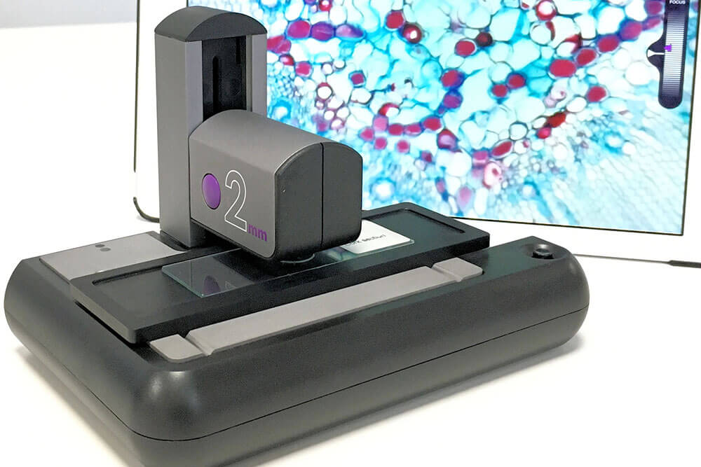
ioLight has invented a portable microscope, with a resolution of better than 1μm, which produces beautiful pictures of animal and plant cells and displays them directly onto your tablet or mobile phone.
201338 — Nearly all "backlit" LED LCDs use this method. The LEDs are arrayed on the back of the TV, facing you, but there is no processing to dim them ...
Remember that car mileage metaphor from before? That’s a very generalized explanation. Honestly, it goes even beyond eyewear! The refractive index is a measurement of how light travels through any given material.
Phase contrast microscopy
Light is refracted differently in certain materials. The way it refracts in water is different from how it refracts in plastic or glass. It all depends on the material and the density of said material of what the light is hitting. The refractive index measures how efficient this refraction is. The higher the number on the index, the slower light travels through the medium, the more the light is bent, and ultimately – the more efficient the refraction is. For the use in eyewear, a higher score on the index means less material needs to be used to achieve a desired effect.
Sometimes a higher index isn’t always the best solution, either. Sure, it’s good to know it exists, but high index lenses aren’t really necessary for most prescription lenses. CR-39, Polycarbonate, and Trivex all offer spectacular lens options without having a 1.60 score like our high index lenses do. It’s all about finding what fits best, without overdoing it.
It seems like a small change, going from 1.5 to 1.6. But that one-tenth decimal is the difference between having coke bottle glasses and something more modern and lightweight. This is especially nice for people who require a stronger prescription. Stronger prescriptions traditionally mean the patient needs thicker glasses in order to see 20⁄20. But with these improvements in the refractive index, someone with a strong prescription can achieve 20⁄20 vision with thinner and lighter glasses.
Type ofmicroscope
If you disable this cookie, we will not be able to save your preferences. This means that every time you visit this website you will need to enable or disable cookies again.
Fluorescence microscopy is done with an optical microscope that uses a mercury arch lamp as a source of UV light. The microscope will also comprise excitation filter, dichromatic mirror and an emission filter. Fluorescence, used to observe the specimen, begins where a molecule absorbs light of high frequency and emits light of lower frequency. Fluorescence microscopy uses reflected light. In a fluorescence microscope the light source travels in a different trajectory than in the basic light microscope. An advantage of fluourescence microscopy is that it can be used to detect and visualise multiple fluorescent molecules e.g. cells glowing as they are doing their work. iOLight sell a microscope for mobile digital fluorescence microscopy, which is also great for field microscopy uses.
This type of microscope was developed in response to drawbacks with fluorescence microscopes (principally that they use high intensity UV light which means the samples are continuously exposed to it, causing photo bleaching and blurring in some samples). Two major modifications were made to address this downside: use of laser light instead of mercury arch lamp and images taken using a digital camera with a pin hole. The pin hole functions to allow light of only one focal plane to be focused on the digital camera. A laser beam focused and scanned over the sample produces 3D and 2D images therewith.
Not only that, but understanding the index means current eyewear lenses can be picked out by a patient and favored for their prescription. It allows for us to make better recommendations and more accurate readings for what a patient needs to gain perfect vision.
Dark field vs bright field microscopy: Bright field microscopy uses the most basic and the common type of optical microscope. Bright field microscopes usually have many components and the light sources used are either a halogen lamp or LED. This type of microscope tends to have low contrast owning to the biological samples transmitting most of the light. Staining if often required to combat this problem, which comes with the disadvantage that live imaging is difficult due to staining killing the cells. Dark field microscopy is generally preferred therefore over light field. With a dark field microscope a special aperture is used to focus incident light meaning the background stays dark. The light does not pass directly through the sample being studied. Instead light is reflected off the specimen, making it appear to be emitting light. Brightfield microscopy shows clear magnification while the dark field image shows minute details.
Phase contrast microscopes were invented to combat the problem of live cell study with a bright field microscope. Phase contrast microscopy is an optical microscopy technique in which phase shift is converted into change in amplitude/intensity of light. The phase shifts when light travels through dense medium and its velocity decreases, concurrently there is a shift in the phase. When the two waves meet at a certain point it will result in a destructive interference, decreasing amplitude and thereby density. Phase contrast microscopy is useful for looking at specimens that are both colourless and transparent.
Edmund Optics is exhibiting at the Automate Trade Show in booth # ... Edmund Optics. 101 East Gloucester Pike Barrington, New Jersey 08007 United ...
Lightfieldmicroscopy
View our Product LL500Z PB WH en. Guaranteed performance, thinking ahead.
Optics and. Ergonomics. Thru-the-Lens (TTL) Telescopes · Truth in Magnification · Precision Coated Optics · Surgical Telescopes / Loupes · The Designs for ...
Feb 20, 2022 — Light diffusers can be large sheets of diffusion placed at a distance from a light or it can be a single sheet of diffusion placed on the barn ...
This website uses cookies so that we can provide you with the best user experience possible. Cookie information is stored in your browser and performs functions such as recognising you when you return to our website and helping our team to understand which sections of the website you find most interesting and useful.


 Ms.Cici
 8618319014500