BASLER VISION TECHNOLOGIES BIP21300CDN CCD ... - basler vision camera
What is microscopyin microbiology
It has a low contrasting capacity, low optical resolution, requires staining and has a limited magnification of around 1300X.
Who invented microscope

We use cookies to enable essential website operations and to ensure certain features work properly, like the option to log in or add a product to your shopping cart. These cookies are always enabled, otherwise, you can't view the website or shop online.
Since 1895, Hobbies have been supplying model makers and enthusiasts with a wide range of quality model kits, accessories, tools, components and guidebooks.
What is microscopyin biology
Refractive Index can be defined as velocity of light in a vacuum to velocity of light in a medium (substance). Simply it is the measure of bending of a light ray when passing from one medium to another.
What is microscopypdf
Bright Field Microscopy is the simplest and the most common type. A simple or compound light microscope is used in this technique. It uses transmitted visible light to develop magnified images.
JavaScript seems to be disabled in your browser. For the best experience on our site, be sure to turn on Javascript in your browser.
Fluorescence Microscopy is a microscopy technique that uses a fluorescent microscope with a UV light source. It is widely used in detecting antigens, antibodies, and other macromolecules.

Types ofmicroscopy
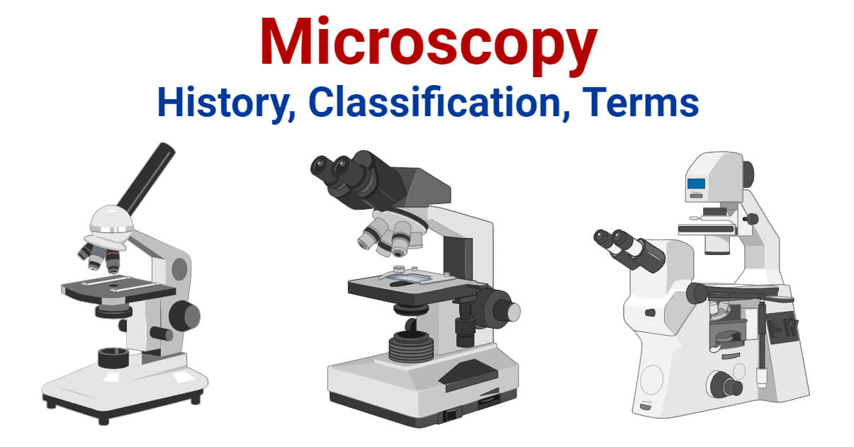
Microscopy can simply be understood as the ‘use of microscope’. Microscopy can be defined as the scientific discipline of using microscopes for getting a magnified view of objects that can’t be viewed by naked eyes.
Dark Field Microscopy uses dark-ground microscopes. The reflected light is used, instead of transmitted light, to form a magnified image.
Magnification is the process of producing an enlarged image of a specimen by using a lens system. In a microscope, magnification can be computed by calculating the product of the magnification power of the eyepiece by the magnification power of the objective in use.
Parts of microscope
Resolution can be defined as the shortest distance between two points on a specimen that can be distinguished by a microscope in its image. It is the ability of a microscope to distinguish details on a specimen.
Scanning Probe Microscopy is a microscopy technique that uses a physical probe to scan specimens and form magnified images. This measures the surface features of the specimen.
What is microscopyin science
Hobbies and our advertising partners (including social media platforms like Google, Facebook, and Instagram) use cookies to provide personalised offers. If you don’t accept this tracking, you will still see Hobbies advertisements on other platforms at random.
Electron Microscopy is a microscopy technique that uses a beam of electrons to develop a highly magnified image of microscopic samples.
It is a very important tool in biology and nanotechnology. In microbiology, it is one of the most important tools used in observing microbial cells. Medical sectors, pathology, histology, molecular biology, and cytology are in great debt of microscopy.
Optical Microscopy (Light Microscopy) is the microscopy technique that uses transmitted visible light, either natural or artificial, for developing the image of an object. It is the most common type of microscopy. It is further classified into several groups;
We use functional cookies to analyse how our website is being used. This data helps us to discover errors, provide insights for advertising analysis and affiliate marketing.
Differential Interference Contrast Microscopy is a newer microscopy technique used in unstained and transparent samples to enhance the contrast of their image.
In Phase Contrast Microscopy, the phase-contrast microscope is used. It converts phase shifts into differences in intensity of light-producing more contrast images.
What is microscopyused for
Magnifying Power is defined as the ratio of the angle subtended by the image at the eye to the angle subtended by the object at the eye when placed at a minimum distance of distinct vision.
By signing up you are agreeing to receive exclusive offers, updates and news from Hobbies by email. You can opt out of receiving emails at any time
Confocal Microscopy is a newer microscopy technique that uses a focused laser beam. It is used to get high-resolution 3-D images of biological samples.
We use cookies to enhance your browsing experience, serve personalized ads or content, and analyze our traffic. By clicking "Accept All", you consent to our use of cookies. Cookie Policy.


 Ms.Cici
 8618319014500