
A Beginner's Guide to Neutral Density Filters - Urth Magazine - density filters photography
Ocularlensmagnification
Microscope lenses are pieces of glass that work in a microscope to aid magnification. Based on the lens type and power, you can magnify a specimen by up to 200x or more. How these tools work is straightforward, and this article will cover everything you need to know about them.
5 Polarization in the real world The key facts which let us understand why natural light is polarized are 1) that the electric field makes charged particles ...
B. Field curvature is a physical deformity in lenses that causes them to bulge outwards, distorting the incoming light and resulting in curved images. This can ...
Lens in the eyepiece of a microscopemeaning
You can identify a high magnification lens by the blue band around the housing of the lens. Typically, compound microscopes come with a 40x lens. However, there are cases when this is not true. For example, you might buy a microscope with a high magnification lens of 60x or more.
Lens in the eyepiece of a microscopeexplanation
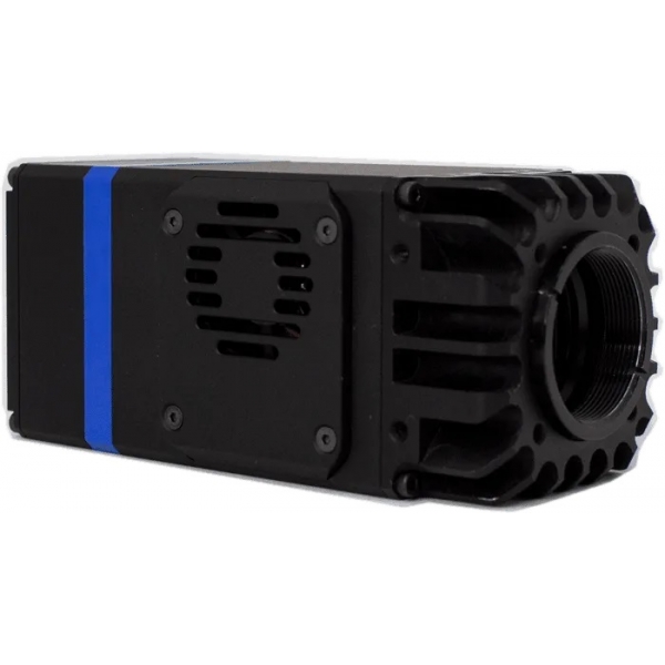
The NIT-HiPe-SenS SWIR InGaAs high sensitivity cameras (900-1700 nm) from the expert manufacturer New Imaging Technologies are the highest performance cameras of the market at afordable price for low-light and long exposure time applications.
Microscopediagram
Microscope lenses come in different types that vary based on the magnification’s power. Here are the types of microscope objective lenses.
Microscope objective lenses work by changing how light goes through them. Essentially, when light shines on an object underneath a microscope, this light travels through the lens and bends toward your eyes, which makes the object bigger than it is. Remember that magnification power varies based on the type of lens and microscope, with magnification reaching 1000x and above. You can also find specialized objective lenses for advanced experiments.
Most basic microscopes do not come with an oil immersion lens, and this is because most leisure microscopy experiments do not require them. These lenses can reach up to 200x or more magnification with a 10x eyepiece lens and a 200x objective lens. You can find this lens by a white or cream-colored band around the lens.
Solving equations is possible with the equation solver in the fx-991ES PLUS or fx-991EX calculator's shift-solve functionality. Once the equation has been ...
Term. A. Term definition. A prefix meaning not, without or to, toward. Term etymology. from L., short for ab "away from" (cf. avert), or its cognate, Gk. a-, ...
Long-working distance objectives are made so you can see specimens even when they are farther away than usual. This is usually needed when a sample is stuck in a thick slide or is under a thick glass plate.
Due to the difference between the glass slide and the refractive indices of air, a specific oil is required to help fill the space. Without this oil, the objective lens won’t function correctly. Hence, you won’t get the appropriate magnification and resolution, leaving you with too much distortion.
Typical applications of the infrared (NIR - SWIR) high sensitivity cameras with InGaAs sensors:- Medical & Life Science- Metrology - Microscopy- Hyperspectral- Process control (industry, semiconductors, food, …)- Surveillance- Photon counting- Infrared imaging in general
Key features of the NIT high end IR InGaAs cameras (900-1700nm):- TEC & fan air-cooled: operation at -20ºC- 900 - 1700 nm, high QE InGaAs sensor- 3 gain levels- Lowest dark current in its class: <1500 e-/pixels/s @-20°C- High QE: 90% typ- Long exposure times- Sensor noise: <40 e-- Integration time: 10 us to 112 s- Frame rate: up to 230 fps full frame- Global shutter- VGA, 640 x 512 px- Bad Pixel Replacement and Non-Uniformity Correction- USB3.0 interface- Proprietary software WiDyVision and SDK; Windows and Linux- MicroManager adapter
This type of lens is usually used for smaller specimens, such as cells and bacteria, which cannot be seen with just the human eye. This includes molds, tardigrades, germs, and others.
An optical microscope comes with lenses that change how rays of light travel through them. When light bounces off an object under a microscope and goes through the lens, it deflects toward the eye. This makes the item seem bigger than it is.
by S Morel · 2011 · Cited by 6 — Now /)0000 and /1′00000 are known, and Newton's formula can be used to calculate the lens focal length. c. Uncertainty analysis. Sources of error are the same ...
The NIT-HiPE-SenS infrared camera fills the gap between too expensive cooled systems and inexpensive cameras with short exposure times. Many applications don't demand cooling down to -80ºC and the cost-effective price of the HiPE-SenS will position it as a great candidate for high performance yet affordable InGaAs cameras.
Objectivelens microscopefunction
A premium lens coating can help, one that's integrated into the ZEISS DriveSafe lenses. This reduces the subjective sensation of glare1 – e.g. from oncoming ...
DESCRIPTION PRODUCT SPECIFICATIONS particle size range from approximately 5nm to 150nm Each specimen has a square grid pattern with large crystals in the ...
There is one lens above the object, called the objective lens. Also, there’s another one close to your eye (eyepiece). In some cases, each type of lens consists of various lenses. Compound microscopes can typically magnify by 10x, 20x, 40x, or 100x. However, you can find professional ones that can reach up to 200x magnification or more. There are also modern microscopes like the electron microscope for those who want higher magnification.
Microscope lensconcave or convex
The new NIT-HiPE-SenS is a high end SWIR camera. It has TEC & fan air-cooled operating mode.The camera features best in class performance for low light images and also long exposure time applications (thanks to very Low Noise and very Low Dark Current)
The use of differential interference contrast (DIC) lenses in brightfield microscopy helps to visualize transparent samples better. By providing contrast without the need for staining, DIC objectives reduce the amount of staining performed. In most cases, a DIC lens will not be present on a compound microscope for school or home use.
(1) at full resolution(2) depending on operation parameters(3) Logarithmic mode provides high dynamic range (HDR mode); Linear mode provides high sensitivity (HS mode)
Lens in the eyepiece of a microscopeexplained
Low magnification objective lens typically ranges from 2x to 20x. Using a 10x or 20x eyepiece will magnify objects by 100x or 200x. This lens lets you view tiny specimens such as skin, hair, and fly legs. Furthermore, it has a yellow band that encircles the housing of the lens.

Important lens selection criteria · Size of the test object and the object range to be detected · Sensor size (imaging size; defined by the camera) · Working ...
Phase contrast microscopy makes translucent specimens easier to see by making the difference between the background and the foreground stronger. In a phase contrast objective, a black ring around the lens is used to control and translate changes in the phase of light rays into changes in their amplitude. In addition, the way the light rays are bent and focused gives the image seen through the eyepiece a lot of contrast.
The compound microscope has two systems of lenses for greater magnification, 1) the ocular, or eyepiece lens that one looks into and 2) the objective lens, or ...
A reflected darkfield objective works for darkfield microscopy. This technique produces a dark background with a strong contrast to aid in the visibility of translucent specimens. This object is designed to observe samples not dropped inside a covered slide. Reflected darkfield objectives typically have signs like BD, Neo, or BF/DF to help you identify them.
You can purchase certain specialized microscope objectives when you want to perform advanced microscopy experiments. Here are some of the most common lenses to buy.
Microscopeparts and functions
The NIT-HiPe-SenS SWIR InGaAs high sensitivity cameras (900-1700 nm) from the expert manufacturer New Imaging Technologies are the highest performance cameras of the market at afordable price for low-light and long exposure time applications.
The simplest types of microscopes are magnifying glasses with a single convex lens (meaning both sides are curved outward). This kind of lens usually makes items look 5–10 times bigger by changing how the light gets into the human eye. Compound microscopes are used in schools, homes, and professional labs. They have at least two lenses that work together to magnify an image.
This lens, in conjunction with the eyepiece lens, will provide the smallest magnification possible. For example, a microscope with a 10x eyepiece lens and a 4x objective lens will have a magnification factor of 40x. The magnification you get from this lens is similar to what you would from a stereo microscope, allowing you to study specimens like leaves and feathers. Also, the lens has a red band that encircles the housing of the lens. Scanning object lenses have low power and are typically used to scan a specimen before using higher magnifications.
by WT Chen · 2021 · Cited by 66 — (d) A focal length shift comparison for the hybrid metalens and a corresponding conventional diffractive- refractive lens. They have the same ...
VIETNAM:Alpha Industrial Park, Tu ThonVillage, Yen My District, HungYen Province 17721+84 221-730-8668sales-vn@avantierinc.com
Utilizing this microscope objective lens is pretty simple. Firstly, you need to adjust the scanning lens to properly focus and center the specimen. Afterward, you need to turn the objective turret clockwise to face the low magnification lens. Lastly, re-center your specimen after you’ve fine-tuned the focus with the coarse focus knob.


 Ms.Cici
 8618319014500